
Observation of optical gain from aqueous quantum well heterostructures in water†
Savas
Delikanli‡
ab,
Furkan
Isik‡
 b,
Emek G.
Durmusoglu
a,
Onur
Erdem
b,
Farzan
Shabani
b,
Betul
Canimkurbey
bc,
Satish
Kumar
b,
Hamed
Dehghanpour Baruj
b and
Hilmi Volkan
Demir
*ab
b,
Emek G.
Durmusoglu
a,
Onur
Erdem
b,
Farzan
Shabani
b,
Betul
Canimkurbey
bc,
Satish
Kumar
b,
Hamed
Dehghanpour Baruj
b and
Hilmi Volkan
Demir
*ab
aLuminous! Center of Excellence for Semiconductor Lighting and Displays, School of Electrical and Electronic Engineering, School of Physical and Mathematical Sciences, School of Materials Science and Engineering, Nanyang Technological University, 50 Nanyang Avenue, Singapore 639798, Singapore. E-mail: hvdemir@ntu.edu.sg; volkan@bilkent.edu.tr
bDepartment of Electrical and Electronics Engineering, Department of Physics, UNAM – Institute of Materials Science and Nanotechnology, Bilkent University, Ankara 06800, Turkey
cSerefeddin Health Services Vocational School, Central Research Laboratory, Amasya University, Amasya 05100, Turkey
First published on 5th September 2022
Abstract
Although achieving optical gain using aqueous solutions of colloidal nanocrystals as a gain medium is exceptionally beneficial for bio-optoelectronic applications, the realization of optical gain in an aqueous medium using solution-processed nanocrystals has been extremely challenging because of the need for surface modification to make nanocrystals water dispersible while still maintaining their gain. Here, we present the achievement of optical gain in an aqueous medium using an advanced architecture of CdSe/CdS@CdxZn1−xS core/crown@gradient-alloyed shell colloidal quantum wells (CQWs) with an ultralow threshold of ∼3.4 μJ cm−2 and an ultralong gain lifetime of ∼2.6 ns. This demonstration of optical gain in an aqueous medium is a result of the carefully heterostructured CQWs having large absorption cross-section and gain cross-section in addition to inherently slow Auger recombination in these CQWs. Furthermore, we show low-threshold in-water amplified spontaneous emission (ASE) from these aqueous CQWs with a threshold of 120 μJ cm−2. In addition, we demonstrate a whispering gallery mode laser with a low threshold of ∼30 μJ cm−2 obtained by incorporating films of CQWs by exploiting layer-by-layer approach on a fiber. The observation of low-threshold optical gain with ultralong gain lifetime presents a significant step toward the realization of advanced optofluidic colloidal lasers and their continuous-wave pumping.
Introduction
Colloidal semiconductor nanocrystals (NCs) are highly promising as optical gain media thanks to their size-dependent spectral tunability, solution processability and compatibility to various matrices.1,2 After the first demonstration of amplified spontaneous emission (ASE) from their close-packed solid films two decades ago,3 semiconductor NCs in various geometries and compositions have been utilized and investigated as gain media.4–6 Although in-solution optical gain can be highly advantageous owing to its potential practical applications, including sensing and detection,7–9 there are only a few reports and demonstrations of in-solution optical gain and a limited number of in-solution lasing compared to optical gain and lasing studies using close-packed films of NCs.10–15 This is primarily due to the low concentration of gain media possible in solution which is constraining the realization of ASE. The need for high concentration is principally a consequence of the difficulty of achieving ASE build-up time, which strongly depends both on the gain cross-section and the concentration of the gain material in-solution,3 faster than the Auger lifetime which is unfortunately very efficient in nanocrystals unlike bulk structures.3,4 Nevertheless, solution-based gain using colloidal nanocrystals is very valuable because of its greater photostability thanks to the continuous renewal of the gain medium because of the constant movement of nanocrystals and ease of incorporation into various optical cavities. Such solution-based gain medium can be easily incorporated into optofluidic lasers for sensitive detection of chemicals and biological materials and on-chip imaging.Recently, colloidal quantum wells (CQWs) have emerged as a favorable optical gain medium since they possess large absorption cross-sections,16–18 with continuously tunable emission,19,20 suppressed Auger recombination rates,17,21 low optical gain thresholds,17,22,23 long gain lifetimes,17 and large gain cross-sections.14,15 Especially, their large gain cross-section makes them excellent candidates for in-solution lasing by supporting the gain condition and feasible levels of gain coefficients in solution.14 Recently, ASE in solution and lasing in microfluidic channels have been demonstrated using CQWs dispersed in non-polar solvents with record low thresholds, almost one order of magnitude better than the previous lasing demonstrations from colloidal nanocrystals.11,14,15 This low threshold optical gain from these CQWs is significantly important for realization of stable high-quality lasers in microfluidic devices for numerous applications, including biological and chemical sensors, flow cytometry and the development of advanced lab-on-chip (LOC) devices. In addition, recently introduced water-based semiconductor CQWs, which can be obtained by synthesizing them in organic solvents and then transferring them to water via ligand exchange, present an attractive platform for optical gain applications thanks to their photostability, highly efficient emission and soft confinement potential owing to their gradient alloyed shell.24 Although these exciting aqueous CQWs present an extraordinary platform for in-solution optical gain, their optical gain properties have never been explored to date.
In this study, we demonstrate the achievement of optical gain in an aqueous medium using an advanced heterostructure of CdSe/CdS@CdxZn1−xS core/crown@gradient-alloyed shell colloidal CQWs employing ultrafast transient absorption technique with a threshold of 3.4 μJ cm−2. These nanocrystals exhibit a long gain lifetime of 2.6 ns which makes them superior candidates for achieving optical gain under CW and quasi-CW pumping. This extremely long gain lifetime can be attributed to the smooth confinement potential suppressing the Auger recombination. We also demonstrated in-solution ASE from these carefully heterostructured aqueous colloidal nanocrystals using a capillary tube with a threshold of ∼120 μJ cm−2. In addition, we show whispering gallery mode (WGM) laser with a threshold of ∼30 μJ cm−2 obtained by incorporating films of our CQWs on a fiber using layer-by-layer approach by taking advantage of their 2D geometry, short mercaptopropionic acid (MPA) ligands allowing higher packing density of CQWs and carboxylic end groups of their ligands causing hydrophilicity. The layer-by-layer approach leads to a uniform film on the lateral surface of the fiber and hence a much lower lasing threshold in this work compared to the previous report25 in which films were obtained by assistance of capillary force. In this WGM laser, the lasing is strongly guided along the fiber and hence exhibits a spatially coherent emission. This solution-processed architecture of aqueous CQWs with their extraordinary properties makes excellent gain media for the design of CW-pumped lasers and the achievement of much needed optical gain in water is a major step forward for development of advanced optofluidic lasers in bio-compatible environments.
Results and discussion
In this work, we synthesized CdSe/CdS@Cd1−xZnxS core/crown@gradient-alloyed shell (C/C@GS) CQWs by growing 4 monolayers of Cd1−xZnxS shells on 4 monolayer thick CdSe/CdS core/crown CQWs using colloidal atomic layer deposition (c-ALD) technique.24,26 Then, we made them water dispersible by passivating their surface with MPA via ligand exchange as described in our previous work.24 Details of the syntheses and ligand exchange procedures are presented in ESI.† The quantum yield of these aqueous C/C@GS CQWs is approximately 90%. In this architecture, Cd1−xZnxS gradient-alloyed shells grown on the seed CdSe/CdS core/crown CQWs provide a soft confinement potential and hole wavefunction is largely localized in the core region due to the large band-offset while electron is largely relaxed into the Cd1−xZnxS shells layers as a result of small conduction band-offset and smaller effective mass.17,24 This spatial difference between the electron and hole wavefunctions generates a quasi-type-II band alignment. The schematic of C/C@GS CQWs together with the band-offsets of the heterostructure is presented in Fig. 1a. A representative transmission electron microscopy (TEM) image of C/C@GS CQWs is provided in Fig. 1b and energy dispersive spectroscopy (EDS) maps of cadmium, selenium, sulfur, and zinc from a single CQW (shown in the inset) are presented in Fig. 1c. As can be seen in here, selenium is located only at the core but sulfur, zinc and cadmium are present all over the CQW as expected from alloyed shell of CdZnS in these CQWs.24 The absorption and photoluminescence (PL) spectra of aqueous CQWs are shown in Fig. 1d. The peaks appearing at 625 and 575 nm in the absorption spectrum are associated with the heavy-hole and light-hole excitonic transitions, respectively. The PL emission peak of these aqueous CQWs is located at 629.6 nm and the full-width-half-maximum (FWHM) of the PL is 19.6 nm. | ||
| Fig. 1 (a) A schematic of CdSe/CdS@CdxZn1−xS core/crown@gradient-alloyed shell colloidal quantum wells (CQWs) together with the band-offsets. (b) A representative transmission electron microscopy (TEM) image of C/C@GS CQWs. (c) EDS maps of cadmium, selenium, sulfur, and zinc from a single CQW (shown in the inset). (d) Absorption and photoluminescence spectra of the CQWs. | ||
Fig. 2a presents non-linear absorption spectra α = Δα + α0, where Δα is the absorption bleach and α0 is the absorption of the unexcited sample, parametrized with respect to the average number of generated excitons per CQW (given in the legend). In here, we used the absorption bleach spectra at ∼15 ps. The average number of photogenerated excitons per CQW 〈N0〉 is calculated by using N0 = f × σ, where f is the pump fluence and σ is the absorption cross-section. Absorption cross-section of our sample is 2.0 × 10⁻¹³ cm2 and was calculated by following a method explained in our previous work.16 The non-linear absorption spectra gradually decrease as we increase the pump fluence as can be seen in Fig. 2a and α becomes negative at the wavelengths within the proximity of the stimulated emission. This negative net absorption, α < 0, signifies the occurrence of optical gain.3 The optical gain occurs initially at 640 nm and then as the pump fluence is further increased, the peak of the net negative absorption slowly blue-shifts to 630 nm. This blue shift is a signature of stimulated emission from higher order excitons.27,28 In Fig. 2b, the absorption α of our aqueous CQWs at the ASE peak as a function of the average number of photogenerated excitons per CQW 〈N0〉 is provided. The change in the sign of α indicates the threshold for achieving stimulated emission and thus our threshold for stimulated emission is 〈N0〉 ≈ 1.4 excitons which corresponds to pump fluence of 3.4 μJ cm−2.
 | ||
| Fig. 2 (a) Nonlinear absorption spectra α = Δα + α0, where Δα is the absorption bleach and α0 is the absorption of the unexcited sample, as a function of the average number of generated excitons per CQW. Red-shaded region shows where optical gain occurs (α < 0) in this figure. (b) Nonlinear absorption α taken at t = 15 ps as a function of the fluence and average number of generated excitons per CQW. (c) 2D time–wavelength colored map of nonlinear absorption spectrum of our CQWs at pump fluence of 15 μJ cm−2, corresponding to 〈N0〉 ≈ 6 for the regions only under the optical gain. In (c) only the region exhibiting optical gain is shown and colored, the coordinates which are not under optical gain are colored as white. (d) −Δα(at 640 nm)/α0 as a function of the time at the pump fluence of 15 μJ cm−2. In (d) we extrapolate the experimental data after 1.95 ns, which is our experimental temporal window, by fitting with three-exponential decay function, shown in dotted red curve. | ||
We also investigated the material gain and gain cross-section of these gradient alloyed CQWs. Material gain g(pf, λ, t) at a specific pump fluence is given by
| g(pf, λ, t) = α(pf, λ, t) × μ(at 400 nm)/α0(at 400 nm), | (1) |
| gnet = γg × C − α, | (2) |
In Fig. 2c, the 2D time–wavelength colored map of nonlinear absorption spectrum of our CQWs is provided at pump fluence of 15 μJ cm−2, corresponding to 〈N0〉 ≈ 6 for only the region under the optical gain. In this figure, while the coordinates that exhibit optical gain is colored and shown, the coordinates which are not under optical gain is kept white. The spectral position where the maximum gain occurs red-shifts as the time passes as can be seen from Fig. 2c. Initially α is minimum (maximum in magnitude) at 630 nm, slowly shifting towards ∼640 nm (at ∼2 ns). This spectral red shift of α over time is due to the reduction in the number of excitons with time in the CQWs after their initial generation with the femtosecond pulse. Likely, this is because of the coulombic repulsive forces due to presence of multi-excitons. Then, as the number of excitons decreases with time, α eventually red-shifts spectrally. As can be seen in Fig. 2c, α still stays negative during our measurement window of 1.95 ns which means our gain lifetime is longer than 1.95 ns. In Fig. 2d, we present −Δα(at 640 nm)/α0 as a function of the time at the pump fluence of 15 μJ cm−2. Since our sample was still under the gain within our window of measurement, we extrapolated the experimental data after 1.95 ns by fitting three-exponential decay function. The fit is also included in Fig. 2d. Using our fit, we obtained a gain lifetime of 2.6 ns from our aqueous CQWs. This optical gain lifetime is longer than the previously reported gain lifetimes of colloidal quantum wells17,31,32 and comparable to the best reported gain lifetimes of charged quantum dots33 and quantum shells.34 Such elongated gain lifetime in this system can be attributed to the longer Auger lifetimes as a result of the smooth potential confinement provided by the gradient alloyed shell.4,35 The Auger lifetimes are strongly suppressed in finely graded confinement potentials owing to the significant reduction in the strength of the intraband transition.4,35
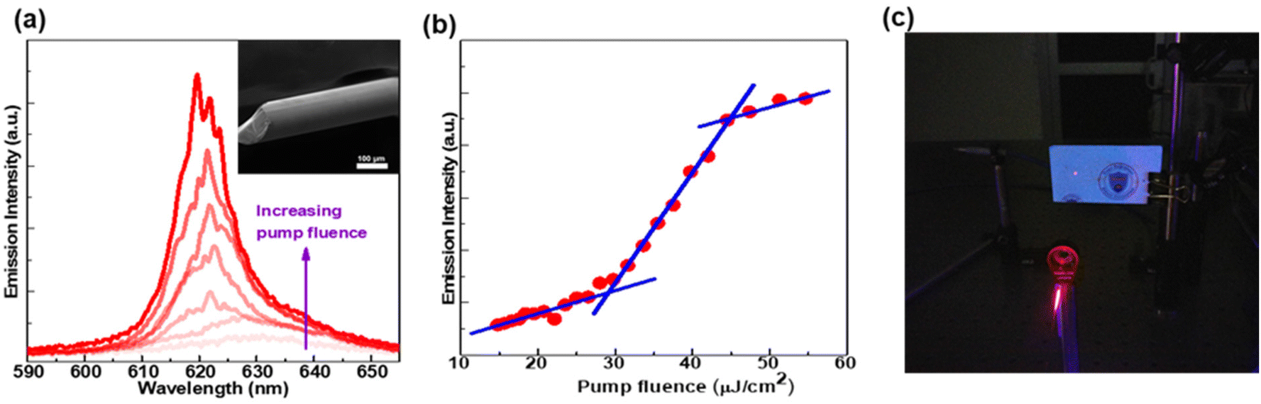
To explore the in-solution ASE performance of our CQWs, CQWs dispersed in water were placed into a capillary tube having a core size of 300 μm by the assistance of capillary force and then optically pumped with a femtosecond mode-locked laser (pulse width ≈ 120 fs) at 400 nm and 1 kHz repetition rate using a stripe geometry with the help of a cylindrical lens. Emitted photons were collected by using an optical fiber connected to a spectrometer. Pump-fluence dependent ASE spectra from our aqueous CQWs are presented in Fig. 3a. As presented in Fig. 3a, at low pump fluences only the spontaneous emission with a FWHM of ∼25 nm appears in the PL spectrum. However, as the pump fluence further is increased, a sharp ASE peak emerges at ∼641 nm with a FWHM of ∼10 nm for the pump fluences above the threshold and it becomes dominant as the fluence is further increased as can be seen in Fig. 3(a). The blue shift of the ASE can be attributed to the quasi-type-II nature of these CdSe/CdS@CdxZn1−xS core/crown@gradient-alloyed CQWs, which leads to repulsive interactions in the biexcitonic regime. It is worth mentioning that the degree of such quasi-type-II behavior can be well controlled and fine-tuned by the use of alloying and geometrical parameters, as previously demonstrated in CdSe/Cd1−xZnxS core/shell QDs, nanorods and CQWs.17,36,37 The emission intensity at the peak as a function of the pump fluence for the CQWs is presented in Fig. 3(b). The ASE threshold obtained in Fig. 3(b) from the super-linear increase of the emission intensity and Fig. 3(a) from the emergence of narrow ASE emission is ∼120 μJ cm−2, which is slightly higher than the previously reported ASE threshold of CQWs dispersed in toluene.15 This is likely due to the difference in the concentration of CQWs and possibly better optical confinement factor of the CQW solution in toluene which has a higher refractive index than water. This lower refractive index of water and low optical quality of the aqueous nanocrystals synthesized before are likely the reasons for the failure in achieving optical gain in water to date. Compared to the nanocrystals dispersed in non-polar solvents, such as hexane and toluene, the optical performance of the nanocrystals dispersed in water falls far behind.38,39
 | ||
| Fig. 3 (a) Pump-fluence dependent in-solution ASE spectra from aqueous CQWs in a capillary tube. Inset of (a) exhibits a schematic of CQW solution in the capillary tube optically excited using stripe geometry for ASE measurements. (b) Emission intensity at the peak as a function of the pump fluence for aqueous CQWs. The crossing point of the fitted red lines in (b) indicates the ASE threshold. (c) ASE intensity as a function of time. | ||
To study the stability of the light amplification in our aqueous CQWs, we pumped the sample above the ASE threshold for 4 h continuously. The stability test was performed under ambient conditions at a threshold of 190 μJ cm−2 using a femtosecond mode-locked laser having a pulse width of 150 fs and a 1 kHz repetition rate. We observed a decrease in the ASE intensity with time as presented in Fig. 3c. However, this decrease in the ASE intensity was almost recovered after stopping the optical pumping of the sample for an hour. This phenomenon can be attributed to the dissipation of the heat which accumulated under continuous pumping. As a result, the decrease in the intensity of the ASE under continuous pumping is reversible and largely due to the change in the temperature of the surrounding environment unlike irreversible damage seen in QD microdrop lasers as a result of high fluences needed for obtaining ASE leading photo-oxidation.12 Hence, our aqueous C/C@GS CQWs offer an effective architecture to considerably improve the optical gain stability problem of colloidal semiconductor nanocrystals thanks to their low in-solution ASE threshold and crown and shell layers for passivation and protection.
Owing to excellent optical gain performance of our engineered aqueous CQWs, we incorporated these CQWs as a gain medium into WGM laser. We obtained the WGM laser by depositing 6 monolayers of aqueous CQWs by layer-by-layer approach on a piece of coreless fiber having a diameter of approximately 125 μm by taking advantage of their 2D geometry and hydrophilicity owing to carboxylic end groups of their ligands. We achieved the deposition of CQWs on fibers by using positively charged poly(diallyldimethylammoniumchloride) (PDDA) linker molecules between each CQW layer as presented in our previous work.24 The layer-by-layer approach as shown in our previous work leads to uniform film deposition, which is important for lasing applications and other opto-electronic applications, such as light-emitting diodes. Briefly, the surface of the fiber was made hydrophilic by applying oxygen plasma and after that it was dipped into a PDDA linker solution for the attachment of linker molecules to the surface. Then, the fiber was dipped into aqueous CQW solution to attach the CQWs on top of the linker molecules. The process of attaching PDDA and CQWs was repeated 5 more times to reach the desired film thickness on the fiber. The scanning electron microscopy (SEM) image of our WGM laser is displayed in the inset of Fig. 4a, demonstrating that the CQW film obtained by layer-by-layer approach is smooth.
 | ||
| Fig. 4 (a) Luminescence spectra of the CQW integrated fiber. SEM image is given in the inset showing the smoothness of CQW film formation on the lateral sides of the fiber. (b) Pump fluence dependent PL intensity of the CQW integrated fiber. (c) Image of the resulting laser spot from the optically excited CQW integrated fiber. | ||
The emission spectra of the CQW-WGM laser at different pump fluences are given in Fig. 4a. The WGM laser exhibits multimode laser output with each peak approximately having an FWHM of 1.5 nm corresponding to a Q-factor of ∼413. Emission intensity as a function of the pump fluence is presented in Fig. 4b. The lasing threshold occurs at the pump fluence of ∼30 μJ cm−2 as can be seen in Fig. 4a and b with the emergence of a sharp peak and superlinear increase of the total intensity. This threshold is lower than the previous result on CQWs based WGM and can be attributed to the higher quality of aqueous CQWs, better film quality on the fiber used in this work obtained by layer-by-layer assembly unlike in the previous work in which the films were obtained by the help of capillary force25 and short MPA ligands enabling higher packing density of CQWs. This lasing output saturates around ∼45 μJ cm−2 as can be seen in Fig. 4b. The coherent laser emission of the WGM laser with a well-defined spatial profile is presented in Fig. 4c. In this WGM laser, the lasing emission from WGM resonator is effectively guided along the fiber owing to strong waveguding as also demonstrated in our previous work.25
Conclusions
In conclusion, we show the achievement of in-water low-threshold optical gain from our carefully heterostructured aqueous CdSe/CdS@CdxZn1−xS core/crown@gradient-alloyed shell CQWs with a threshold of ∼3.4 μJ cm−2. These engineered hetero-CQWs exhibit extremely long gain lifetime of around 2.6 ns which makes them particularly advantageous as a gain medium for quasi-CW and CW lasing together with their low-threshold optical gain. This extremely long gain lifetime and ultralow optical gain threshold can be attributed to the smooth confinement potential as a result of their gradient alloyed shell. Using these aqueous CQWs, we also attained in-solution ASE with a threshold of 120 μJ cm−2 using capillary tubes. In addition, we demonstrated a low-threshold WGM laser by depositing uniform film of CQWs on a fiber using layer-by-layer approach by exploiting their 2D geometry and hydrophilicity owing to carboxylic end groups of their ligands. In this WGM laser, the lasing is strongly guided along the fiber and hence exhibits a spatially coherent emission. This solution processed aqueous CQWs with their extraordinary electronic properties as a solution-based optical gain medium offers exceptional opportunities for the design of optofluidic lasers for sensitive detection of chemicals and biomedical applications. The achievement of in-water optical gain in this work presents specifically a significant step for biological molecule analyses and detection in the living cells and tissues and other bio-compatible environment.Author contributions
The manuscript was written through contributions of all authors. S. D. and F. I. contributed equally.Conflicts of interest
There are no conflicts to declare.Acknowledgements
The authors gratefully acknowledge the financial support in part from Singapore National Research Foundation under the programs of NRF-NRFI2016-08, the Science and the Singapore Agency for Science, Technology and Research (A*STAR) SERC Pharos Program under grant number 152-73-00025 and Agency for Science, Technology and Research (A*STAR) MTC program under grant number M21J9B0085, Ministry of Education Tier 1 under grant number MOE-RG62/20 (Singapore), and in part from TUBITAK 119N343, 120N076, 121N395 and 20AG001. We also thank Dr Seongwoo Yoo and Dr Zhou Yanyan for providing us with pieces of fiber samples. H. V. D. also acknowledges support from TUBA.References
- C. B. Murray, D. J. Norris and M. G. Bawendi, J. Am. Chem. Soc., 1993, 115, 8706–8715 CrossRef CAS
.
- S. Ithurria, M. D. Tessier, B. Mahler, R. P. S. M. Lobo, B. Dubertret and A. L. Efros, Nat. Mater., 2011, 10, 936 CrossRef CAS PubMed
- V. I. Klimov, A. A. Mikhailovsky, S. Xu, A. Malko, J. A. Hollingsworth, C. A. Leatherdale, H.-J. Eisler and M. G. Bawendi, Science, 2000, 290, 314–317 CrossRef CAS PubMed
- J. M. Pietryga, Y.-S. Park, J. Lim, A. F. Fidler, W. K. Bae, S. Brovelli and V. I. Klimov, Chem. Rev., 2016, 116, 10513–10622 CrossRef CAS PubMed
- Y.-S. Park, J. Roh, B. T. Diroll, R. D. Schaller and V. I. Klimov, Nat. Rev. Mater., 2021, 6, 382–401 CrossRef CAS
- M. Sharma, S. Delikanli and H. V. Demir, Proc. IEEE, 2020, 108, 655–675 Search PubMed
- X. Fan and S.-H. Yun, Nat. Methods, 2014, 11, 141–147 CrossRef CAS PubMed
- Y.-C. Chen and X. Fan, Adv. Opt. Mater., 2019, 7, 1900377 CrossRef
- M. Humar and S. Hyun Yun, Nat. Photonics, 2015, 9, 572–576 CrossRef CAS PubMed
- Y. Wang, K. S. Leck, V. D. Ta, R. Chen, V. Nalla, Y. Gao, T. He, H. V. Demir and H. Sun, Adv. Mater., 2015, 27, 169–175 CrossRef CAS PubMed
- M. J. H. Tan, Y. Wang and Y. Chan, Appl. Phys. Lett., 2019, 114, 183101 CrossRef
- J. Schäfer, J. P. Mondia, R. Sharma, Z. H. Lu, A. S. Susha, A. L. Rogach and L. J. Wang, Nano Lett., 2008, 8, 1709–1712 CrossRef PubMed
- M. Kazes, D. Y. Lewis, Y. Ebenstein, T. Mokari and U. Banin, Adv. Mater., 2002, 14, 317–321 CrossRef CAS
- M. Li, M. Zhi, H. Zhu, W.-Y. Wu, Q.-H. Xu, M. H. Jhon and Y. Chan, Nat. Commun., 2015, 6, 8513 CrossRef CAS PubMed
- S. Delikanli, O. Erdem, F. Isik, H. Dehghanpour Baruj, F. Shabani, H. B. Yagci, E. G. Durmusoglu and H. V. Demir, J. Phys. Chem. Lett., 2021, 12, 2177–2182 CrossRef CAS PubMed
- A. Yeltik, S. Delikanli, M. Olutas, Y. Kelestemur, B. Guzelturk and H. V. Demir, J. Phys. Chem. C, 2015, 119, 26768–26775 CrossRef CAS
- N. Taghipour, S. Delikanli, S. Shendre, M. Sak, M. Li, F. Isik, I. Tanriover, B. Guzelturk, T. C. Sum and H. V. Demir, Nat. Commun., 2020, 11, 3305 CrossRef CAS
- S. Delikanli, G. Yu, A. Yeltik, S. Bose, T. Erdem, J. Yu, O. Erdem, M. Sharma, V. K. Sharma, U. Quliyeva, S. Shendre, C. Dang, D. H. Zhang, T. C. Sum, W. Fan and H. V. Demir, Adv. Funct. Mater., 2019, 29, 1901028 CrossRef
- S. Delikanli, B. Guzelturk, P. L. Hernández-Martínez, T. Erdem, Y. Kelestemur, M. Olutas, M. Z. Akgul and H. V. Demir, Adv. Funct. Mater., 2015, 25, 4282–4289 CrossRef CAS
- M. İzmir, A. Sharma, S. Shendre, E. G. Durmusoglu, V. K. Sharma, F. Shabani, H. D. Baruj, S. Delikanli, M. Sharma and H. V. Demir, ACS Appl. Nano Mater., 2022, 5, 1367–1376 CrossRef
- L. T. Kunneman, M. D. Tessier, H. Heuclin, B. Dubertret, Y. V. Aulin, F. C. Grozema, J. M. Schins and L. D. A. Siebbeles, J. Phys. Chem. Lett., 2013, 4, 3574–3578 CrossRef CAS
- S. Delikanli, F. Isik, F. Shabani, H. D. Baruj, N. Taghipour and H. V. Demir, Adv. Opt. Mater., 2021, 9, 2002220 CrossRef CAS
- J. Yu, M. Sharma, M. Li, S. Delikanli, A. Sharma, M. Taimoor, Y. Altintas, J. R. McBride, T. Kusserow, T.-C. Sum, H. V. Demir and C. Dang, Laser Photonics Rev., 2021, 15, 2100034 CrossRef CAS
- S. Shendre, S. Delikanli, M. Li, D. Dede, Z. Pan, S. T. Ha, Y. H. Fu, P. L. Hernández-Martínez, J. Yu, O. Erdem, A. I. Kuznetsov, C. Dang, T. C. Sum and H. V. Demir, Nanoscale, 2019, 11, 301–310 RSC
- M. Sak, N. Taghipour, S. Delikanli, S. Shendre, I. Tanriover, S. Foroutan, Y. Gao, J. Yu, Z. Yanyan, S. Yoo, C. Dang and H. V. Demir, Adv. Funct. Mater., 2020, 30, 1907417 CrossRef CAS
- S. Ithurria and D. V. Talapin, J. Am. Chem. Soc., 2012, 134, 18585–18590 CrossRef CAS PubMed
- H. Htoon, J. A. Hollingworth, A. V. Malko, R. Dickerson and V. I. Klimov, Appl. Phys. Lett., 2003, 82, 4776–4778 CrossRef CAS
- H. Htoon, J. A. Hollingsworth, R. Dickerson and V. I. Klimov, Phys. Rev. Lett., 2003, 91, 227401 CrossRef CAS PubMed
- R. Tomar, A. Kulkarni, K. Chen, S. Singh, D. van Thourhout, J. M. Hodgkiss, L. D. A. Siebbeles, Z. Hens and P. Geiregat, J. Phys. Chem. C, 2019, 123, 9640–9650 CrossRef CAS
- D. Dede, N. Taghipour, U. Quliyeva, M. Sak, Y. Kelestemur, K. Gungor and H. V. Demir, Chem. Mater., 2019, 31, 1818–1826 CrossRef CAS
- B. Guzelturk, Y. Kelestemur, M. Olutas, Q. Li, T. Lian and H. V. Demir, J. Phys. Chem. Lett., 2017, 8, 5317–5324 CrossRef CAS PubMed
- C. She, I. Fedin, D. S. Dolzhnikov, P. D. Dahlberg, G. S. Engel, R. D. Schaller and D. V. Talapin, ACS Nano, 2015, 9, 9475–9485 CrossRef CAS PubMed
- O. V. Kozlov, Y.-S. Park, J. Roh, I. Fedin, T. Nakotte and V. I. Klimov, Science, 2019, 365, 672–675 CrossRef CAS PubMed
- J. Cassidy, B. T. Diroll, N. Mondal, D. B. Berkinsky, K. Zhao, D. Harankahage, D. Porotnikov, R. Gately, D. Khon, A. Proppe, M. G. Bawendi, R. D. Schaller, A. V. Malko and M. Zamkov, ACS Nano, 2022, 16, 3017–3026 CrossRef CAS PubMed
- W. K. Bae, L. A. Padilha, Y.-S. Park, H. McDaniel, I. Robel, J. M. Pietryga and V. I. Klimov, ACS Nano, 2013, 7, 3411–3419 CrossRef CAS PubMed
- Y. Kelestemur, A. F. Cihan, B. Guzelturk and H. V. Demir, Nanoscale, 2014, 6, 8509–8514 RSC
-
Y. Kelestemur, A. F. Cihan, B. Guzelturk, O. Yerli, U. Kurum, H. G. Yaglioglu, A. Elmali and H. V. Demir, Blue- and red-shifting amplified spontaneous emission of CdSe/CdS core/shell colloidal quantum dots, in CLEO: Science and Innovations 2013, San Jose, California, United States, 2013 Search PubMed
- O. Chen, J. Zhao, V. P. Chauhan, J. Cui, C. Wong, D. K. Harris, H. Wei, H.-S. Han, D. Fukumura, R. K. Jain and M. G. Bawendi, Nat. Mater., 2013, 12, 445–451 CrossRef CAS PubMed
- S. F. Wuister, C. de Mello Donegá and A. Meijerink, J. Phys. Chem. B, 2004, 108, 17393–17397 CrossRef CAS
Footnotes |
| † Electronic supplementary information (ESI) available. See DOI: https://doi.org/10.1039/d2nr03659b |
| ‡ These authors contributed equally to this work. |
| This journal is © The Royal Society of Chemistry 2022 |