
Emergence of magnetic nanoparticles in photothermal and ferroptotic therapies
Aurore
Van de Walle
 *a,
Albert
Figuerola
bc,
Ana
Espinosa
d,
Ali
Abou-Hassan
ef,
Marta
Estrader
bc and
Claire
Wilhelm
*a
*a,
Albert
Figuerola
bc,
Ana
Espinosa
d,
Ali
Abou-Hassan
ef,
Marta
Estrader
bc and
Claire
Wilhelm
*a
aLaboratory Physical Chemistry Curie (PCC), UMR168, Curie Institute and CNRS, 75005 Paris, France. E-mail: aurore.van-de-walle@curie.fr; claire.wilhelm@cnrs.fr
bDepartament de Química Inorgànica i Orgànica, Secció de Química Inorgànica, Universitat de Barcelona, Martí i Franqués 1, E-08028 Barcelona, Spain
cInstitute of Nanoscience and Nanotechnology of the University of Barcelona (IN2UB), Martí i Franques 1, E-08028 Barcelona, Spain
dInstituto de Ciencia de Materiales de Madrid, Consejo Superior de Investigaciones Científicas, calle Sor Juana Inés de la Cruz 3, 28049-Madrid, Spain
eSorbonne Université, UMR CNRS 8234, Physico-chimie des Électrolytes et Nanosystèmes Interfaciaux (PHENIX), F-75005, Paris, France
fInstitut Universitaire de France (IUF), 75231 Cedex 05, Paris, France
First published on 14th September 2023
Abstract
With their distinctive physicochemical features, nanoparticles have gained recognition as effective multifunctional tools for biomedical applications, with designs and compositions tailored for specific uses. Notably, magnetic nanoparticles stand out as first-in-class examples of multiple modalities provided by the iron-based composition. They have long been exploited as contrast agents for magnetic resonance imaging (MRI) or as anti-cancer agents generating therapeutic hyperthermia through high-frequency magnetic field application, known as magnetic hyperthermia (MHT). This review focuses on two more recent applications in oncology using iron-based nanomaterials: photothermal therapy (PTT) and ferroptosis. In PTT, the iron oxide core responds to a near-infrared (NIR) excitation and generates heat in its surrounding area, rivaling the efficiency of plasmonic gold-standard nanoparticles. This opens up the possibility of a dual MHT + PTT approach using a single nanomaterial. Moreover, the iron composition of magnetic nanoparticles can be harnessed as a chemotherapeutic asset. Degradation in the intracellular environment triggers the release of iron ions, which can stimulate the production of reactive oxygen species (ROS) and induce cancer cell death through ferroptosis. Consequently, this review emphasizes these emerging physical and chemical approaches for anti-cancer therapy facilitated by magnetic nanoparticles, combining all-in-one functionalities.
 Aurore Van de Walle | Aurore Van de Walle earned her PhD in Biomedical Engineering in 2015 from the University of Florida. Her doctoral research focused on engineering vascular tissues using perinatal-derived bioscaffolds and cells. She then started a postdoctoral stay at the Université Paris Cité focused on employing magnetic nanoparticles as tools for the remote manipulation of cells and the construction of tissues. Subsequently, she investigated the fate of these nanoparticles in stem cells and observed that they are mainly biodegraded in the intracellular environment, yet under specific conditions they can be biomineralized de novo. Being a tenured researcher at CNRS since 2020, she continues to center her work on the use of magnetic nanoparticles for biomedical applications and on seeking to unravel the biomineralization processes of these nanoparticles within human systems. |
 Albert Figuerola | Albert Figuerola received his PhD in Chemistry in 2005 from the University of Barcelona (UB). From 2005 until 2011 he worked as a postdoctoral researcher in the Nanochemistry facility at the Italian Institute of Technology (IIT). In 2011 he was awarded with a highly recognized Ramón y Cajal researcher position in Spain. He is currently Associate Professor at UB and co-founder of the Laboratory for Nanostructured and Nanocomposite Materials (LM2N) focused on the colloidal synthesis of semiconductor and magnetic nanocrystals, with special emphasis on the rational design of compositionally complex systems of interest in biomedicine and energy-related fields. |
 Ana Espinosa | She received her PhD in Materials Physics in 2010 from the Universidad Autónoma de Madrid (Spain). Her doctoral research focused on heterostructures and thin films based on wide band gap oxides for applications in Information Technologies. After her PhD, she worked as a postdoctoral fellow at the Magnetism and Magnetotransport Laboratory (CSIC, Spain) leading the magnetic nanoparticle research for biomedical applications and spintronic devices. In 2014, she was awarded a Marie Curie Intra European Fellowship at the Laboratoire MSC (Paris, France) to study combined thermal nanotherapies for cancer treatment. At IMDEA Nanoscience (Spain), she led a research based on multimodal hyperthermia-based methods through the use of magnetic, photo- and X-ray-activated nanomaterials. In 2022, she joined the ICMM-CSIC institute as a Ramón y Cajal fellow to develop novel strategies based on thermo- and X-ray-activated nanoagents and their translation into the biological environment. |
 Ali Abou-Hassan | Ali Abou-Hassan is full professor of chemistry at Sorbonne University and junior member of the Institut Universitaire de France since 2022. He obtained his thesis in 2009 from Pierre & Marie Curie University. In 2009–2010, he worked as a postdoc with Profs. Dayang Wang and Helmut Möhwald at Max Planck Institute for Colloids and Interfaces (MPIKG), Germany. His current scientific interests are the elaboration of optimized nanostructures including theranostics, the study of their structure-properties-function relationship in different milieu including biological ones using physical-chemistry, microfluidics and materials science approaches. |
 Marta Estrader | Marta Estrader is a senior Ramón y Cajal scientist in the Department of Inorganic and Organic Chemistry at the University of Barcelona (UB). She obtained her PhD from UB working on coordination chemistry and molecular magnetism (2008). She then carried postdoctoral stages in the fields of magnetic nanoparticle synthesis and nanomagnetism: Catalan Institute of Nanoscience and Nanotechnology (Prof. J. Nogués; Barcelona), Stockholm University (Dr G. Salazar-Álvarez) and Laboratoire de Physique et Chimie des Nano-objets (Dr K. Soulantica and Dr B. Chaudret; Toulouse). Her current fields of research include synthesis of hybrid magnetic-based nanostructures, magneto/optical-structural correlations and bio/energy-environmental applications. |
 Claire Wilhelm | Claire Wilhelm, biophysicist, has oriented her research to the biomedical field since obtaining her PhD in 2003 in soft matter physics. She used her knowledge of materials science to advance the field of nanomedicine, by adopting the materials angle from the outset. The goal has been to provide the fullest possible picture of the modes of action and fates of nanoparticles in their biological target environments. Her 2014 ERC consolidator project MaTissE led to the development of magnetic-based methods to manipulate living cells, explore tissue mechanics, and provide magnetic artificial tissue replacements. Her next 2019 ERC consolidator grant NanoBioMade was founded on previous works proposing combined cancer solutions by applying multiple stimuli to the same nanoparticle, introducing the use of magnetic nanoparticles as photothermal tools, and developing extracellular vesicles engineering with nanoparticles and drugs as bio-camouflaged vectors. One bridging theme between these two areas of expertise has been whether and how nanoparticles properties can be affected once achieving their therapeutic mission, as they journey within their cellular. Claire Wilhelm was appointed CNRS research director in 2013 and senior research director in 2018. She received the CNRS bronze medal in 2011, the Louis Ancel prize in 2014, and the CNRS silver medal in 2022. She has co-authored 190+ publications (18 000+ citations, h-index 70), 7 patents, and she delivered 60+ invited lectures. |
Wider impactMagnetic nanoparticles have emerged as a fascinating area of study in the field of nanomedicine, offering unique multifunctionalities for cancer therapy. They can be stimulated remotely with the application of magnetic fields (magnetic hyperthermia) or lasers (photothermia) and generate heat that can effectively eliminate cancer cells while minimizing damage to healthy tissue. Moreover, their iron composition allows to produce pro-oxidative iron ions that can induce cell death by the iron-dependent cell death pathway known as ferroptosis. The promise of magnetic nanoparticles extends beyond cancer treatment, encompassing diagnostic capabilities facilitated by their physical features. Researchers are exploring their potential in imaging but also tissue engineering, regenerative medicine, biosensing, and targeted therapy for various diseases. The control and comprehension of their mode of action thus transcend the boundaries of cancer treatment and the insights gained from studies on iron oxide nanoparticles are instrumental in advancing materials science. They pave the way for the development of nanomaterials and their translation into clinical practice. |
Introduction
Nanoparticles (NPs) are at the forefront of the rapidly developing field of nanomedicine. These nanoscale materials exhibit original properties stemming from the reduction of their size, altering the physical behavior of their bulk counterparts, and leading to the emergence of novel diagnosis and therapeutic strategies. Magnetic iron oxide NPs have long been the prime candidates in the development of such nanomedicine-oriented approaches, as the multiple functions driven from their magnetic core are unique for both medical diagnostics and therapeutics. These functions include their use as contrast agents for magnetic resonance imaging (MRI)1 and as heating mediators for magnetic hyperthermia (MHT),2–4 both of which have long been studied in clinical settings. Another advantage of magnetic nanoparticles is their capacity for magnetic remote manipulation, enabling emerging applications5,6 in biology, bioengineering, regenerative medicine, and therapy, such as magnetic transfection,7 magnetogenetic manipulations,8 magnetic tissue engineering,9,10 or magnetic drug targeting.11 The all-iron composition of these NPs offers advantages in terms of biocompatibility, as iron is naturally processed by the human metabolism that possess proteins dedicated to its oxidation, reduction, transport, and storage.12The development of anticancer nanotherapies using magnetic nanoparticles has primarily focused on MHT since its inception a few decades ago.13,14 MHT delivers thermal therapy through a high-frequency magnetic field that causes the nanoparticles to convert magnetic energy into heat, which can destroy cancer cells. The magnetic field can be precisely localized and is biologically inert, offering significant advantages in limiting potential side effects. MHT not only enables non-invasive means of heating cancer cells at therapeutic levels using localized NPs but has also been exploited to target and trigger drug release by generating hot-spots surrounding the nanoparticles cores.15–18 Additionally, it can act in synergy with other therapies such as chemotherapy by sensitizing the tumor environment.19
From the start, magnetic iron oxide NPs were thus the first-in-class examples of a single concept opening up to multiple therapeutic functions. In recent years, magnetic nanoparticles gained novel features to fight cancer with the advent of photothermal therapy (PTT)20 and ferroptosis.21 This review first proposes an overview of the chemical approaches best adapted to the production of magnetic nanoparticles for nanomedicine applications, along with a reminder of their use as MHT agents. Their potential for heating upon infrared laser irradiation for PTT is then introduced and discussed, along with the concept of a combined magneto-photo-thermal therapy encompassing both MHT and PTT. Finally, the concept of combined therapy can be extended even further thanks to the iron composition of these particles. Iron has been considered to trigger reactive oxygen species (ROS) production via Fenton and Fenton-like reactions and also to trigger ferroptosis – a cell death pathway relying on iron and first described in 2012.22 This adds a chemotherapeutic function to the physical ones. These emerging additional assets are addressed, with a focus on the central role of iron, particularly the iron delivered by the nanoparticles, in triggering ferroptosis.
Synthesis of magnetic nanoparticles for nanomedicine applications – critical parameters for improved biomedical performance
The performance of iron oxide nanoparticles (NPs) as therapeutic agents is strongly bound to the chemical stability and physical properties showed upon the application of an external stimulus, either light, radiation, or magnetic field. As it is well known, quantum confinement and surface effects do govern these issues at the nanoscale. Consequently, the control over size, size distribution, structure and crystallinity, shape, degree of aggregation, and surface capping of iron oxide nanoparticles becomes critical when trying to understand and optimize their therapeutic activity. In the following lines, we will briefly offer some paradigmatic examples on how these parameters can be controlled through the choice of an appropriate synthetic method, and how they do affect the performance of iron oxide nanoparticles as thermal or chemical therapeutic agents. Corresponding summaries are proposed in Tables 1 and 2. For further insights into the details of the synthetic methods and their correlation with their biomedical performance, the reader is redirected to more specialized excellent reviews on the topic.3,23| MHT | PTT | Ferroptosis | |
|---|---|---|---|
| Size & size distrib. | • Optimal size for every specific material | • Larger sized or clustered nanostructures | • Faster release of Fe2+ ions from small NPs |
| • Narrow size distribution required | • Hydro/solvothermal methods mostly used | • Thermal decomposition and co-precipitation methods mostly used | |
| • Thermal decomposition methods mostly used | |||
| Struct. & crystallinity | • Magnetite (Fe3O4) and maghemite (γ-Fe2O3) with highest saturation magnetization | • Mixed valence magnetite (Fe3O4) with highest NIR absorption | • Magnetite (Fe3O4) as an Fe2+-containing magnetic iron oxide |
| • High crystallinity and absence of defects required | • High crystallinity and absence of defects required | ||
| • Thermal decomposition and hydrothermal methods mostly used | • Thermal decomposition and hydrothermal methods mostly used | ||
| Shape | • Shape anisotropy as an additional source for magnetic heating | • Shape effect on PTT performance not fully described yet | |
| • Thermal decomposition and hydrothermal methods mostly used | • Thermal decomposition and hydrothermal methods mostly used | ||
| Aggregation | • Multi-core or aggregated iron oxide NPs with less magnetic surface disorder as efficient heating enhancers | • Multi-core or aggregated iron oxide NPs remain efficient heating enhancers | |
| • Co-precipitation and polyol methods mostly used | • Co-precipitation and polyol methods mostly used | ||
| Surface coating | • Convenience of surface coating-mediated aggregation procedures for enhanced magnetic heating | ||
| Chemical modification | • Ferrites (MFe2O4, M2+) and lanthanide-doped iron oxide nanoparticles as magnetic anisotropy enhancers for improved performance | • Magnetooptically active hybrid nanostructures for multitherapeutic approaches | • Ferrites (MFe2O4, M2+) as potential cytotoxic cationic carriers |
| • Magnetooptically active hybrid nanostructures for multitherapeutic approaches |
| MHT | PTT | Ferroptosis | |
|---|---|---|---|
| Thermal decomposition | • Provides high control over size and narrow size distribution for optimal performance | • Often provides high crystallinity and absence of defects | • Access to ultrasmall iron oxide nanoparticles with faster Fe2+ ions release |
| • Often provides high crystallinity and absence of defects | • Allows for elongated shapes, which effect is not conclusive yet | ||
| • Provides high shape control for increasing magnetic anisotropy | |||
| Hydro/solvothermal | • Ensures phase purity and high saturation magnetization | • Ensures magnetite phase purity and NIR absorption | |
| • Provides high crystallinity and absence of defects | • Provides larger sized and clustered nanostructures for higher photoabsorption | ||
| •Provides high shape control for increasing magnetic anisotropy | • Provides high crystallinity and absence of defects | ||
| • Allows for elongated shapes, which effect is not conclusive yet | |||
| Co-Precipitation | • Promotes controlled clustering of nanoparticles for improved magnetic performance | • Promotes controlled clustering of nanoparticles for enhanced optical cross-section | • Access to ultrasmall iron oxide nanoparticles with faster Fe2+ ions release |
Size and size distribution
Magnetic hyperthermia seems to be by far the most demanding therapy in terms of size control and narrow size distribution of the nanoheaters. The dependence of their heating efficiency or specific loss power (SLP) (or specific absorption rate (SAR)) on these both parameters has been theoretically and experimentally studied.24,25 These studies concluded that maximum heat is dissipated by samples containing NPs with a specific average diameter, depending on every material, and the narrowest size distribution possible, experiencing a drastic drop of SLP with increasing standard deviation. As a result, the synthetic method should be carefully chosen to obtain the finest control possible over these parameters. The thermal decomposition method has offered some of the best examples of size control and homogeneity for iron oxide NPs, such as the work published by Salas et al. in which uniform spherical NPs with sizes in the range 9–22 nm were synthesized through the thermolysis of an iron oleate complex in 1-octadecene.26 The work highlights the strong correlation between size, size dispersion, and heating capacity. On the other hand, Guardia et al. described the synthesis of homogeneous iron oxide polyhedral NPs with an exquisite control of the size between 14 and 100 nm and particularly low standard deviation values, which are difficult to achieve at this size range.27 Hydro or solvothermal methods have also proved useful for the synthesis of monodisperse iron oxide NPs, often leading to larger sized structures or clustered systems, which might be of interest for photoinduced therapies like PTT, where size dispersion does not seem to be as critic as in MHT.28,29 On the contrary, co-precipitation method usually delivers iron oxide NPs within the superparamagnetic lower sized regime, but with significantly wider size distributions compared to thermal decomposition methods.30,31 Ultrasmall iron oxide NPs (<∼10 nm) often perform more efficiently in triggering the generation of hydroxyl radicals than larger NPs. This is due to the quicker release of Fe2+ ions associated with small NPs, because of their higher surface-to-volume ratio and stronger interaction with the surrounding medium. Thus, small superparamagnetic iron oxide NPs are promising candidates for chemodynamic and ferroptosis-based therapy. Although co-precipitation and thermal decomposition methods are the best strategies that access this ultrasmall size range for iron oxide NPs,21,32,33 the hydrothermal method is also viable for the synthesis of ferroptosis-active 2, 4 and 10 nm Fe3O4 NPs, as demonstrated by Tian et al.34Structure and crystallinity
Among the different iron oxide phases, magnetite (Fe3O4) and maghemite (γ-Fe2O3) are the two best performing heating agents for MHT, considering their high thermodynamic stability, chemical resistance, and bulk saturation magnetization, which is of ca. 92 and 76 emu g−1, respectively. Syntheses can be optimized to reach even higher saturation values.35–37 Iron carbide NPs have recently drawn much attention as they exhibit large bulk saturation magnetization. For example, Fe2.2C NPs have achieved ca. 198 emu g−1.38 Furthermore, their biocompatibility is comparable to the best of the iron oxide nanoparticles, showing also very low cytotoxicity.39 Additionally, the existence of mixed oxidation states in the magnetite crystal produces a d–d type charge transfer, which in turn gives rise to an absorption in the NIR-II region at 1000–1350 nm, which reveals crucial regarding its photo-induced heating capacities in PTT.40 Last but not least, the availability of Fe2+ ions in the magnetite phase offers the possibility to exploit these NPs in chemodynamic and ferroptosis therapies as well, in which Fe2+ is indeed responsible for the formation of ROS.41,42 All in all, synthetic approaches must be oriented to avoid the nucleation of other phases and, if possible, to deliver pure magnetite NPs due to its slightly superior magnetization, optical properties, and biochemical activity. Kemp et al. reported the development of a scalable thermal decomposition procedure for synthesizing pure magnetite NPs with diameter ranging from 15 to about 35 nm.43 The method uses iron(III) oleate as precursor and requires the flow of a controlled amount of O2 during the synthesis. Thermal decomposition procedures must pay attention to the use of Fe(CO)5 as precursor, which often leads to reduced metallic cores or oxygen deficient phases in oxide NPs if oxidizing conditions are not optimized.44 Ferrimagnetic magnetite NPs of 39 nm in diameter have also been prepared by a combination of co-precipitation and hydrothermal methods, being the latter indispensable to isolate a pure mixed valence oxide, instead of a mixture of magnetite and maghemite phases.45 Indeed, hydro and solvothermal approaches have long been considered as effective methods to prepare highly crystalline Fe3O4 NPs with high magnetization values.46–48Besides the crystal phase, a high degree of crystallinity and absence of crystal defects is a must in order to avoid the degradation of magnetic nanoparticles and preserve their optical properties. Levy et al. studied a series of highly monodisperse iron oxide NP samples with different average sizes prepared by thermal decomposition methods via a seeded growth approach.49 The authors detected a big discrepancy between the crystal volume and the effective magnetic volume of the NPs, which had a severe detrimental impact on the SLP by MHT. The presence of surface and internal defects resulted in lower crystallinity and the growth of a magnetic disorder volume at the interface between the seed and the growing nanocrystal layer.50 Therefore, the use of the seeded growth method to synthesize larger nanocrystals starting from small seeds might not be an appropriate alternative for biomedical applications. On the other hand, highly crystallized iron oxide NPs proved to be specially efficient for PTT.51
Shape
Deviation from spherical shapes is often translated into a significant improvement in terms of heating capacity in MHT for iron oxide NPs. Such an improvement is related to the increase of magnetic anisotropy in the crystals, such as shape anisotropy, which might help at better aligning the magnetization of the particle in a specific direction, and at increasing the squareness of the hysteresis loop.52 Thermal decomposition and solvothermal approaches are the preferred methods when the anisotropic growth of NPs is required. Among the different shapes studied, iron oxide nanocubes show, so far, the best heating performance reported for MHT. Guardia et al. described their synthesis following thermal decomposition methods through a one-pot approach: iron acetylacetonate and decanoic acid were used as precursor and surfactant, respectively, and were mixed in dibenzyl ether under reflux, obtaining cube-shaped NPs in a size range between 13 and 40 nm, with optimal MHT performance for 19 nm NPs.53 Iron oxide nanorods have shown their superiority as contrast agents with respect to spherical NPs,54 while the tunability of their aspect ratio in order to improve their heating performance in MHT has been recently the focus of some studies.55,56 Uniform magnetite nanorods stabilized by oleic acid can be synthesized using a solvothermal process reported by Si et al. with a tuneable length of 58–250 nm and a width of 8–64 nm,57 or alternatively by an hydrothermal route obtaining first β-FeOOH nanorods that are reduced in a second step.58 In the latter case, the authors studied the dependence of the SLP value on the aspect ratio of the Fe3O4 nanorods. In-depth analysis of the methods used for the synthesis of shaped iron oxide NPs can be found in a recent review by Roca et al.59Degree of aggregation
The study of multi-core monocrystalline or aggregated iron oxide NPs as heating agents has been the focus of some works during the last decade, suggesting that this kind of controlled architectures are highly convenient for both MHT and PTT and more efficient than individual or isolated NPs. Magnetic clusters might show less magnetic surface disorder and enhance thermal losses under an alternating current (AC) magnetic field,60 while in the case of photothermia, different effects associated with aggregation have been reported.61–63 On one hand, few examples have been included here where the primary NPs are in a very close contact, substantially fused together and sharing an inorganic interface which provides them with effective magnetic exchange. On the other hand, procedures to induce the controlled assembly through surface capping interactions will be briefly mentioned in the following surface capping paragraph. Co-precipitation method represents a valuable, easy, and cheap strategy to obtain clustered iron oxide NPs in a controlled manner, as reported by Dutz et al. when fabricating carboxymethyldextran-coated multicore iron oxide NPs.64 The co-precipitation route can be adapted to more sophisticated surfactant-assisted methods in higher boiling point polar solvents such as ethylene glycol, becoming what is known as the polyol method. Many reported works proof the suitability of the polyol method for the formation of controlled nanoclusters or nanoflowers of iron oxide. Indeed, the polyol synthesis can lead to such assembled structures both at atmospheric pressure, as reported by Hugounenq et al., Lartigue et al., Ge et al., Hemery et al., and Barick et al.,36,60,65–67 or under solvothermal conditions, as described by Shen et al., Gavilán et al. and Li et al.37,61,68 More recently, Bertuit et al. set up two protocols, one in solution69 and one in microfluidics.70 While few works have revealed the efficiency of clustered iron oxide NPs as heating agents for MHT,37,60,64,66 others have studied their performance as PTT agents,61 MRI contrast agents,60,67 or even their role in ferroptosis-mediated and chemodynamic therapy (CDT).68Surface coating
The surface coating does also play a critical role in the heating, and thus therapeutic performance of iron oxide NPs, in particular regarding its ability to tailor their controlled aggregation. First, individual NPs are obtained, either in water or in organic media, and then they are aggregated in a second step in a way that internal dipolar interactions are established between NPs within the assembled structure. One way to achieve this is by using a combination of two immiscible liquids with a specific ratio, and amphiphilic molecules that allow the formation of microemulsions and the assembly of magnetic NPs inside the stabilized droplets.71 Some of the organized clusters obtained in this way show promising properties for MRI and photodynamic therapy (PDT), as reported by Yan et al.72 Other methods take advantage of the copolymer-assisted encapsulation of magnetic NPs to force their physical proximity in clusters. The heating efficiency under an AC magnetic field was assessed for individually coated iron oxide nanocubes and soft colloidal nanoclusters made of small ensembles of nanocubes arranged in different geometries and capped with poly(styrene-co-maleic anhydride) cumene-terminated polymer.73 The results showed how the heating efficiency is highly dependent on the type and size of assembly formed. Yildirim et al. reported the size-controlled clustering of iron oxide NPs within fluorescent polymer nanogels through the lower critical solution temperature-driven self-assembly,74 while Paquet et al. studied the formation of clusters of iron oxide NPs encapsulated in a pH-responsive hydrogel and their potential as MRI contrast agents.75 Alternatively, the encapsulation of magnetic NPs into liposomes, both produced synthetically or by magnetotactic bacteria, results also in a potential strategy for enhanced therapeutic heating.76–78 Nevertheless, it is important to note that the establishment of dipolar interactions between iron oxide NPs can have positive but also negative effects on the heating performance depending on several parameters.79,80Chemical modification
The modification of the chemical composition with respect to that of pure iron oxide is another strategy to tune magnetic and optical properties and enhance in this way the heating efficiency of iron oxide-based NPs. Many different synthetic methods have been used to control the chemistry and stoichiometry of magnetic NPs. Here just some examples will be mentioned to briefly illustrate the explored possibilities. One of the most widespread alternatives to pure iron oxide are transition metal ferrites with formula MFe2O4, where M usually represents any divalent transition metal ion (M2+), displaying a spinel structure. Differences in magnetic moment and magnetic anisotropy might represent an improvement in MHT performance as indicated for instance by Pellegrino and coworkers with CoFe2O4 NPs,81 by Albarqi et al. with mixed CoMn ferrites,82 or by van Lierop and coworkers who performed a comparative study of Co, Mn and Ni ferrite NPs as MHT heating agents.83 In a similar way, relatively low amounts of lanthanide ions can be used to dope iron oxide NPs: their large magnetic moments and intrinsic magnetic anisotropy might boost the performance of iron oxide-based nanostructures as contrast agents for MRI and heating agents for MHT, as reviewed by Lah et al.84 Many other approaches deal with the exploitation of hybrid nanocrystals, where two different inorganic domains with distinct physical properties share an interface. Among the endless examples of heterostructures, those in which magnetically and/or optically-active domains are coupled together are those with the highest impact in terms of thermal therapy, i.e. Fe@Fe3O4 core@shell NPs, Fe@iron carbide core@shell NPs, Ag–Fe3O4 nanoflowers and Au@Fe3O4 core@shell nanohybrids to mention a few.85–89Magnetic nanoparticles for cancer therapy
Magnetic hyperthermia was the first modality implemented for the treatment of tumors using iron oxide nanoparticles; the heat produced by exposure to a high frequency alternative magnetic field being exploited to trigger cancer cell death. Numerous studies have now explored this possibility, mostly on solid tumors, and clinical trials as well as clinically approved treatments have been implemented (Nanotherm® – MagForce).90,91 The intrinsic magnetic features of these nanoparticles can be exploited to add therapeutic and even theranostic prospects to this hyperthermia treatment. They can for instance be used for magnetic-guided delivery and enhanced targeting at the tumor site, or they can serve as MRI contrast agent for the real-time monitoring of treatment progress.Remarkably, tumor ablation with iron-based nanoparticles is not limited to MHT alone; these nanoparticles can also produce heat upon NIR light activation, making them photothermal agents for PTT. Additionally, they can deliver reactive oxygen species and thus be effective agents for chemodynamic therapy (CDT) and ferroptosis. This section details the combined possibilities of delivering MHT, PTT, CDT, and ferroptotic effects with a single iron-based structure and composition.
Magnetic hyperthermia
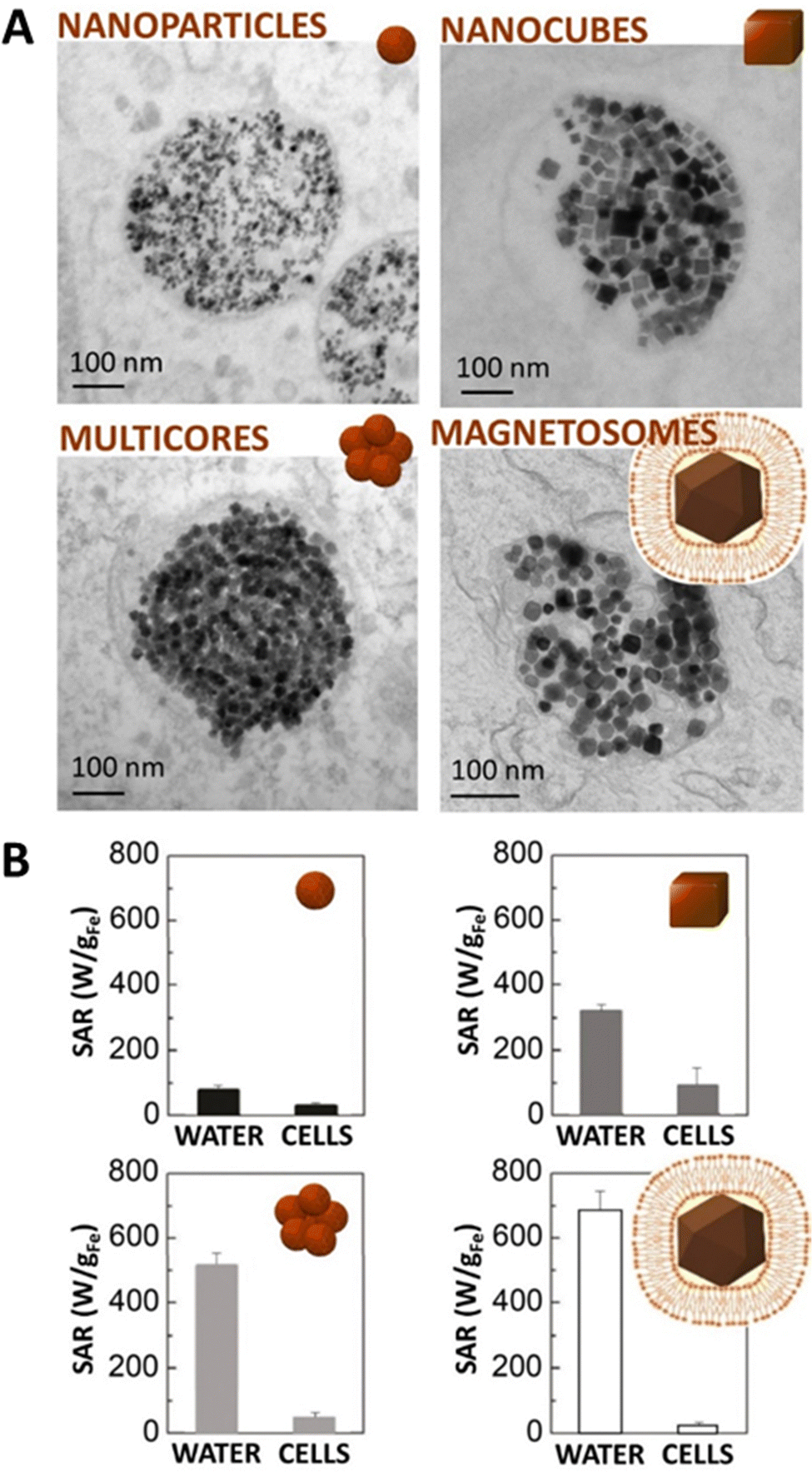
For long, iron oxide nanoparticles have been the prime candidates for magnetic hyperthermia. Nanoparticle design optimisation, advantages, and challenges associated to their use for MHT has been documented in a number of quality reviews.3,14,92–94 Briefly, in MHT, application of a high-frequency (hundreds of kHz) alternating magnetic field excites the fluctuations of the magnetic moment of nanoparticles, and the magnetic energy released is dissipated as heat. The size and shape of the NPs are then crucial and can enhance the heat-generating capacity, generally quantified with the specific absorption rate in Watts per gram of iron. Differences in anisotropy between spherical nanoparticles (SAR 10–200 W g−1) and nanocubes (SAR 500–1500 W g−1) or magnetosomes (SAR 1000–1500 W g−1) as well as cooperative effects in multicore flower-like nanoparticles (SAR 500–1500 W g−1) were exploited to increase heat generation.36,37,69,73,95,96Yet, in nanomedicine applications, nanoparticles are generally internalized via endocytosis97 and end up confined within intracellular endosomes, in very close contact with one another (Fig. 1A). This confinement is likely to affect their heating efficiency.
 | ||
| Fig. 1 Intracellular confinement of magnetic nanoparticles within endosomes decreases their heating efficiency via magnetic hyperthermia. (A) Transmission electron microscopy (TEM) images of magnetic nanoparticles confined in endosomal compartments following their internalization in cells. (B) A decrease in generated heat upon exposure to a 18 mT–470 kHz magnetic field is observed for magnetic nanoparticles confined into cellular endosomes (extracted with permission from ref. 96). | ||
Indeed, regardless of the type of iron oxide NPs tested, it has been revealed that their internalization in cells reduces heat generation, probably due to magnetic interactions or steric frustrations76,96,98 (Fig. 1B). Two distinct behaviors were evidenced: when the magnetic core is superparamagnetic and magnetic relaxation is governed by Néel mechanism, the decrease is not as pronounced, being less than two-fold. In contrast, when the magnetic core is blocked, relaxation is governed by Brownian motion and inhibited by strong intracellular confinement. This confinement highly affects the heating leading to a ten-fold or even higher decrease in overall heat.76,96,98 Unfortunately, this scenario corresponds to the performance of the most efficient nano-heaters in water.
As stated before, syntheses have been developed to counter this loss of efficiency in the intracellular environment; however, large quantities of iron oxide NPs are still necessary to reach sufficient heat production. For this reason, mixed ferrites doped with metal ions such as Co, Zn or Mn are explored.99,100In vivo studies demonstrated tumor inhibition at safe MHT conditions, once their toxicity limits have been accurately defined.81,101 Other strategies have been investigated using heterostructures including iron-based elements such as metallic iron or wüstite in core@shell configurations that proved effective for triggering cancer cell death via MHT.102 For instance, Fe2+-deficient Fe3O4 nanocubes obtained from a phase transformation of FeO@Fe3O4 nanomaterials resulted in a singular structure that preserved their magneto-thermal losses in the intracellular environment.102
Magnetic nanoparticles, emerging candidates for photothermal therapy, thus revealed as combined agents for magneto-photo-thermia
More recently, it was revealed that magnetic nanoparticles are also efficient heaters for photothermia in the near-infrared (NIR) window.20,51,61–63,69,103,104 This revelation has led to the exploration of the optical properties of these nanoparticles, less visited than the magnetic ones. In PTT, it is the optical excitation of absorbing nanoparticles by a laser light that causes localized heating in the surrounding. Initially, NPs used in photothermia were plasmonic ones, generally gold-based, for which the oscillating electromagnetic field of light engenders a collective coherent oscillation of free electrons at the surface of the nanoparticles. The frequency at which the oscillation is maximal is named localized surface plasmon resonance (LSPR), and depends on metal type, size, shape, and structure.105,106 Plasmonic nanoparticles with LSPR in the NIR region were preferred to offer deeper laser penetration within tissues, while keeping the absorbance minimal on distant organs.For iron oxide NPs, it was reported that it is the magnetite crystal phase that significantly enhances the photothermal heating.69,107 For instance, with magnetite nanocubes20 and magnetite magnetosomes,108–110 a very efficient heating was obtained in the NIR spectrum, increasing with concentration (Fig. 2). Heating powers were very high, easily reaching the 1–10 kW gFe−1 range, which was impressive considering that the maximum threshold reached for MHT with magnetic nanoparticles is in the 1000 W gFe−1 range. These two objects, magnetic nanocubes and magnetosomes, hitherto the experimental forerunners for MHT, were in fact 100 to 1000 times more effective for PTT in the intracellular environment than for MHT.108 This is due to a similar heat generation when the nanoparticles are in water or confined in the cellular endosomes with PTT, contrary to MHT that suffers a decrease in heating up to 10-fold. Magnetic nanoparticles were then becoming competitors to the state-of-the-art plasmonic gold nanoparticles intended for PTT.96,111
 | ||
| Fig. 2 (A) Magnetosomes (45 nm in diameter) and (B) magnetite nanocubes (20 nm in diameter) are efficient nanoheaters under MHT and, more remarkably, under PTT. When subjected to PTT, these nanomaterials reach similar heating temperatures than (C) gold nanostars (25 nm in diameter) when at sufficiently high nanoparticle dosages ((A) is adapted with permission from ref. 108; (B) and (C) are adapted with permission from ref. 96). (D)–(G) “Magnetic” photothermia was also exploited after in vivo injection. (D) Magnetosomes were injected intravenously and, once in cells, no heat is produced via MHT, while a high temperature increase is obtained via PTT. (E) Magnetite nanocubes were injected intratumorally. (F) Tagging of the magnetosomes with RGD made possible a targeted PTT after intravenous injection. (G) The intratumoral injection of the magnetite nanocubes and their exposure to dual (MHT + PTT) excitation drove total tumor ablation. ((D) and (F) are adapted with permission from ref. 108; (E) and (G) are adapted with permission from ref. 20). | ||
Hence, photothermal heating with magnetic nanoparticles is now recognized as a viable alternative to MHT.112 Interestingly, both PTT and MHT modalities have different windows of applicability. PTT is highly effective at low concentrations of nanoparticles, but it is difficult to reach high temperature increments at acceptable (clinically approved) laser power due to heat saturation. Indeed, due to sample adsorption, heating saturates as the concentration increases (calculation details can be found in ref. 108 and 113). By contrast, MHT needs much larger doses of nanoparticles, but suffers no saturation at all with various designs, enabling it to reach high temperatures, provided that the local concentration of nanoparticles is sufficient.89,96
Both magnetosomes and nanocubes were investigated for cancer therapy in preclinical in vivo settings.20,108 Magnetic nanocubes were found efficient for tumor regression with PTT after intratumoral injection. Furthermore, it was possible to move one step ahead and merge MHT and PTT in a single magneto-photo-thermal modality mediated by the same magnetic nanocubes, thereby yielding cumulative heat generation. This antitumoral nanotherapeutic concept based on combined magnetic and photo-induced hyperthermia solely with iron oxide cores provided localized temperature increase leading to complete cancer cell destruction in vitro and complete tumor ablation in vivo20 (Fig. 2E and G). Concerning the magnetosomes, they were tagged with arginine-glycine-aspartic acid (RGD) and could home to model tumors after systemic administration in mice.108 They were then able to generate a local cytotoxic heating effect under laser illumination with PTT, while it could hardly be considered with MHT (Fig. 2F). This was one proof of concept that a thermal therapy mediated by magnetic nanoparticles could be efficient for tumor growth inhibition after intravenous injection. Other works reporting PTT in vivo, combined or not with other strategies, are referenced in Table 3.51,61,85,104,108,114–125
| Nanoparticle type, size | In vivo tumor model, administration mode (dose) | Laser wavelength, power, duration | Ref. |
|---|---|---|---|
| Clusters of Fe3O4 nanoparticles (15 nm), 225 nm | A549 subcutaneous tumor-bearing Balb/c mice, Intratumoral injection (25 μL at 2 mg mL−1) | 808 nm, 5 W cm−2, 3 min | 61 |
| Monodisperse Fe3O4 nanoparticles, 10–310 nm; anticancer doxorubicin loading | ICR mice bearing S180 tumors, intratumoral injection (200 μL at 5 mg mL−1) | 808 nm, 1.5 W cm−2, 3 min & 3 irradiations | 117 |
| Iron oxide nanostructures with benzene-1,3,5-tricarboxylic acid and sodium citrate as co-coordinating agents, 440 nm | KB subcutaneous tumors, intratumoral injection (0.5 mg kg−1) | 808 nm, 2 W cm−2, 10 min | 118 |
| Assemblies of magnetic nanoparticles (4 nm) and tannic acid, 76 nm | HepG2-subcutaneous-tumor-bearing BALB/c nude mice, intratumoral injection (10 mg kg−1) | 808 nm, 1 W cm−2, 10 min | 119 |
| Hyaluronan-coated iron oxide nanoparticles | MDA-MB-231 cells subcutaneous tumors in BALB/c nude mice, intravenous injection (20 mg kg−1) | 808 nm, 2 W cm−2, 10 min every 24 h for 8 days | 120 |
| Iron oxide nanoparticles polysiloxane coated, 25 nm | SUM-159 tumor-bearing BALB/c mice, intravenous injection (20 mg kg−1) | 885 nm, 2.5 W cm−2, 10 min | 51 |
| Fe3O4 nanoparticles with carboxyl-terminated PEG-phospholipid coating, 9 nm | Eca-109 subcutaneous tumors in BALB/c nude mice, intratumoral injection (70 μL at 8 mg mL−1 magnetite) | 808 nm, 20 min, every 24 h for 24 days | 104 |
| Fe3O4 functionalized with carboxymethyl chitosan, 177 nm | BALB/c mice bearing S180 subcutaneous tumors, intravenous injection (200 μL at 10 mg mL−1) | 808 nm, 1.5 W cm−2, 5 min | 121 |
| RGD conjugated PEGylated Fe@Fe3O4 nanoparticles, 38 nm | U87MG subcutaneous tumor-bearing BALB/c nude mice, intravenous injection (10 mg kg−1) | 808 nm, 0.5 W cm−2, 5 min (3 repeated injections & treatments) | 122 |
| PEGylated Fe@Fe3O4 core/shell nanoparticles, 100 nm | HeLa subcutaneous tumor-bearing nude mice, intravenous injection (1.46 g[Fe] kg−1) | 808 nm, 0.31 W cm−2, 10 min | 85 |
| Azo-functionalized Fe3O4 nanoparticles, 160 nm | BALB/c mice bearing S180 subcutaneous tumors, intravenous injection, injected (100 mg kg−1) | 808 nm, 2 W cm−2, 5 min every 24 h for 10 days. | 123 |
| Magnetite magnetosomes linked to RGD peptide, 45 nm | NMRI Nude mice bearing PC3 subcutaneous tumors, intravenous injection (200 μL at 3 g l−1 Fe). | 808 nm, 1.5 W cm−2, 30 min | 108 |
| Nanogels loaded with hydrophobic magnetic nanoparticles (20 nm) and anticancer drug HCPT, 200 nm | Balb/c mice bearing MCF-7 & 4T1 solid tumors, intravenous injection, (3 mg (HCPT) kg−1) | 808 nm, 3 W cm−2, 10 min | 124 |
| Imiquimod loaded iron oxide nanoparticles, 20 nm | C57BL/6 mice bearing orthotopic pancreatic Panc02-H7 tumor, intravenous injection. | 805 nm, 1 W, 10 min | 125 |
| PLGA nanocapsules of Fe@FeO core–shell (8 nm@5 nm) (DOX-ICG@Fe/FeO-PPP), 220 nm | KB tumor (subcutaneous)-bearing nude mice, intravenous injection (20 mg kg−1) | 808 nm, 0.3 W cm−2, 5 min, 2 irradiations | 149 |
Even if the iron oxide composition of magnetic nanoparticles can already ensure a potential PTT functionality, plasmonic nanoparticles remain more potent for photothermal conversion in the NIR.126,127 Magnetic hybrid nanoplatforms should thus continue to be considered for cancer theranostics. The rationale is generally to decorate iron oxide cores with plasmonic NPs, such as gold and silver, to deliver an enhanced hybrid with both sets of magnetic and photoexcitable properties.128,129 Controlled morphologies are built mostly via seed-mediated growth. For this method, metal ions (here gold) are reduced to form small metal nuclei, and these nuclei serve as seeds for the growth of larger NPs in the presence of shape-directing additives.130 It allows the production of gold nanorods,131,132 nanotriangles,133 nanostars,134 among others. Multi-core iron oxide nanoflowers have this way been combined with a gold-branched shell, which amplified the heat generation at the tumor region.135 Other designs based on silver are also explored, such as Ag@Fe3O4 nanoflowers that present optimized heating efficiencies in solution.87 Additional example include gold coating of magnetic nanoparticles that resulted in significant apoptosis with PTT136 and also magnetic-hollow gold nanospheres that were applied to targeted MRI and photoacoustic imaging of cancer cells, and in addition that exhibited a high PTT effect under NIR laser irradiation.137 The use of Janus gold-iron oxide nanoparticles was also investigated as efficient heat generator platform when subjected to stand-alone or to combined magnetic and optical treatments, producing a cooperative cytotoxic effect on cancer cells.89 Moreover, the magnetic part was also utilized to improve NPs cellular internalization via magnetic targeting. It led to a bimodal treatment: magnetically enhanced PTT that headed to tumor growth inhibition.
The control of temperature during hyperthermia therapies is a key determinant toward successful use in clinical practice. In situ thermal tracking approaches are needful to evaluate the magneto- or/and photothermal effects at the target tumor tissue and safeguarding healthy tissues.138,139 Fiber-optic thermometers can for instance be employed to measure the released heat, but they need to be inserted invasively. Other techniques are being developed such as infrared thermometry and, more recently, thermal nanoprobes. In this regard, the local temperature produced by iron oxide nanoparticles under an AC magnetic field or laser light were probed with luminescent nanoparticles,140–143 Ag2S quantum dots,144,145 and fluorescent proteins.35,146 On the other hand, nanoscale photothermal effects were examined using X-ray spectroscopy to report a higher local heating at the nanoparticle scale.147,148
Combination of thermal and chemical therapies (chemodynamic therapy, ferroptosis) with magnetic nanoparticles only
Other modalities were also explored leveraging the ionic composition of iron oxide NPs (Fig. 3). When magnetic nanoparticles are internalized by cells, they can undergo gradual degradation,150,151 leading to the release of iron-based species within the intracellular environment. This degradation has been shown for various cell types, including cancer cells, and its rate can be tracked in situ by measuring the cell magnetism.152 Over their biodegradation, magnetic NPs release iron ions, which have been shown to tilt iron metabolism, engaging iron storage and limited iron import.153 Cellular metabolism adaptation appears generally sufficient to handle iron excess brought by nanoparticle degradation, without impacting cell function.153 The biodegradability feature of magnetic NPs is currently exploited in clinical settings for the treatment of anaemia, and an iron supplement in the form of magnetic NPs has been approved by the Food and Drug Administration (FDA) and is marketed in the United States under the name Feraheme™. | ||
| Fig. 3 Path options for magnetic nanoparticles in the intracellular environment. (left) Without additional trigger and at doses relevant for biological handling (typically, when remaining below 10 pg of iron per cell), magnetic nanoparticles internalized within endosomes are progressively degraded. This degradation engenders iron release, which is stored in ferritin proteins. In specific cases, cells can produce new magnetic nanoparticles from the released iron ions. Transmission electron microscopy images illustrating these steps are extracted with permission from ref. 219 for the top image (endosome with intact nanoparticles, right after internalization),150 for the middle one (degraded nanoparticles), and ref. 218 for the bottom one (biosynthesized nanoparticles). (right) In parallel, the released iron ions, especially Fe2+, can engender the production of ROS and enhance ferroptosis. Ferroptosis is an iron-dependent cell death pathway involving the accumulation of lipid ROS via the inhibition of System Xc− and GPX4. Triggering the intracellular degradation of iron oxide nanoparticles, and maintaining the imbalance created by the Fe2+ excess to promote the formation of ROS, is an additional alternative explored for induction of cancer cell death. (cell structure drawing extracted from https://smart.servier.com). | ||
It is now envisaged that the iron ions released over this degradation can contribute to the anti-cancer therapeutic prospects, this time through chemical means. This emerging approach is sometimes referred to as chemodynamic therapy.42 It is grounded on the production of reactive oxygen species, which are part of the normal cellular metabolism, but can also engender oxidative stress when in excess. The term ROS englobes several oxygen-containing species, such as hydroxyl radical (˙OH), hydrogen peroxide (H2O2), superoxide radical (˙O2−), hypochlorous acid (HOCl) or again singlet oxygen (1O2), that engender varying levels of damage. Chemodynamic agents can for instance convert internal H2O2 into lethal ROS, such as ˙OH described as the most oxidizing ROS, through Fenton or Fenton-like reactions.154,155 This ROS production was shown to induce DNA damage and inactivation of proteins, leading to massive cancer cell death. This is in part possible in cancer cells as they present a unique phenotype, with a higher content of hydrogen peroxide in tumors than in normal tissues.156 However, despite being higher in tumors, the presence of H2O2 remains limited in these cellular environments.42 Strategies are thus explored to trigger the production of H2O2. Conjugating magnetic nanoparticles with glucose oxidase (GOx) has been attempted in this objective. GOx catalyses the decomposition of glucose, promoting cell starvation, and generates H2O2 in the process. The synergy of glucose depletion and ROS production has been shown to significantly suppress mammary tumor growth.157
The release of iron ions and production of ROS could also serve ferroptosis purposes.41,158 Ferroptosis is an iron-dependent cell death path characterized by the accumulation of lipid ROS.22,159–161 At the biochemical level, in cells undergoing ferroptosis, the reduced activity of glutathione peroxidase 4 (GPX4) is generally central to the mechanism.162 This enzyme, GPX4, has a phospholipid peroxidase activity that catalyses the reduction of lipid peroxides.163 When this activity is decreased, lipid peroxides accumulate and initiate ferroptosis. Free iron present in the intracellular pool (for instance Fe2+) are known to drive the transformation of low reactive oxygen species such as H2O2 into much stronger oxidative species such as OH˙ capable to drive the oxidation of most cell components, including nucleic acids and unsaturated lipids.164 In the context of ferroptosis, the role of iron is among the same line. Free iron ions, and especially Fe2+, catalyse the formation of damaging radicals from lipid peroxides, such as lipid alkoxyl (RO˙), via the Fenton reaction. This cascade ultimately leads to ferroptosis.
This new form of cell death, ferroptosis, is explored for cancer treatment. Molecules encouraging this path, such as the ones that inhibit System Xc− or GPX4 are being assessed.165,166 The activity of GPX4 can either be inhibited directly or via the depletion of intracellular glutathione (GSH), an essential cofactor of GPX4. GSH level is regulated by System Xc−, a cystine/glutamate antiporter composed of a light (xCT) and heavy chain.167 The light chain mediates the cystine-glutamate exchange. It exports glutamate and imports cystine within the cells, where it is reduced to cysteine and utilized, with intracellular glutamate, to produce glutathione (GSH). Molecules encouraging ferroptosis are thus classified in four classes: class I agents inhibit System Xc−; class II directly inhibit GPX4; class III indirectly inhibit GPX4; and class IV induce lipid peroxidation by increasing iron levels or iron oxidation168 (Table 4).
| Class | Name | Clinically approved? | Clinical purpose | Mode of action in ferroptosis | Ref. |
|---|---|---|---|---|---|
| I | Sorafenib | Yes | Anti-cancer | Inhibits system Xc− | 169–171 |
| Sulfasalazine | Yes | Anti-inflammatory | Inhibits system Xc− | 172,173 | |
| Erastin | No | — | Inhibits system Xc− and inactivates GPX4 (depletes glutathione) | 162, 174 and 175 | |
| II | Altretamine | Yes | Anti-cancer | Inhibits GPX4 | 176 |
| 1S,3R-RSL3 (RSL3) | No | — | Inhibits GPX4 | 177 | |
| Withaferin A | No | — | Depletes and inactivates GPX4 | 178 | |
| III | Ferroptosis inducer 56 (FIN56) | No | — | Indirectly degrades GPX4 | 179 |
| IV | 1,2-Dioxolane (FINO2) | No | — | Oxidizes iron, induces lipid peroxidation | 180 |
| More | Artemisinin | Yes | Anti-malaria | Ferritin degradation, lipid peroxidation | 181 and 182 |
| Lapatinib | No | — | Tyrosine kinase inhibitor | 183 | |
| Siramesine | No | — | Lysosome disrupting agent | 183 | |
| Cyst(e)inase | No | — | Depletes extracellular cysteine/cystine | 184 | |
| BAY 87-2243 | No | — | inhibits NADH-coenzyme Q oxidoreductase (complex I) | 185 | |
| (+)-JQ1 | No | — | Inhibits BRD4 | 186 | |
Ferroptosis has been evidenced without external input of iron within cells. On the other hand, it was suggested that the anticancer activity of some iron-based nanoparticles are related to ferroptosis induction, following their degradation and release of ferrous or ferric ions in acidic lysosomes.41 This has for instance been confirmed on iron oxide NPs coated with gallic acid and polyacrylic acid as their exposure to glioblastoma, neuroblastoma, and fibrosarcoma cells was shown to efficiently induce ferroptosis, with a dose-dependent effect.187 Triggering the release of additional iron ions derived from magnetic nanoparticles is explored to intensify this effect,188 such as laser irradiation that has been applied to accelerate nanoparticle degradation and provoke the rapid release of a large pool of iron ions.189 Using this laser irradiation technique borrowed to PTT approaches, the role of nanoparticle composition in triggering ferroptosis was assessed. Two nanoparticle variants were examined: magnetite NPs, composed primarily of Fe2+, and maghemite, their oxidized counterparts that possess a diminished Fe2+ content. Results underscore that the non-oxidized magnetite NPs, characterized by an elevated Fe2+ content, induced ferroptosis to a significantly greater extent.189 More complex structures are also built to increase ferroptotic potential, such as vesicles incorporating iron oxide nanocubes into their shell and ascorbic acid into their core.190 When exposed to a circularly polarized magnetic field, the vesicle shell can be destroyed, and the released ascorbic acid reduces ferric iron derived from IONC into ferrous. Results showed that these hybrid vesicles induced significant tumor suppression, due to ferroptosis-like cell-death. Another platform consisted in sorafenib (for deactivation of GPX4) and iron oxide NPs loaded into the mesopores and onto the surface of mesoporous polydopamine NPs. This nanoplatform was efficient, in particular upon NIR laser irradiation that offered moderate heat and boosted the ferroptosis effect.191 The direct loading of ferric ions and sulfasalazine in polydopamine nanoparticles was also attempted.192 In this case, iron ions engendered Fenton reaction, sulfasalazine restrains xCT signaling (the functional subunit of system Xc−) and deactivates GPX4, and the synergistic effect led to ferroptotic death. This effect was intensified with irradiation via near-infrared light and with the acidic tumor microenvironment.
Domino effect of magnetic nanoparticle-based thermal and chemical therapies
An additional trend in cancer therapy consists in helping or activating the immune system of the patient to prevent, control, and ultimately eliminate cancer cells.193 This cancer immunotherapy or immune-oncology area is encountering clinical success in particular by prolonging survival of patients with rapidly fatal cancers. The therapeutic strategy is grounded on the activation of immune cells such as T cells193 but also on cell reprogramming. For instance, macrophages have two distinct phenotypes, the M1 and M2. A strategy consists in reprogramming tumor-associated macrophages from the M2 phenotype, which promotes tumor progression, to the M1, which suppresses tumor growth.194,195Iron metabolism is interplayed in immune cell phenotypes and has been standing out as target for cancer immunotherapy.196 It is thus interesting to consider magnetic nanoparticles in this approach. First off, because even though anti-cancer treatments relying on magnetic nanoparticles usually aim at cancer cells targeting, injected NPs may not be fully internalized by the cancer cells only. Nanoparticle targeting can be either passive, taking advantage of the enhanced permeability and retention (EPR) effect of tumor tissues due to leaky tumor blood vessels and impaired lymphatic drainage,197 so not being fully specific, or active,198 for instance via cancer-specific antibody ligand attached at the surface of the nanoparticles. Korangath et al. compared the efficiency of active versus passive targeting on systemic delivery of nanoparticles and demonstrated that antibody-labelled nanoparticles were better retained by tumors than plain ones but elicited similar immune responses and tumor growth inhibition.199 Additionally, intratumor retention of antibody-labeled nanoparticles (“active targeting”) was determined by tumor-associated dendritic cells, neutrophils, monocytes, and macrophages and not by antibody-antigen interactions.199 Also, M1 macrophages were shown as more effective at ingesting nanoparticles than M2 macrophages.200 Despite better targeting being elicited by targeted approaches, internalization might thus not be fully specific. In addition, when the nanoparticles are internalized in cancer cells, they are usually biodegraded, and it releases iron ions.151
Magnetic nanoparticles and the iron ions released over their degradation are thus being studied for immune cells activation. Exposure of macrophages to iron has for instance been shown to cause their phenotypic change toward a proinflammatory state.201–203 A study based on 102 non-small cell lung cancer (NSCLC) human tissues obtained from patients indicated that, when iron was accumulated in the tumor microenvironment, higher numbers of M1-like pro-inflammatory TAM were present and patients presented a survival advantage.204 Similar results were obtained with in vivo injection in mice of cross-linked iron oxide (CLIO) magnetic NPs that are more specifically ingested by phagocytic cells such as TAMs than neighbouring tumor cells or leukocytes.205 Following injection, tumor growth was delayed and significantly smaller tumor sizes were measured after 15 days when compared to controls.205 Magnetic NPs have also been shown to orient tumor-associated macrophages (TAM) toward their suppressor phenotype and inhibit tumor growth.194,206,207 This effect is here also grounded on the degradation of the nanoparticles and accumulation of intracellular iron that promotes the transcriptional reprogramming of macrophages phenotypes. Studies indicate that magnetic NPs can induce a variety of transcription factors related to the expression of iron metabolism-related proteins and that their effects may be related to ROS production by Fenton reaction, TLR4 activation and cytokine production. However, exact molecular mechanisms that initiate the reprogramming of macrophages are still not well understood.
All these results comfort in the multifaceted possibilities brought by magnetic nanoparticles, of their capacity to tilt the iron balance within cells, and lead to multiple options for anti-cancer treatment.
Biocompatibility and biological fate of magnetic nanoparticles
It should first be noted that the biocompatibility, metabolism, and excretion pathway of magnetic NPs can vary depending on the type of nanoparticle, the surface coating, size, and on the cell type they are in contact with. Ensuring their biocompatibility involves meticulous surface engineering to minimize adverse reactions with biological entities, prevent cytotoxicity, and mitigate immune response. Chemists have been extensively working on finely tuning the surface properties, such as functionalizing with biocompatible coatings, to aim at optimal biocompatibility and at guaranteeing safe biomedical uses taking advantage on the remarkable capabilities of these nanoparticles. A number of studies and reviews have addressed biocompatibility and fate of magnetic nanoparticles in vitro and in vivo, and can be referred to.153,208–210Within the body, magnetic NPs can interact with biomolecules, such as proteins and lipids, and these interactions will depend on the surface coating and functionalization of the nanoparticles. This can influence their circulation, distribution, clearance, and as such their fate. However, the general path for iron oxide nanoparticles is their excretion through either the renal or hepatic pathway.211 Renal clearance means that the nanoparticles undergo kidney filtration and are subsequently excreted via the urines. This is particularly true for ultra-small NPs with a low propensity for protein opsonization meaning that they present a small hydrodynamic size.212 For the hepatic route, nanoparticles are captured by the mononuclear phagocyte system (MPS) or tissue-resident phagocytes, leading to their accumulation in the liver and spleen, and are then subject to biliary elimination and excretion via the stools.211 This holds especially true for nanoparticles susceptible to opsonization, meaning to the formation of a protein corona, which can trigger recognition and sequestration by the MPS. To avoid recognition, and extend circulation time, antifouling or hydrophilic polymer, such as polyethylene glycol (PEG)213 or zwitterionic polymers214 can be coated onto the surface of the nanoparticles.
When magnetic NPs are internalized within cells, they are mainly endocytosed, and the endosomal compartments in which they are confined will then become lysosomes.151 Lysosomes have an acidic pH favourable to the biodegradation of the nanoparticles that will engender the release of iron in the form of ions (Fig. 3). These iron ions can then be metabolized by the cells that possess a set of proteins capable of storing, excreting, importing, and changing the valence state of these iron ions. The released iron is thus integrated within the natural iron metabolism of the cells, as confirmed by tracking the fate of nanoparticles synthesized using exogeneous iron isotopes (e.g.59Fe, 57Fe) that could then be monitored and differentiated from the endogenous iron (mainly 56Fe).215,216
Interestingly, quantification of the biodegradation of magnetic NPs in a close in vitro system composed of cell-spheroids indicated that they are dissolved rapidly, resulting in an all-or-nothing signal obtained by magnetometry: contribution of small entities derived from the initial nanoparticles is not detectable.217 This might have implications for its applicative potential, as the initial physicochemical properties might either be fully conserved or absent. Moreover, under specific conditions, human cells might be able to use the iron ions released over the biodegradation for the production of new magnetic nanoparticles, fully biological218,219 that appear to arise upon weeks of cell culture.220 This biomineralization of magnetic nanoparticles could become advantageous for repeated treatment due to their potential long-term persistence. A protective coating avoiding the dissolution of the magnetic core could also be considered.217
Conclusion
Iron is one of the few metallic elements placed at the heart of capital biological functions, making its presence essential to life. Iron oxide nanoparticles combine this extraordinary elemental structure with unique size and physicochemical properties. First of all, the nanometric size of iron oxide NPs allows their circulation in the blood stream, penetration within tissues, and entry inside cells. By their size again are brought unique superparamagnetic features, via which they can be “activated” at a distance. Not only can they be directed magnetically, providing a solution for targeting upon injection in the bloodstream, by attracting them using an external magnet. They can also be visualized in vivo via MRI, so used as contrast agents. It is actually the first application these nano-objects obtained regulatory approval for. Again, and of main interest for cancer therapy, they can generate heat when subjected either to an alternative magnetic field (magnetic hyperthermia) or, shown more recently, to light (photothermia). Morphological, structural, and chemical parameters have been identified as responsible factors controlling their heating efficiency in both magnetic and thermal therapies.This generation of heat is being exploited for anti-cancer therapy, to provoke cancer cell death and ultimately tumor ablation. Some clinical trials are already accomplished or underway for magnetic hyperthermia, notably with MagForce (NanoTherm®)90,91 that has been approved by regulatory boards in the European Union in 2011 for treating brain tumors. The key limitation for MHT remains the poor heating rate, meaning that clinical efficacy is only reached by injecting very large amounts of nanoparticles directly into the tumor. The recent reveal of magnetic nanoparticles as efficient heaters for photothermia in the first and second biological windows has paved the way to the exploration of the optical properties less visited than the magnetic ones,20 which could overcome this limitation.
A question always subjacent to the use of these nano-objects in the body remains their biocompatibility. It is all the more important to consider as magnetic nanoparticles can be degraded by human cells, and the iron ions released over this degradation could engender the production of ROS. Interestingly, cytocompatibility studies show that no adverse effect is observed even at high doses of internalized NPs (up to 10 pg of iron per cell),153 and this dose can be much higher depending on cell and nanoparticle type, with sometimes reaching up to 80 pg of iron per cell without provoking cell damage.221 The released iron is absorbed by the natural metabolism of cells, adapted to handle this metallic ion. It goes in accordance with the fact that the production of ROS coming from the degradation of magnetic nanoparticles could be used as chemical trigger of cell death, via chemodynamic therapy or again ferroptosis, but studies indicate that nanoparticle degradation alone is not sufficient for such applications, additional agents are needed to sufficiently imbalance iron homeostasis.
Finally, the micromachinery of cancer cells could be either an asset or challenging considering all these applications. Cancer cells have a unique way of managing iron,222 and as such the biodegradation products of magnetic NPs. They have an increased dependence to iron in comparison to healthy cells,222 but ferroptosis events also regulate their dissemination capacity.223,224 Despite having more H2O2 than normal cells,225 the presence of magnetic nanoparticles in their intracellular environment in not sufficient to trigger ferroptosis. Moreover, cancer cells seem able to store iron in the form of magnetic nanoparticles,226,227 believed as less reactive, in addition to the more conventional iron storage as non-magnetic iron (ferrihydrite) inside the ferritin protein. The progressive understanding of this very specific microenvironment for iron could help in delineating adequate parameters for optimal therapy.
Conflicts of interest
There are no conflicts to declare.Acknowledgements
This work was supported by the European Union (ERC-2019-CoG project NanoBioMade 865629). M. E. and A. F. acknowledge support from the Spanish Ministerio de Ciencia e Innovación (MICINN) through grant PID2019-106165GB-C22 funded by MCIN/AEI/10.13039/501100011033. M. E. acknowledges RYC2018-024396-I grant funded by MCIN/AEI/10.13039/501100011033 and by “ESF Investing in your future’’. A. F. is a Serra Húnter fellow. A. E. acknowledges support from MICINN (PID2021-127033OB-C21 project and RYC2020-029282-I grant) and Comunidad de Madrid (ASAP-CM – S2022/BMD-7434 project).References
- A. Avasthi, C. Caro, E. Pozo-Torres, M. P. Leal and M. L. García-Martín, Top. Curr. Chem. (Z), 2020, 378, 40 CrossRef CAS PubMed
.
- H. Gavilán, K. Simeonidis, E. Myrovali, E. Mazarío, O. Chubykalo-Fesenko, R. Chantrell, L. Balcells, M. Angelakeris, M. P. Morales and D. Serantes, Nanoscale, 2021, 13, 15631–15646 RSC
- H. Gavilán, S. K. Avugadda, T. Fernández-Cabada, N. Soni, M. Cassani, B. T. Mai, R. Chantrell and T. Pellegrino, Chem. Soc. Rev., 2021, 50, 11614–11667 RSC
- J.-H. Lee, J.-T. Jang, J.-S. Choi, S. H. Moon, S.-H. Noh, J.-W. Kim, J.-G. Kim, I.-S. Kim, K. I. Park and J. Cheon, Nat. Nanotechnol., 2011, 6, 418–422 CrossRef CAS PubMed
- E. M. Materón, C. M. Miyazaki, O. Carr, N. Joshi, P. H. S. Picciani, C. J. Dalmaschio, F. Davis and F. M. Shimizu, Appl. Surf. Sci. Adv., 2021, 6, 100163 CrossRef
- V. F. Cardoso, A. Francesko, C. Ribeiro, M. Bañobre-López, P. Martins and S. Lanceros-Mendez, Adv. Healthcare Mater., 2018, 7, 1700845 CrossRef PubMed
- A. Fouriki and J. Dobson, Materials, 2013, 6, 255–264 CrossRef CAS PubMed
- F. Etoc, D. Lisse, Y. Bellaiche, J. Piehler, M. Coppey and M. Dahan, Nat. Nanotechnol., 2013, 8, 193–198 CrossRef CAS PubMed
- G. Mary, A. Van de Walle, J. E. Perez, T. Ukai, T. Maekawa, N. Luciani and C. Wilhelm, Adv. Funct. Mater., 2020, 30, 2002541 CrossRef CAS
- A. Van de Walle, J. E. Perez and C. Wilhelm, Bioprinting, 2023, 30, e00265 CrossRef
- G. Mikhaylov, U. Mikac, A. A. Magaeva, V. I. Itin, E. P. Naiden, I. Psakhye, L. Babes, T. Reinheckel, C. Peters, R. Zeiser, M. Bogyo, V. Turk, S. G. Psakhye, B. Turk and O. Vasiljeva, Nat. Nanotechnol., 2011, 6, 594–602 CrossRef CAS PubMed
- S. Dev and J. L. Babitt, Hemodial. Int., 2017, 21, S6–S20 CrossRef PubMed
- X. Liu, Y. Zhang, Y. Wang, W. Zhu, G. Li, X. Ma, Y. Zhang, S. Chen, S. Tiwari, K. Shi, S. Zhang, H. M. Fan, Y. X. Zhao and X.-J. Liang, Theranostics, 2020, 10, 3793–3815 CrossRef CAS PubMed
- I. Rubia-Rodríguez, A. Santana-Otero, S. Spassov, E. Tombácz, C. Johansson, P. De La Presa, F. J. Teran, M. Del Puerto Morales, S. Veintemillas-Verdaguer, N. T. K. Thanh, M. O. Besenhard, C. Wilhelm, F. Gazeau, Q. Harmer, E. Mayes, B. B. Manshian, S. J. Soenen, Y. Gu, Á. Millán, E. K. Efthimiadou, J. Gaudet, P. Goodwill, J. Mansfield, U. Steinhoff, J. Wells, F. Wiekhorst and D. Ortega, Materials, 2021, 14, 706 CrossRef PubMed
- J. C. Bear, P. S. Patrick, A. Casson, P. Southern, F.-Y. Lin, M. J. Powell, Q. A. Pankhurst, T. Kalber, M. Lythgoe, I. P. Parkin and A. G. Mayes, Sci. Rep., 2016, 6, 34271 CrossRef CAS PubMed
- J. F. Liu, B. Jang, D. Issadore and A. Tsourkas, Wiley Interdiscip. Rev.: Nanomed. Nanobiotechnol., 2019, 11, e1571 Search PubMed
- E. Cazares-Cortes, A. Espinosa, J.-M. Guigner, A. Michel, N. Griffete, C. Wilhelm and C. Ménager, ACS Appl. Mater. Interfaces, 2017, 9, 25775–25788 CrossRef CAS PubMed
- D. Mertz, O. Sandre and S. Bégin-Colin, Biochim. Biophys. Acta, Gen. Subj., 2017, 1861, 1617–1641 CrossRef CAS PubMed
- M. Li, W. Bu, J. Ren, J. Li, L. Deng, M. Gao, X. Gao and P. Wang, Theranostics, 2018, 8, 693–709 CrossRef CAS PubMed
- A. Espinosa, R. Di Corato, J. Kolosnjaj-Tabi, P. Flaud, T. Pellegrino and C. Wilhelm, ACS Nano, 2016, 10, 2436–2446 CrossRef CAS PubMed
- X. Liang, M. Chen, P. Bhattarai, S. Hameed, Y. Tang and Z. Dai, ACS Nano, 2021, 15, 20164–20180 CrossRef CAS PubMed
- S. J. Dixon, K. M. Lemberg, M. R. Lamprecht, R. Skouta, E. M. Zaitsev, C. E. Gleason, D. N. Patel, A. J. Bauer, A. M. Cantley, W. S. Yang, B. Morrison and B. R. Stockwell, Cell, 2012, 149, 1060–1072 CrossRef CAS PubMed
- S. Laurent, D. Forge, M. Port, A. Roch, C. Robic, L. Vander Elst and R. N. Muller, Chem. Rev., 2008, 108, 2064–2110 CrossRef CAS PubMed
- J.-P. Fortin, C. Wilhelm, J. Servais, C. Ménager, J.-C. Bacri and F. Gazeau, J. Am. Chem. Soc., 2007, 129, 2628–2635 CrossRef CAS PubMed
- B. Mehdaoui, A. Meffre, J. Carrey, S. Lachaize, L.-M. Lacroix, M. Gougeon, B. Chaudret and M. Respaud, Adv. Funct. Mater., 2011, 21, 4573–4581 CrossRef CAS
- G. Salas, C. Casado, F. J. Teran, R. Miranda, C. J. Serna and M. P. Morales, J. Mater. Chem., 2012, 22, 21065–21075 RSC
- P. Guardia, A. Riedinger, S. Nitti, G. Pugliese, S. Marras, A. Genovese, M. E. Materia, C. Lefevre, L. Manna and T. Pellegrino, J. Mater. Chem. B, 2014, 2, 4426–4434 RSC
- H. Deng, X. Li, Q. Peng, X. Wang, J. Chen and Y. Li, Angew. Chem., Int. Ed., 2005, 44, 2782–2785 CrossRef CAS PubMed
- J. Gao, X. Ran, C. Shi, H. Cheng, T. Cheng and Y. Su, Nanoscale, 2013, 5, 7026–7033 RSC
- Z. Li, B. Tan, M. Allix, A. I. Cooper and M. J. Rosseinsky, Small, 2008, 4, 231–239 CrossRef CAS PubMed
- A. P. LaGrow, M. O. Besenhard, A. Hodzic, A. Sergides, L. K. Bogart, A. Gavriilidis and N. T. K. Thanh, Nanoscale, 2019, 11, 6620–6628 RSC
- T. Hyeon, S. S. Lee, J. Park, Y. Chung and H. B. Na, J. Am. Chem. Soc., 2001, 123, 12798–12801 CrossRef CAS PubMed
- A. Lassenberger, T. A. Grünewald, P. D. J. van Oostrum, H. Rennhofer, H. Amenitsch, R. Zirbs, H. C. Lichtenegger and E. Reimhult, Chem. Mater., 2017, 29, 4511–4522 CrossRef CAS PubMed
- X. Tian, L. Ruan, S. Zhou, L. Wu, J. Cao, X. Qi, X. Zhang and S. Shen, ACS Appl. Bio Mater., 2022, 5, 1692–1699 CrossRef CAS PubMed
- J. G. Ovejero, I. Armenia, D. Serantes, S. Veintemillas-Verdaguer, N. Zeballos, F. López-Gallego, C. Grüttner, J. M. de la Fuente, M. del Puerto Morales and V. Grazu, Nano Lett., 2021, 21, 7213–7220 CrossRef CAS PubMed
- P. Hugounenq, M. Levy, D. Alloyeau, L. Lartigue, E. Dubois, V. Cabuil, C. Ricolleau, S. Roux, C. Wilhelm, F. Gazeau and R. Bazzi, J. Phys. Chem. C, 2012, 116, 15702–15712 CrossRef CAS
- H. Gavilán, E. H. Sánchez, M. E. F. Brollo, L. Asín, K. K. Moerner, C. Frandsen, F. J. Lázaro, C. J. Serna, S. Veintemillas-Verdaguer, M. P. Morales and L. Gutiérrez, ACS Omega, 2017, 2, 7172–7184 CrossRef PubMed
- J. M. Asensio, J. Marbaix, N. Mille, L.-M. Lacroix, K. Soulantica, P.-F. Fazzini, J. Carrey and B. Chaudret, Nanoscale, 2019, 11, 5402–5411 RSC
- A. Bordet, R. F. Landis, Y. Lee, G. Y. Tonga, J. M. Asensio, C.-H. Li, P.-F. Fazzini, K. Soulantica, V. M. Rotello and B. Chaudret, ACS Nano, 2019, 13, 2870–2878 CrossRef CAS PubMed
- J. Tang, M. Myers, K. A. Bosnick and L. E. Brus, J. Phys. Chem. B, 2003, 107, 7501–7506 CrossRef CAS
- S. Wang, J. Luo, Z. Zhang, D. Dong, Y. Shen, Y. Fang, L. Hu, M. Liu, C. Dai, S. Peng, Z. Fang and P. Shang, Am. J. Cancer Res., 2018, 8, 1933–1946 CAS
- Q. Sun, Z. Wang, B. Liu, F. He, S. Gai, P. Yang, D. Yang, C. Li and J. Lin, Coord. Chem. Rev., 2022, 451, 214267 CrossRef CAS
- S. J. Kemp, R. M. Ferguson, A. P. Khandhar and K. M. Krishnan, RSC Adv., 2016, 6, 77452–77464 RSC
- R. Hufschmid, H. Arami, R. M. Ferguson, M. Gonzales, E. Teeman, L. N. Brush, N. D. Browning and K. M. Krishnan, Nanoscale, 2015, 7, 11142–11154 RSC
- T. J. Daou, G. Pourroy, S. Bégin-Colin, J. M. Grenèche, C. Ulhaq-Bouillet, P. Legaré, P. Bernhardt, C. Leuvrey and G. Rogez, Chem. Mater., 2006, 18, 4399–4404 CrossRef CAS
- J. Wang, J. Sun, Q. Sun and Q. Chen, Mater. Res. Bull., 2003, 38, 1113–1118 CrossRef CAS
- S. Ge, X. Shi, K. Sun, C. Li, C. Uher, J. R. Baker, M. M. Banaszak Holl and B. G. Orr, J. Phys. Chem. C, 2009, 113, 13593–13599 CrossRef CAS PubMed
- X. Liang, X. Wang, J. Zhuang, Y. Chen, D. Wang and Y. Li, Adv. Funct. Mater., 2006, 16, 1805–1813 CrossRef CAS
- M. Levy, A. Quarta, A. Espinosa, A. Figuerola, C. Wilhelm, M. García-Hernández, A. Genovese, A. Falqui, D. Alloyeau, R. Buonsanti, P. D. Cozzoli, M. A. García, F. Gazeau and T. Pellegrino, Chem. Mater., 2011, 23, 4170–4180 CrossRef CAS
- A. Espinosa, A. Muñoz-Noval, M. García-Hernández, A. Serrano, J. Jiménez De La Morena, A. Figuerola, A. Quarta, T. Pellegrino, C. Wilhelm and M. A. García, J. Nanopart. Res., 2013, 15, 1514 CrossRef
- H. Chen, J. Burnett, F. Zhang, J. Zhang, H. Paholak and D. Sun, J. Mater. Chem. B, 2014, 2, 757–765 RSC
- R. Krahne, G. Morello, A. Figuerola, C. George, S. Deka and L. Manna, Phys. Rep., 2011, 501, 75–221 CrossRef CAS
- P. Guardia, R. Di Corato, L. Lartigue, C. Wilhelm, A. Espinosa, M. Garcia-Hernandez, F. Gazeau, L. Manna and T. Pellegrino, ACS Nano, 2012, 6, 3080–3091 CrossRef CAS PubMed
- J. Mohapatra, A. Mitra, H. Tyagi, D. Bahadur and M. Aslam, Nanoscale, 2015, 7, 9174–9184 RSC
- Y. Yang, M. Huang, J. Qian, D. Gao and X. Liang, Sci. Rep., 2020, 10, 8331 CrossRef CAS PubMed
- P. J. Sugumaran, Y. Yang, Y. Wang, X. Liu and J. Ding, ACS Appl. Bio Mater., 2021, 4, 4809–4820 CrossRef CAS PubMed
- J.-C. Si, Y. Xing, M.-L. Peng, C. Zhang, N. Buske, C. Chen and Y.-L. Cui, CrystEngComm, 2013, 16, 512–516 RSC
- S. Geng, H. Yang, X. Ren, Y. Liu, S. He, J. Zhou, N. Su, Y. Li, C. Xu, X. Zhang and Z. Cheng, Chem. – Asian J., 2016, 11, 2996–3000 CrossRef CAS PubMed
- A. G. Roca, L. Gutiérrez, H. Gavilán, M. E. Fortes Brollo, S. Veintemillas-Verdaguer and M. D. P. Morales, Adv. Drug Delivery Rev., 2019, 138, 68–104 CrossRef CAS PubMed
- L. Lartigue, P. Hugounenq, D. Alloyeau, S. P. Clarke, M. Lévy, J.-C. Bacri, R. Bazzi, D. F. Brougham, C. Wilhelm and F. Gazeau, ACS Nano, 2012, 6, 10935–10949 CrossRef CAS PubMed
- S. Shen, S. Wang, R. Zheng, X. Zhu, X. Jiang, D. Fu and W. Yang, Biomaterials, 2015, 39, 67–74 CrossRef CAS PubMed
- C. Lozano-Pedraza, E. Plaza-Mayoral, A. Espinosa, B. Sot, A. Serrano, G. Salas, C. Blanco-Andujar, G. Cotin, D. Felder-Flesch, S. Begin-Colin and F. J. Teran, Nanoscale Adv., 2021, 3, 6490–6502 RSC
- S. Nemec, S. Kralj, C. Wilhelm, A. Abou-Hassan, M.-P. Rols and J. Kolosnjaj-Tabi, Appl. Sci., 2020, 10, 7322 CrossRef CAS
- S. Dutz, J. H. Clement, D. Eberbeck, T. Gelbrich, R. Hergt, R. Müller, J. Wotschadlo and M. Zeisberger, J. Magn. Magn. Mater., 2009, 321, 1501–1504 CrossRef CAS
- J. Ge, Y. Hu, M. Biasini, W. P. Beyermann and Y. Yin, Angew. Chem., Int. Ed., 2007, 46, 4342–4345 CrossRef CAS PubMed
- G. Hemery, C. Genevois, F. Couillaud, S. Lacomme, E. Gontier, E. Ibarboure, S. Lecommandoux, E. Garanger and O. Sandre, Mol. Syst. Des. Eng., 2017, 2, 629–639 RSC
- K. C. Barick, M. Aslam, Y.-P. Lin, D. Bahadur, P. V. Prasad and V. P. Dravid, J. Mater. Chem., 2009, 19, 7023–7029 RSC
- Y. Li, J. Yang, G. Gu, X. Guo, C. He, J. Sun, H. Zou, H. Wang, S. Liu, X. Li, S. Zhang, K. Wang, L. Yang, Y. Jiang, L. Wu and X. Sun, Nano Lett., 2022, 22, 963–972 CrossRef CAS PubMed
- E. Bertuit, E. Benassai, G. Mériguet, J.-M. Greneche, B. Baptiste, S. Neveu, C. Wilhelm and A. Abou-Hassan, ACS Nano, 2022, 16, 271–284 CrossRef CAS PubMed
- E. Bertuit, S. Neveu and A. Abou-Hassan, Nanomaterials, 2022, 12, 119 CrossRef CAS PubMed
- R. Turcu, V. Socoliuc, I. Craciunescu, A. Petran, A. Paulus, M. Franzreb, E. Vasile and L. Vekas, Soft Matter, 2015, 11, 1008–1018 RSC
- L. Yan, A. Amirshaghaghi, D. Huang, J. Miller, J. M. Stein, T. M. Busch, Z. Cheng and A. Tsourkas, Adv. Funct. Mater., 2018, 28, 1707030 CrossRef PubMed
- D. Niculaes, A. Lak, G. C. Anyfantis, S. Marras, O. Laslett, S. K. Avugadda, M. Cassani, D. Serantes, O. Hovorka, R. Chantrell and T. Pellegrino, ACS Nano, 2017, 11, 12121–12133 CrossRef CAS PubMed
- T. Yildirim, M. Pervez, B. Li and R. K. O’Reilly, J. Mater. Chem. B, 2020, 8, 5330–5335 RSC
- C. Paquet, H. W. de Haan, D. M. Leek, H.-Y. Lin, B. Xiang, G. Tian, A. Kell and B. Simard, ACS Nano, 2011, 5, 3104–3112 CrossRef CAS PubMed
- R. Di Corato, A. Espinosa, L. Lartigue, M. Tharaud, S. Chat, T. Pellegrino, C. Ménager, F. Gazeau and C. Wilhelm, Biomaterials, 2014, 35, 6400–6411 CrossRef CAS PubMed
- D. Gandia, L. Gandarias, I. Rodrigo, J. Robles-García, R. Das, E. Garaio, J. Á. García, M.-H. Phan, H. Srikanth, I. Orue, J. Alonso, A. Muela and M. L. Fdez-Gubieda, Small, 2019, 15, e1902626 CrossRef PubMed
- M. Theodosiou, E. Sakellis, N. Boukos, V. Kusigerski, B. Kalska-Szostko and E. Efthimiadou, Sci. Rep., 2022, 12, 8697 CrossRef CAS PubMed
- C. Martinez-Boubeta, K. Simeonidis, A. Makridis, M. Angelakeris, O. Iglesias, P. Guardia, A. Cabot, L. Yedra, S. Estradé, F. Peiró, Z. Saghi, P. A. Midgley, I. Conde-Leborán, D. Serantes and D. Baldomir, Sci. Rep., 2013, 3, 1652 CrossRef PubMed
- G. Salas, J. Camarero, D. Cabrera, H. Takacs, M. Varela, R. Ludwig, H. Dähring, I. Hilger, R. Miranda, M. del, P. Morales and F. J. Teran, J. Phys. Chem. C, 2014, 118, 19985–19994 CrossRef CAS
- P. B. Balakrishnan, N. Silvestri, T. Fernandez-Cabada, F. Marinaro, S. Fernandes, S. Fiorito, M. Miscuglio, D. Serantes, S. Ruta, K. Livesey, O. Hovorka, R. Chantrell and T. Pellegrino, Adv. Mater., 2020, 32, 2003712 CrossRef CAS PubMed
- H. A. Albarqi, L. H. Wong, C. Schumann, F. Y. Sabei, T. Korzun, X. Li, M. N. Hansen, P. Dhagat, A. S. Moses, O. Taratula and O. Taratula, ACS Nano, 2019, 13, 6383–6395 CrossRef CAS PubMed
- Ç. E. Demirci Dönmez, P. K. Manna, R. Nickel, S. Aktürk and J. van Lierop, ACS Appl. Mater. Interfaces, 2019, 11, 6858–6866 CrossRef PubMed
- S. S. Laha, N. D. Thorat, G. Singh, C. I. Sathish, J. Yi, A. Dixit and A. Vinu, Small, 2022, 18, 2104855 CrossRef CAS PubMed
- Z. Zhou, Y. Sun, J. Shen, J. Wei, C. Yu, B. Kong, W. Liu, H. Yang, S. Yang and W. Wang, Biomaterials, 2014, 35, 7470–7478 CrossRef CAS PubMed
- A. Meffre, B. Mehdaoui, V. Kelsen, P. F. Fazzini, J. Carrey, S. Lachaize, M. Respaud and B. Chaudret, Nano Lett., 2012, 12, 4722–4728 CrossRef CAS PubMed
- R. Das, N. Rinaldi-Montes, J. Alonso, Z. Amghouz, E. Garaio, J. A. García, P. Gorria, J. A. Blanco, M. H. Phan and H. Srikanth, ACS Appl. Mater. Interfaces, 2016, 8, 25162–25169 CrossRef CAS PubMed
- M. Rincón-Iglesias, I. Rodrigo, L. B. Berganza, E. S. A. Serea, F. Plazaola, S. Lanceros-Méndez, E. Lizundia and J. Reguera, ACS Appl. Mater. Interfaces, 2022, 14, 7130–7140 CrossRef PubMed
- A. Espinosa, J. Reguera, A. Curcio, Á. Muñoz-Noval, C. Kuttner, A. Van de Walle, L. M. Liz-Marzán and C. Wilhelm, Small, 2020, 16, 1904960 CrossRef CAS PubMed
- M. Johannsen, U. Gneveckow, B. Thiesen, K. Taymoorian, C. H. Cho, N. Waldöfner, R. Scholz, A. Jordan, S. A. Loening and P. Wust, Eur. Urol., 2007, 52, 1653–1661 CrossRef PubMed
- K. Maier-Hauff, F. Ulrich, D. Nestler, H. Niehoff, P. Wust, B. Thiesen, H. Orawa, V. Budach and A. Jordan, J. Neurooncol., 2011, 103, 317–324 CrossRef PubMed
- E. A. Périgo, G. Hemery, O. Sandre, D. Ortega, E. Garaio, F. Plazaola and F. J. Teran, Appl. Phys. Rev., 2015, 2, 041302 Search PubMed
- A. B. Salunkhe, V. M. Khot and S. H. Pawar, Curr. Top. Med. Chem., 2014, 14, 572–594 CrossRef CAS PubMed
- M. Peiravi, H. Eslami, M. Ansari and H. Zare-Zardini, J. Indian Chem. Soc., 2022, 99, 100269 CrossRef CAS
- R. Le Fèvre, M. Durand-Dubief, I. Chebbi, C. Mandawala, F. Lagroix, J.-P. Valet, A. Idbaih, C. Adam, J.-Y. Delattre, C. Schmitt, C. Maake, F. Guyot and E. Alphandéry, Theranostics, 2017, 7, 4618–4631 CrossRef PubMed
- A. Espinosa, J. Kolosnjaj-Tabi, A. Abou-Hassan, A. Plan Sangnier, A. Curcio, A. K. A. Silva, R. Di Corato, S. Neveu, T. Pellegrino, L. M. Liz-Marzán and C. Wilhelm, Adv. Funct. Mater., 2018, 28, 1803660 CrossRef
- D. Manzanares and V. Ceña, Pharmaceutics, 2020, 12, 371 CrossRef CAS PubMed
- D. Cabrera, A. Coene, J. Leliaert, E. J. Artés-Ibáñez, L. Dupré, N. D. Telling and F. J. Teran, ACS Nano, 2018, 12, 2741–2752 CrossRef CAS PubMed
- J. Jang, H. Nah, J.-H. Lee, S. H. Moon, M. G. Kim and J. Cheon, Angew. Chem., Int. Ed., 2009, 48, 1234–1238 CrossRef CAS PubMed
- G. Kasparis, A. P. Sangnier, L. Wang, C. Efstathiou, A. P. LaGrow, A. Sergides, C. Wilhelm and N. T. K. Thanh, J. Mater. Chem. B, 2023, 11, 787–801 RSC
- J. Xie, C. Yan, Y. Yan, L. Chen, L. Song, F. Zang, Y. An, G. Teng, N. Gu and Y. Zhang, Nanoscale, 2016, 8, 16902–16915 RSC
- A. Lak, M. Cassani, B. T. Mai, N. Winckelmans, D. Cabrera, E. Sadrollahi, S. Marras, H. Remmer, S. Fiorito, L. Cremades-Jimeno, F. J. Litterst, F. Ludwig, L. Manna, F. J. Teran, S. Bals and T. Pellegrino, Nano Lett., 2018, 18, 6856–6866 CrossRef CAS PubMed
- M. E. Sadat, M. Kaveh Baghbador, A. W. Dunn, H. P. Wagner, R. C. Ewing, J. Zhang, H. Xu, G. M. Pauletti, D. B. Mast and D. Shi, Appl. Phys. Lett., 2014, 105, 091903 CrossRef
- M. Chu, Y. Shao, J. Peng, X. Dai, H. Li, Q. Wu and D. Shi, Biomaterials, 2013, 34, 4078–4088 CrossRef CAS PubMed
- G. Mie, Ann. Phys., 1908, 330, 377–445 CrossRef
- P. K. Jain, K. S. Lee, I. H. El-Sayed and M. A. El-Sayed, J. Phys. Chem. B, 2006, 110, 7238–7248 CrossRef CAS PubMed
- S. Cabana, A. Curcio, A. Michel, C. Wilhelm and A. Abou-Hassan, Nanomaterials, 2020, 10, 1548 CrossRef CAS PubMed
- A. Plan Sangnier, S. Preveral, A. Curcio, A. K. A. Silva, C. T. Lefèvre, D. Pignol, Y. Lalatonne and C. Wilhelm, J. Controlled Release, 2018, 279, 271–281 CrossRef CAS PubMed
- C. Chen, S. Wang, L. Li, P. Wang, C. Chen, Z. Sun and T. Song, Biomaterials, 2016, 104, 352–360 CrossRef CAS PubMed
- G. Jiang, D. Fan, J. Tian, Z. Xiang and Q. Fang, Adv. Healthcare Mater., 2022, 11, e2200841 CrossRef PubMed
- A. Paściak, R. Marin, L. Abiven, A. Pilch-Wróbel, M. Misiak, W. Xu, K. Prorok, O. Bezkrovnyi, Ł. Marciniak, C. Chanéac, F. Gazeau, R. Bazzi, S. Roux, B. Viana, V.-P. Lehto, D. Jaque and A. Bednarkiewicz, ACS Appl. Mater. Interfaces, 2022, 14, 33555–33566 CrossRef PubMed
- S. K. Shaw, J. Kailashiya, A. Gangwar, S. K. Alla, S. K. Gupta, C. L. Prajapat, S. S. Meena, D. Dash, P. Maiti and N. K. Prasad, Appl. Surf. Sci., 2021, 560, 150025 CrossRef CAS
- A. Plan Sangnier, R. Aufaure, S. Cheong, L. Motte, B. Palpant, R. D. Tilley, E. Guenin, C. Wilhelm and Y. Lalatonne, Chem. Commun., 2019, 55, 4055–4058 RSC
- X. Yang, J. Xiao, L. Jiang, L. Ran, Y. Fan, M. Zhang, Y. Xu, C. Yao, B. An, Y. Yang, C. Yang, G. Tian, G. Zhang and Y. Zhang, ACS Appl. Mater. Interfaces, 2022, 14, 28514–28526 CrossRef CAS PubMed
- R. Amatya, S. Hwang, T. Park, K. A. Min and M. C. Shin, Pharmaceutics, 2021, 13, 871 CrossRef CAS PubMed
- L. Chen, Y. Wu, H. Wu, J. Li, J. Xie, F. Zang, M. Ma, N. Gu and Y. Zhang, Acta Biomater., 2019, 96, 491–504 CrossRef CAS PubMed
- X. Guo, W. Li, L. Luo, Z. Wang, Q. Li, F. Kong, H. Zhang, J. Yang, C. Zhu, Y. Du and J. You, ACS Appl. Mater. Interfaces, 2017, 9, 16581–16593 CrossRef CAS PubMed
- M.-Y. Liao, P.-S. Lai, H.-P. Yu, H.-P. Lin and C.-C. Huang, Chem. Commun., 2012, 48, 5319–5321 RSC
- X.-R. Song, S.-H. Li, J. Dai, L. Song, G. Huang, R. Lin, J. Li, G. Liu and H.-H. Yang, Small, 2017, 13, 1603997 CrossRef PubMed
- R.-M. Yang, C.-P. Fu, J.-Z. Fang, X.-D. Xu, X.-H. Wei, W.-J. Tang, X.-Q. Jiang and L.-M. Zhang, Int. J. Nanomed., 2017, 12, 197–206 CrossRef CAS PubMed
- S. Shen, F. Kong, X. Guo, L. Wu, H. Shen, M. Xie, X. Wang, Y. Jin and Y. Ge, Nanoscale, 2013, 5, 8056–8066 RSC
- J. Wang, H. Zhao, Z. Zhou, P. Zhou, Y. Yan, M. Wang, H. Yang, Y. Zhang and S. Yang, ACS Appl. Mater. Interfaces, 2016, 8, 19872–19882 CrossRef CAS PubMed
- L. Wu, L. Chen, F. Liu, X. Qi, Y. Ge and S. Shen, Colloids Surf., B, 2017, 152, 440–448 CrossRef CAS PubMed
- W. Li, B. Xue, K. Shi, Y. Qu, B. Chu and Z. Qian, Appl. Mater. Today, 2019, 14, 84–95 CrossRef
- M. Wang, Y. Li, M. Wang, K. Liu, A. R. Hoover, M. Li, R. A. Towner, P. Mukherjee, F. Zhou, J. Qu and W. R. Chen, Acta Biomater., 2022, 138, 453–462 CrossRef CAS PubMed
- T. He, C. Jiang, J. He, Y. Zhang, G. He, J. Wu, J. Lin, X. Zhou and P. Huang, Adv. Mater., 2021, 33, 2008540 CrossRef CAS PubMed
- A. Plan Sangnier, A. Van de Walle, R. Aufaure, M. Fradet, L. Motte, E. Guénin, Y. Lalatonne and C. Wilhelm, Adv. Biosyst., 2020, 4, 1900284 CrossRef CAS PubMed
- C. de la Encarnación, D. Jimenez de Aberasturi and L. M. Liz-Marzán, Adv. Drug Delivery Rev., 2022, 189, 114484 CrossRef PubMed
- M. A. M. Tarkistani, V. Komalla and V. Kayser, Nanomaterials, 2021, 11, 1227 CrossRef CAS PubMed
- L. M. Liz-Marzán and M. Grzelczak, Science, 2017, 356, 1120–1121 CrossRef PubMed
- A. Sánchez-Iglesias, N. Winckelmans, T. Altantzis, S. Bals, M. Grzelczak and L. M. Liz-Marzán, J. Am. Chem. Soc., 2017, 139, 107–110 CrossRef PubMed
- G. González-Rubio, V. Kumar, P. Llombart, P. Díaz-Núñez, E. Bladt, T. Altantzis, S. Bals, O. Peña-Rodríguez, E. G. Noya, L. G. MacDowell, A. Guerrero-Martínez and L. M. Liz-Marzán, ACS Nano, 2019, 13, 4424–4435 CrossRef PubMed
- L. Scarabelli, M. Coronado-Puchau, J. J. Giner-Casares, J. Langer and L. M. Liz-Marzán, ACS Nano, 2014, 8, 5833–5842 CrossRef CAS PubMed
- D. Jimenez de Aberasturi, A. B. Serrano-Montes, J. Langer, M. Henriksen-Lacey, W. J. Parak and L. M. Liz-Marzán, Chem. Mater., 2016, 28, 6779–6790 CrossRef CAS
- A. Espinosa, M. Bugnet, G. Radtke, S. Neveu, G. A. Botton, C. Wilhelm and A. Abou-Hassan, Nanoscale, 2015, 7, 18872–18877 RSC
- M. Cândido, P. Vieira, A. Campos, C. Soares and L. Raniero, Pharmaceutics, 2023, 15, 100 CrossRef PubMed
- L.-Y. Bai, X.-Q. Yang, J. An, L. Zhang, K. Zhao, M.-Y. Qin, B.-Y. Fang, C. Li, Y. Xuan, X.-S. Zhang, Y.-D. Zhao and Z.-Y. Ma, Nanotechnology, 2015, 26, 315701 CrossRef PubMed
- R. Oliveira-Silva, R. A. Pereira, F. M. Silva, V. M. Gaspar, A. Ibarra, Á. Millán, F. L. Sousa, J. F. Mano and N. J. O. Silva, Mater. Horiz., 2019, 6, 524–530 RSC
- H. F. Rodrigues, G. Capistrano and A. F. Bakuzis, Int. J. Hyperthermia, 2020, 37, 76–99 CrossRef CAS PubMed
- D. H. Ortgies, F. J. Teran, U. Rocha, L. de la Cueva, G. Salas, D. Cabrera, A. S. Vanetsev, M. Rähn, V. Sammelselg, Y. V. Orlovskii and D. Jaque, Adv. Funct. Mater., 2018, 28, 1704434 CrossRef
- I. Castellanos-Rubio, A. Barón, O. Luis-Lizarraga, I. Rodrigo, I. G. de Muro, I. Orue, V. Martínez-Martínez, A. Castellanos-Rubio, F. López-Arbeloa and M. Insausti, ACS Appl. Mater. Interfaces, 2022, 14, 50033–50044 CrossRef CAS PubMed
- Y. Gu, R. Piñol, R. Moreno-Loshuertos, C. D. S. Brites, J. Zeler, A. Martínez, G. Maurin-Pasturel, P. Fernández-Silva, J. Marco-Brualla, P. Téllez, R. Cases, R. N. Belsué, D. Bonvin, L. D. Carlos and A. Millán, ACS Nano, 2023, 17, 6822–6832 CrossRef CAS PubMed
- M. Vinícius-Araújo, N. Shrivastava, A. A. Sousa-Junior, S. A. Mendanha, R. C. D. Santana and A. F. Bakuzis, ACS Appl. Nano Mater., 2021, 4, 2190–2210 CrossRef
- D. Ruiz, B. del Rosal, M. Acebrón, C. Palencia, C. Sun, J. Cabanillas-González, M. López-Haro, A. B. Hungría, D. Jaque and B. H. Juarez, Adv. Funct. Mater., 2017, 27, 1604629 CrossRef
- E. Ximendes, R. Marin, Y. Shen, D. Ruiz, D. Gómez-Cerezo, P. Rodríguez-Sevilla, J. Lifante, P. X. Viveros-Méndez, F. Gámez, D. García-Soriano, G. Salas, C. Zalbidea, A. Espinosa, A. Benayas, N. García-Carrillo, L. Cussó, M. Desco, F. J. Teran, B. H. Juárez and D. Jaque, Adv. Mater., 2021, 33, 2100077 CrossRef CAS PubMed
- P. L. Silva, O. A. Savchuk, J. Gallo, L. García-Hevia, M. Bañobre-López and J. B. Nieder, Nanoscale, 2020, 12, 21647–21656 RSC
- A. Espinosa, G. R. Castro, J. Reguera, C. Castellano, J. Castillo, J. Camarero, C. Wilhelm, M. A. García and Á. Muñoz-Noval, Nano Lett., 2021, 21, 769–777 CrossRef CAS PubMed
- R. López-Méndez, J. Reguera, A. Fromain, E. S. A. Serea, E. Céspedes, F. J. Terán, F. Zheng, A. Parente, M. Á. García, E. Fonda, J. Camarero, C. Wilhelm, Á. Muñoz-Noval and A. Espinosa, Adv. Healthcare Mater., 2023, 2301863, DOI:10.1002/adhm.202301863
- Z. Wang, Y. Ju, Z. Ali, H. Yin, F. Sheng, J. Lin, B. Wang and Y. Hou, Nat. Commun., 2019, 10, 4418 CrossRef PubMed
- F. Mazuel, A. Espinosa, N. Luciani, M. Reffay, R. Le Borgne, L. Motte, K. Desboeufs, A. Michel, T. Pellegrino, Y. Lalatonne and C. Wilhelm, ACS Nano, 2016, 10, 7627–7638 CrossRef CAS PubMed
- A. Van de Walle, J. Kolosnjaj-Tabi, Y. Lalatonne and C. Wilhelm, Acc. Chem. Res., 2020, 53, 2212–2224 CrossRef CAS PubMed
- A. Van de Walle, A. Plan Sangnier, A. Fromain, C. Wilhelm and Y. Lalatonne, J. Vis. Exp., 2021, 168, e61106, DOI:10.3791/61106
- A. Van de Walle, J. E. Perez, A. Abou-Hassan, M. Hémadi, N. Luciani and C. Wilhelm, Mater. Today Nano, 2020, 11, 100084 CrossRef
- Y. Wang, F. Gao, X. Li, G. Niu, Y. Yang, H. Li and Y. Jiang, J. Nanobiotechnol., 2022, 20, 69 CrossRef CAS PubMed
- Z. Wang, Z. Li, Z. Sun, S. Wang, Z. Ali, S. Zhu, S. Liu, Q. Ren, F. Sheng, B. Wang and Y. Hou, Sci. Adv., 2020, 6, eabc8733 CrossRef CAS PubMed
- C. M. Doskey, V. Buranasudja, B. A. Wagner, J. G. Wilkes, J. Du, J. J. Cullen and G. R. Buettner, Redox Biol., 2016, 10, 274–284 CrossRef CAS PubMed
- Y. Zhang, Y. Wan, Y. Liao, Y. Hu, T. Jiang, T. He, W. Bi, J. Lin, P. Gong, L. Tang and P. Huang, Sci. Bull., 2020, 65, 564–572 CrossRef CAS PubMed
- Y.-J. He, X.-Y. Liu, L. Xing, X. Wan, X. Chang and H.-L. Jiang, Biomaterials, 2020, 241, 119911 CrossRef CAS PubMed
- Y. Xie, W. Hou, X. Song, Y. Yu, J. Huang, X. Sun, R. Kang and D. Tang, Cell Death Differ., 2016, 23, 369–379 CrossRef CAS PubMed
- H. Yan, T. Zou, Q. Tuo, S. Xu, H. Li, A. A. Belaidi and P. Lei, Signal Transduction Targeted Ther., 2021, 6, 1–16 CrossRef PubMed
- J. Y. Cao and S. J. Dixon, Cell. Mol. Life Sci., 2016, 73, 2195–2209 CrossRef CAS PubMed
- W. S. Yang, R. SriRamaratnam, M. E. Welsch, K. Shimada, R. Skouta, V. S. Viswanathan, J. H. Cheah, P. A. Clemons, A. F. Shamji, C. B. Clish, L. M. Brown, A. W. Girotti, V. W. Cornish, S. L. Schreiber and B. R. Stockwell, Cell, 2014, 156, 317–331 CrossRef CAS PubMed
- K. Weaver and R. Skouta, Biomedicines, 2022, 10, 891 CrossRef CAS PubMed
- S. J. Dixon and B. R. Stockwell, Nat. Chem. Biol., 2014, 10, 9–17 CrossRef CAS PubMed
- S. Zuo, J. Yu, H. Pan and L. Lu, Biomarker Res., 2020, 8, 50 CrossRef PubMed
- P. Lei, T. Bai and Y. Sun, Front. Physiol., 2019, 10, 139, DOI:10.3389/fphys.2019.00139
- M.-R. Liu, W.-T. Zhu and D.-S. Pei, Invest. New Drugs, 2021, 39, 1123–1131 CrossRef CAS PubMed
- Q. Nie, Y. Hu, X. Yu, X. Li and X. Fang, Cancer Cell Int., 2022, 22, 12 CrossRef CAS PubMed
- E. Lachaier, C. Louandre, C. Godin, Z. Saidak, M. Baert, M. Diouf, B. Chauffert and A. Galmiche, Anticancer Res., 2014, 34, 6417–6422 CAS
- C. Louandre, I. Marcq, H. Bouhlal, E. Lachaier, C. Godin, Z. Saidak, C. François, D. Chatelain, V. Debuysscher, J.-C. Barbare, B. Chauffert and A. Galmiche, Cancer Lett., 2015, 356, 971–977 CrossRef CAS PubMed
- X. Sun, X. Niu, R. Chen, W. He, D. Chen, R. Kang and D. Tang, Hepatology, 2016, 64, 488–500 CrossRef CAS PubMed
- M. Lo, Y.-Z. Wang and P. W. Gout, J. Cell. Physiol., 2008, 215, 593–602 CrossRef CAS PubMed
- T. Sehm, Z. Fan, A. Ghoochani, M. Rauh, T. Engelhorn, G. Minakaki, A. Dörfler, J. Klucken, M. Buchfelder, I. Y. Eyüpoglu and N. Savaskan, Oncotarget, 2016, 7, 36021–36033 CrossRef PubMed
- S. J. Dixon, D. N. Patel, M. Welsch, R. Skouta, E. D. Lee, M. Hayano, A. G. Thomas, C. E. Gleason, N. P. Tatonetti, B. S. Slusher and B. R. Stockwell, eLife, 2014, 3, e02523 CrossRef PubMed
- Y. Zhang, H. Tan, J. D. Daniels, F. Zandkarimi, H. Liu, L. M. Brown, K. Uchida, O. A. O’Connor and B. R. Stockwell, Cell Chem. Biol., 2019, 26, 623–633 CrossRef CAS PubMed
- J. H. Woo, Y. Shimoni, W. S. Yang, P. Subramaniam, A. Iyer, P. Nicoletti, M. Rodríguez Martínez, G. López, M. Mattioli, R. Realubit, C. Karan, B. R. Stockwell, M. Bansal and A. Califano, Cell, 2015, 162, 441–451 CrossRef CAS PubMed
- X. Sui, R. Zhang, S. Liu, T. Duan, L. Zhai, M. Zhang, X. Han, Y. Xiang, X. Huang, H. Lin and T. Xie, Front. Pharmacol., 2018, 9 DOI:10.3389/fphar.2018.01371
- B. Hassannia, B. Wiernicki, I. Ingold, F. Qu, S.
V. Herck, Y. Y. Tyurina, H. Bayır, B. A. Abhari, J. P. F. Angeli, S. M. Choi, E. Meul, K. Heyninck, K. Declerck, C. S. Chirumamilla, M. Lahtela-Kakkonen, G. V. Camp, D. V. Krysko, P. G. Ekert, S. Fulda, B. G. D. Geest, M. Conrad, V. E. Kagan, W. V. Berghe, P. Vandenabeele and T. V. Berghe, J. Clin. Invest., 2018, 128, 3341–3355 CrossRef PubMed
- Y. Sun, N. Berleth, W. Wu, D. Schlütermann, J. Deitersen, F. Stuhldreier, L. Berning, A. Friedrich, S. Akgün, M. J. Mendiburo, S. Wesselborg, M. Conrad, C. Berndt and B. Stork, Cell Death Dis., 2021, 12, 1–14 CrossRef PubMed
- M. M. Gaschler, A. A. Andia, H. Liu, J. M. Csuka, B. Hurlocker, C. A. Vaiana, D. W. Heindel, D. S. Zuckerman, P. H. Bos, E. Reznik, L. Ye, Y. Y. Tyurina, A. J. Lin, M. S. Shchepinov, A. Y. Chan, E. Peguero-Pereira, M. A. Fomich, J. D. Daniels, A. V. Bekish, V. V. Shmanai, V. E. Kagan, L. K. Mahal, K. A. Woerpel and B. R. Stockwell, Nat. Chem. Biol., 2018, 14, 507–515 CrossRef CAS PubMed
- N. Eling, L. Reuter, J. Hazin, A. Hamacher-Brady and N. R. Brady, Oncoscience, 2015, 2, 517–532 CrossRef PubMed
- G.-Q. Chen, F. A. Benthani, J. Wu, D. Liang, Z.-X. Bian and X. Jiang, Cell Death Differ., 2020, 27, 242–254 CrossRef CAS PubMed
- S. Ma, E. S. Henson, Y. Chen and S. B. Gibson, Cell Death Dis., 2016, 7, e2307 CrossRef CAS PubMed
- M. A. Badgley, D. M. Kremer, H. C. Maurer, K. E. DelGiorno, H.-J. Lee, V. Purohit, I. R. Sagalovskiy, A. Ma, J. Kapilian, C. E. M. Firl, A. R. Decker, S. A. Sastra, C. F. Palermo, L. R. Andrade, P. Sajjakulnukit, L. Zhang, Z. P. Tolstyka, T. Hirschhorn, C. Lamb, T. Liu, W. Gu, E. S. Seeley, E. Stone, G. Georgiou, U. Manor, A. Iuga, G. M. Wahl, B. R. Stockwell, C. A. Lyssiotis and K. P. Olive, Science, 2020, 368, 85–89 CrossRef CAS PubMed
- F. Basit, L. M. van Oppen, L. Schöckel, H. M. Bossenbroek, S. E. van Emst-de Vries, J. C. Hermeling, S. Grefte, C. Kopitz, M. Heroult, P. Hgm Willems and W. J. Koopman, Cell Death Dis., 2017, 8, e2716 CrossRef PubMed
- S. Sui, J. Zhang, S. Xu, Q. Wang, P. Wang and D. Pang, Cell Death Dis., 2019, 10, 1–17 CAS
- R. Fernández-Acosta, C. Iriarte-Mesa, D. Alvarez-Alminaque, B. Hassannia, B. Wiernicki, A. M. Díaz-García, P. Vandenabeele, T. Vanden Berghe and G. L. Pardo Andreu, Molecules, 2022, 27, 3970 CrossRef PubMed
- Z. Shen, J. Song, B. C. Yung, Z. Zhou, A. Wu and X. Chen, Adv. Mater., 2018, 30, 1704007 CrossRef PubMed
- A. Fromain, J. E. Perez, A. Van de Walle, Y. Lalatonne and C. Wilhelm, Nat. Commun., 2023, 14, 4637 CrossRef CAS PubMed
- B. Yu, B. Choi, W. Li and D.-H. Kim, Nat. Commun., 2020, 11, 3637 CrossRef CAS PubMed
- Q. Guan, R. Guo, S. Huang, F. Zhang, J. Liu, Z. Wang, X. Yang, X. Shuai and Z. Cao, J. Controlled Release, 2020, 320, 392–403 CrossRef CAS PubMed
- X. Zhu, Q. Chen, L. Xie, W. Chen, Y. Jiang, E. Song and Y. Song, Acta Biomater., 2022, 145, 210–221 CrossRef CAS PubMed
- A. D. Waldman, J. M. Fritz and M. J. Lenardo, Nat. Rev. Immunol., 2020, 20, 651–668 CrossRef CAS PubMed
- C. S. Nascimento, É. A. R. Alves, C. P. de Melo, R. Corrêa-Oliveira and C. E. Calzavara-Silva, Nanomedicine, 2021, 16, 2633–2650 CrossRef CAS PubMed
- C. D. Mills, L. L. Lenz and R. A. Harris, Cancer Res., 2016, 76, 513–516 CrossRef CAS PubMed
- A. DeRosa and A. Leftin, Front. Immunol., 2021, 12, 614294 CrossRef CAS PubMed
- M. A. Subhan, S. S. K. Yalamarty, N. Filipczak, F. Parveen and V. P. Torchilin, J. Pers. Med., 2021, 11, 571 CrossRef PubMed
- D. Rosenblum, N. Joshi, W. Tao, J. M. Karp and D. Peer, Nat. Commun., 2018, 9, 1410 CrossRef PubMed
- P. Korangath, J. D. Barnett, A. Sharma, E. T. Henderson, J. Stewart, S.-H. Yu, S. K. Kandala, C.-T. Yang, J. S. Caserto, M. Hedayati, T. D. Armstrong, E. Jaffee, C. Gruettner, X. C. Zhou, W. Fu, C. Hu, S. Sukumar, B. W. Simons and R. Ivkov, Sci. Adv., 2020, 6, eaay1601 CrossRef CAS PubMed
- A. Sousa-Junior, C.-T. Yang, P. Korangath, R. Ivkov and A. Bakuzis, Int. J. Mol. Sci., 2022, 23, 15664 CrossRef CAS PubMed
- F. Vinchi, M. Costa da Silva, G. Ingoglia, S. Petrillo, N. Brinkman, A. Zuercher, A. Cerwenka, E. Tolosano and M. U. Muckenthaler, Blood, 2016, 127, 473–486 CrossRef CAS PubMed
- A. Sindrilaru, T. Peters, S. Wieschalka, C. Baican, A. Baican, H. Peter, A. Hainzl, S. Schatz, Y. Qi, A. Schlecht, J. M. Weiss, M. Wlaschek, C. Sunderkötter and K. Scharffetter-Kochanek, J. Clin. Invest., 2011, 121, 985–997 CrossRef CAS PubMed
- A. Kroner, A. D. Greenhalgh, J. G. Zarruk, R. Passos dos Santos, M. Gaestel and S. David, Neuron, 2014, 83, 1098–1116 CrossRef CAS PubMed
- C. M. Thielmann, M. Costa da Silva, T. Muley, M. Meister, E. Herpel and M. U. Muckenthaler, Sci. Rep., 2019, 9, 11326 CrossRef PubMed
- M. Costa da Silva, M. O. Breckwoldt, F. Vinchi, M. P. Correia, A. Stojanovic, C. M. Thielmann, M. Meister, T. Muley, A. Warth, M. Platten, M. W. Hentze, A. Cerwenka and M. U. Muckenthaler, Front. Immunol., 2017, 8 DOI:10.3389/fimmu.2017.01479
- K. Li, L. Lu, C. Xue, J. Liu, Y. He, J. Zhou, Z. Xia, L. Dai, Z. Luo, Y. Mao and K. Cai, Nanoscale, 2019, 12, 130–144 RSC
- S. Zanganeh, G. Hutter, R. Spitler, O. Lenkov, M. Mahmoudi, A. Shaw, J. S. Pajarinen, H. Nejadnik, S. Goodman, M. Moseley, L. M. Coussens and H. E. Daldrup-Link, Nat. Nanotechnol., 2016, 11, 986–994 CrossRef CAS PubMed
- N. Senthilkumar, P. K. Sharma, N. Sood and N. Bhalla, Coord. Chem. Rev., 2021, 445, 214082 CrossRef CAS
- H. Nosrati, M. Salehiabar, M. Fridoni, M.-A. Abdollahifar, H. Kheiri Manjili, S. Davaran and H. Danafar, Sci. Rep., 2019, 9, 7173 CrossRef PubMed
- I. V. Zelepukin, A. V. Yaremenko, I. N. Ivanov, M. V. Yuryev, V. R. Cherkasov, S. M. Deyev, P. I. Nikitin and M. P. Nikitin, ACS Nano, 2021, 15, 11341–11357 CrossRef CAS PubMed
- G. H. Zhu, A. B. C. Gray and H. K. Patra, Trends Pharmacol. Sci., 2022, 43, 709–711 CrossRef CAS PubMed
- J. Liu, M. Yu, C. Zhou and J. Zheng, Mater. Today, 2013, 16, 477–486 CrossRef CAS
- J. S. Suk, Q. Xu, N. Kim, J. Hanes and L. M. Ensign, Adv. Drug Delivery Rev., 2016, 99, 28–51 CrossRef CAS PubMed
- G. Lee, J. Song, H. Han, D. Kwon, J. Park, S. Jeon, S. Jeong and S. Kim, Bioconjugate Chem., 2021, 32, 1052–1057 CrossRef CAS PubMed
- R. Weissleder, D. D. Stark, B. L. Engelstad, B. R. Bacon, C. C. Compton, D. L. White, P. Jacobs and J. Lewis, AJR Am J. Roentgenol., 1989, 152, 167–173 CrossRef CAS PubMed
- M. Masthoff, R. Buchholz, A. Beuker, L. Wachsmuth, A. Kraupner, F. Albers, F. Freppon, A. Helfen, M. Gerwing, C. Höltke, U. Hansen, J. Rehkämper, T. Vielhaber, W. Heindel, M. Eisenblätter, U. Karst, M. Wildgruber and C. Faber, Nano Lett., 2019, 19, 7908–7917 CrossRef CAS PubMed
- F. Mazuel, A. Espinosa, G. Radtke, M. Bugnet, S. Neveu, Y. Lalatonne, G. A. Botton, A. Abou-Hassan and C. Wilhelm, Adv. Funct. Mater., 2017, 27, 1605997 CrossRef
- A. Van de Walle, A. Plan Sangnier, A. Abou-Hassan, A. Curcio, M. Hémadi, N. Menguy, Y. Lalatonne, N. Luciani and C. Wilhelm, Proc. Natl. Acad. Sci. U. S. A., 2019, 116, 4044–4053 CrossRef CAS PubMed
- A. Curcio, A. Van de Walle, A. Serrano, S. Preveral, C. Péchoux, D. Pignol, N. Menguy, C. T. Lefevre, A. Espinosa and C. Wilhelm, ACS Nano, 2020, 14, 1406–1417 CrossRef CAS PubMed
- A. Fromain, A. V. de Walle, G. Curé, C. Péchoux, A. Serrano, Y. Lalatonne, A. Espinosa and C. Wilhelm, Nanoscale, 2023, 15, 10097–10109 RSC
- M. Calero, L. Gutiérrez, G. Salas, Y. Luengo, A. Lázaro, P. Acedo, M. P. Morales, R. Miranda and A. Villanueva, Nanomedicine, 2014, 10, 733–743 CrossRef CAS PubMed
- R. A. M. Brown, K. L. Richardson, T. D. Kabir, D. Trinder, R. Ganss and P. J. Leedman, Front. Oncol., 2020, 10, 476 CrossRef PubMed
- C. M. Bebber, F. Müller, L. Prieto Clemente, J. Weber and S. von Karstedt, Cancers, 2020, 12, 164 CrossRef CAS PubMed
- S. J. Dixon and B. R. Stockwell, Annu. Rev. Cancer Biol., 2019, 3, 35–54 CrossRef
- C. Lennicke, J. Rahn, R. Lichtenfels, L. A. Wessjohann and B. Seliger, Cell Commun. Signaling, 2015, 13, 39 CrossRef PubMed
- A. Kobayashi, N. Yamamoto and J. Kirschvink, J. Jpn. Soc. Powder Powder Metall., 1997, 44, 294–300 CrossRef CAS
- F. Brem, A. M. Hirt, C. Simon, H.-G. Wieser and J. Dobson, J. Phys.: Conf. Ser., 2005, 17, 61 CrossRef CAS
| This journal is © The Royal Society of Chemistry 2023 |